
History

Quantitative electrophysiology refers to the use of mathematical and statistical methods to analyze electrical activity measured from the brain, typically obtained through an electroencephalogram (EEG). This form of analysis is generally conducted to obtain objective, numerical descriptions of the complex patterns of neural activity.
When we discuss quantitative electrophysiology in the context of the quantitative EEG (qEEG), we're referring to the techniques used to process and analyze the raw EEG data. The EEG captures the brain's electrical activity, with millions of neurons firing and creating oscillating patterns of electrical potentials. However, interpreting these complex patterns is not straightforward.
The qEEG provides a solution by converting these raw data into a form that can be more readily understood. It applies various computational algorithms to extract meaningful metrics or features from the raw EEG signals. These could include measures like power spectral density (how power is distributed across different frequency bands), coherence (how two signals correlate), or connectivity metrics (how different brain regions interact).
Quantitative electrophysiology, therefore, provides a deeper, more detailed view of the brain's electrical activity, which can be used for diagnostic purposes, monitoring treatment efficacy, and conducting research into various aspects of brain function and dysfunction.
The history of quantitative electrophysiology, specifically quantitative electroencephalography (qEEG), is a story of gradual scientific and technological advances marked by the integration of mathematical models, statistical analyses, and advanced computational tools.
Below is a chronological breakdown of the major developments we will explore in greater detail.
Late 19th Century to Early 20th Century: The Foundations
The underpinnings of electrophysiology are rooted in the late 19th century, with the discovery of electrical activity in the brain. Richard Caton, a British physician, first observed electrical phenomena in the exposed cerebral hemispheres of rabbits and monkeys in 1875, setting the groundwork for future developments (Caton, 1875).
1920s-1930s: Birth of the EEG
The formal discovery of the EEG was by Hans Berger, a German psychiatrist, in the late 1920s. Berger's work led to the publication of the first human EEG in 1929, marking a significant step in the evolution of quantitative electrophysiology (Berger, 1929).
1940s-1960s: Quantitative Analysis and EEG
Quantitative approaches to EEG analysis began to emerge during the mid-20th century. In the late 1950s, Grey Walter developed the toposcope to record and display EEG from a large number of electrodes, pioneering topographic mapping (Walter, 1957).
In the early 1960s, the implementation of Fast Fourier Transform (FFT) enabled the transformation of time-domain EEG signals into frequency-domain signals. This development revolutionized EEG analysis, providing deeper insights into brain dynamics (Cooley & Tukey, 1965).
1970s-1980s: Computer-Aided Analysis and The Birth of the qEEG
The advent of computer technology and digital signal processing in the 1970s allowed for a shift from visual inspection to quantitative analysis of EEG data, thereby leading to the birth of the quantitative EEG (qEEG). Duffy and his colleagues (1994) were among the pioneers in using the qEEG for clinical applications.
1990s-Present: Advanced Signal Processing Techniques and Modern qEEG
The introduction of advanced signal processing techniques like wavelet transformation, independent component analysis (ICA), and machine learning have transformed the landscape of qEEG. These techniques have facilitated the analysis of non-stationary and non-linear features of EEG data, improving the diagnosis and understanding of various neurological disorders (Burrus et al., 1998; Lotte et al., 2007; Makeig et al., 1996).
In recent years, real-time qEEG (also known as live z-score neurofeedback) and LORETA (Low-Resolution Electromagnetic Tomography) are among the advancements in this field. These advances allow for real-time analysis, 3D source localization of EEG activity, and connectivity measurement, providing clinicians and researchers with greater precision in identifying and targeting specific regions of the brain (Congedo et al., 2004; Thatcher et al., 2005).
IQCB Blueprint Coverage
This unit addresses I. History (1 Hour). Basic knowledge of the history of quantitative electrophysiology.

This unit covers:
A. Early Antecedents
B. Network vs. Neuron Theories
C. Hans Berger
D. Berger's German Contemporaries
E. Edgar Douglas Adrian
F. William Grey Walter
G. North American Sleep and Epilepsy Research
H. Neurophysiological Research: Thalamocortical Relationships
I. The EEG Becomes Mainstream
J. The Development of Fast Fourier Transforms
K. Frank Duffy's Contribution to Computerized Brain Mapping
L. E. Roy John
M. Dr. Leslie S. Prichep
N. Roberto Pascual-Marquis
O. Hershel Toomim
P. J. Peter Rosenfeld and Elsa Baehr
Q. Dr. Joel F. Lubar
R. Jay Gunkelman
S. Dr. Robert Thatcher
T. Dr. Thomas F. Collura
U. Dr. Juri Kropotov
Please click on the podcast icon below to hear a full-length lecture.

Early Antecedents
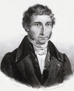
The galvanometer, primarily credited to Leopoldo Nobili in Florence, was further developed by William Thompson in England in 1858, enabling the reliable measurement of ongoing electric currents and their changes in intensity. However, it failed to detect instantaneous electrical events (Niedermeyer & Schomer, 2011). Nobili is pictured below.
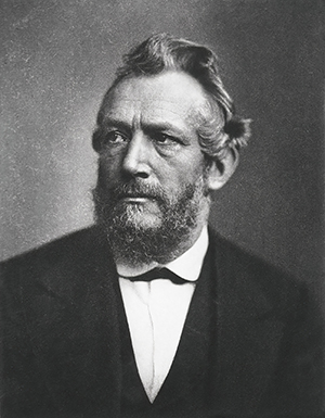
Du Bois-Reymond
Carlo Matteucci in Bologna and Emil Du Bois-Reymond in Berlin spearheaded the development of electrophysiology in studying the nervous system.
Du Bois-Reymond introduced the term negative variation to describe the unexpected drop in current intensity during muscle contraction, a term later used in early EEG research and the concept of contingent negative variation (Niedermeyer & Schomer, 2011).
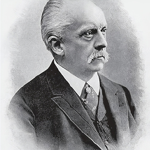
Hermann von Helmholtz
Hermann von Helmholtz accurately gauged nerve conduction speed, correcting previous overestimations. The improvement of nonpolarizable electrodes for physiological research is also attributed to Du Bois-Reymond. The idea of the "action current" was put forward by L. Hermann, providing insight into Du Bois-Reymond’s observed negative variations during muscle contraction.
Julius Bernstein put forth a membrane theory of nerve tissue, eventually expounded upon in the late 1930s and onward by A. L. Hodgkin and A. F. Huxley in England. These advancements set the stage for the first observation of EEG-like electrical brain activity (Niedermeyer & Schomer, 2011).
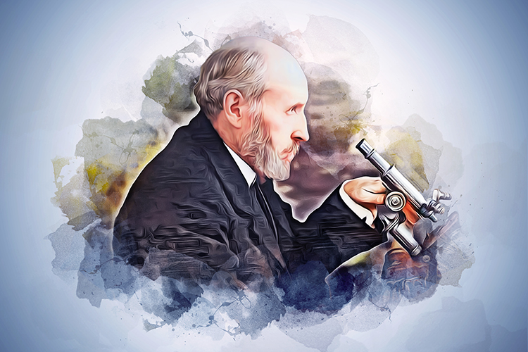
Richard Caton
Richard Caton (1842–1926), a Liverpool-based doctor, developed a strong interest in electrophysiology. The British Medical Association granted him funds to study the electrical activity of rabbit and monkey brains. His initial findings, presented on August 24, 1875, were published as a short report in the British Medical Journal, followed by a more comprehensive report in 1877 on experiments involving over 40 animals.

Caton's experimentation involved the use of a galvanometer, where a beam of light was projected on the galvanometer's mirror and then reflected onto a large wall scale. Through this process, he discovered that weak, varying electrical currents passed through the instrument when electrodes were positioned at two points on the brain's surface or when one was placed on the gray matter and the other on the skull's surface. Although no physical record was made, this discovery is often seen as the birth of the electrophysiologram. It's presumed that EEG phenomena triggered movement in the galvanometer needle. Despite possible interference from other sources, Caton is acknowledged for discovering the changeable potentials that form the EEG.
Caton observed other intriguing phenomena, such as the positive polarity of the gray matter's external surface compared to the deeper cerebral structures. He also noted a connection between the brain's electrical currents and its functions, observing a negative variation in the electric current when the gray matter was functionally active. Consequently, Caton's pioneering work also contributed to understanding evoked potential. However, making firm statements about his findings, such as discovering the "steady potential" or "DC potential" may be premature without supporting evidence. Caton's galvanometer only had a limited frequency response range from 0 to 6 Hz, as pointed out by Geddes (1987).
Caton's ground-breaking research earned him some renown, and he served as the physiology chair at the University College of Liverpool from 1884 to 1891. He later became the dean of the medical faculty and the Lord Mayor of Liverpool in 1907. Though his later career did not primarily focus on brain's electrical activity, his pioneering work in the field remains a significant milestone. More details about Caton's life and work can be found in Mary Brazier's 1961 account (Niedermeyer & Schomer, 2011).
G. Fritsch and Julius Eduard Hitzig
During the era of Caton's pioneering work in 1875 on electrical brain activity, Eastern European scientists also began independently studying the brain's electricity. However, a more transformative discovery came in the 1870s when G. Fritsch and Julius Eduard Hitzig found the human cerebral cortex could be electrically stimulated. This breakthrough was sparked by Fritsch noticing contralateral muscle contractions while tending to a brain wound during the Prussian–Danish War of 1864.
 |
 |
D. Ferrier and G. F. Yeo expanded on this in 1880 by conducting electrical stimulations of the cerebrum in apes and a patient with a brain tumor. These findings shook the scientific community, many of whom believed the cerebrum was a single, uniform organ hosting mental functions.
The discoveries around the cortex's response to electrical stimulation inspired further exploration of its spontaneous electrical activity, particularly in Eastern European laboratories. Despite significant ethnic and national differences, it's worth noting that most of 19th-century Poland was under Czarist Russian rule (Niedermeyer & Schomer, 2011).
Vasili Yakovlevich Danilevsky
Vasili Yakovlevich Danilevsky concluded his brain physiology thesis at 25, studying electrically stimulated and spontaneous brain activity in animals, thus following in Caton's footsteps. Although his aspirations of correlating spontaneous brain activity with psychic and emotional processes were unmet, he remained committed to brain physiology, publishing a comprehensive human physiology textbook in 1915 (Niedermeyer & Schomer, 2011).
Adolf Beck
Adolf Beck's work was also notable. He studied the spontaneous electrical brain activity in rabbits and dogs using nonpolarizable electrodes. Beck noticed that rhythmic oscillations ceased when the eyes were exposed to light, thus laying the groundwork for Berger’s discovery of alpha blocking. His work gained prominence due to its publication in the Centralblatt (Niedermeyer & Schomer, 2011).
Ernst Fleischl von Marxow
In 1883, Ernst Fleischl von Marxow, an Austrian physiologist, left a sealed letter at the Imperial Academy of Sciences, detailing his research on brain electrical activity across animal species. He did not record any rhythmic activity. Despite his assertion of priority over Beck's 1890 data, he overlooked earlier work by Caton and Danilevsky. His research was not top-notch, but his broad skillset, including linguistics, sports, and mountaineering, was noteworthy.
Back in Eastern Europe, innovative research was unfolding. Napoleon Cybulski, Beck's mentor and an esteemed physiology expert in Kraków, used a photographic galvanometer to graphically showcase experimental electroencephalography (EEG) studies, including EEG proof of an epileptic seizure in dogs triggered by electricity.
Two Russian physiologists, Pavel Kaufman and Vladimir Pravdich-Neminsky, continued this research. New technologies emerged, including the d’Arsonval galvanometer and the capillary electrometer. Significantly, Willem Einthoven introduced the string galvanometer in 1903, a sensitive device that revolutionized electrocardiography.
Kaufman theorized that epileptic seizures correlate with abnormal electrical discharges and explored cortical electrical stimulation. He later adopted the name Rostoutsev and primarily worked at the University of Baku during World War I (Niedermeyer & Schomer, 2011).
Vladimir Pravdich-Neminsky
Starting in 1912, Pravdich-Neminsky used the string galvanometer to record animals' brain electrical activity. His 1912 publications, which featured the first EEG images, predated Cybulski's. He recorded the EEG from the brain, dura, or intact dog skull, discovered a regular 12 to 14/sec rhythm, and noted significant slowing under asphyxia. He also introduced the term electrocerebrogram.
The remarkable advancements made by Eastern European neuroscientists in electrophysiologic neurophysiology during the 50 years before World War I are truly awe-inspiring. However, focusing solely on EEG history only scratches the surface. To fully appreciate their neuroscientific institutions, it's crucial to recognize pioneers in related electrophysiologic fields. Ivan Sechenov, considered the founder of this esteemed group of neurophysiologists, explored the electrical activity of the frog's spinal cord and oblongata, paving the way for Pavlovian thought. Nikolai Wedensky, his successor as chair and professor of physiology at St. Petersburg, is known for the Wedensky inhibition concept. Vladimir Larionov, also based in St. Petersburg, conducted excellent studies on the auditory cortex in dogs (Niedermeyer & Schomer, 2011).
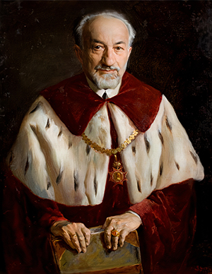
Vladimir Bechterev
The most distinguished Russian neuroscientist and clinical neurologist was Vladimir Bechterev. Holding the psychiatry chair in St. Petersburg, he combined clinical work with rigorous psychophysiological methods. Trained by prominent figures like Du Bois-Reymond, Flechsig, and Wundt and having worked at Charcot’s clinic in Paris, Bechterev made significant contributions to brain functional anatomy, experimental psychology, and clinical neurology. His "associative reflexology" work remains influential.
However, between Bechterev and Nobel laureate Ivan Pavlov, the Soviet regime chose Pavlov. His theory of conditioned reflexes overshadowed all Soviet neurophysiology, aligning closely with the ideology of dialectic materialism. Despite Pavlov's criticism of the regime, his concept dominated and stifled the growth of traditional neurophysiology. This ideopolitically-driven neuroscience approach led to a swift collapse in global leadership in EEG and related fields and a concerning decline in dogmatically governed neurophysiology.
While electroencephalographic (EEG) research thrived in Eastern Europe, it was dormant in Western and Central Europe, which is surprising given the overall good health of neurophysiology in these regions. The work of Ernst Fleischl von Marxow appears like an odd outcrop in this vast field (Niedermeyer & Schomer, 2011).
Network vs. Neuron Theories
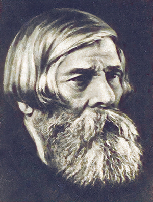
Western neurophysiologists closely followed the debate between network theories, supported by Joseph von Gerlach and Camillo Golgi, and neuron theories, championed by Santiago Ramón y Cajal (pictured below).
Despite resistance from the "reticularists," the neuron theory eventually triumphed. Similarly, proponents of cerebral localization clashed with those opposing it. Researchers such as Friedrich Goltz and H. Rothmann conducted experiments that targeted the entire brain rather than localized areas, contrasting the emerging interest in cortical localization inspired by the work of Fritsch, Hitzig, Ferrier, and Yeo.
During this period, Charles Scott Sherrington's work in Liverpool and Oxford profoundly influenced the development of modern Western reflexology. His groundbreaking book, "The Integrative Action of the Nervous System," covered a range of topics from reflexology to decerebrate rigidity, focusing largely on physical processes and sparingly addressing mental functions.
His concept of inhibition was a major contribution. Yet, this neurophysiology master had little connection to electrophysiology, focusing primarily on ablation techniques and largely ignoring EEG methods. Sherrington introduced the terms neuron and synapse. He contributed significantly to understanding muscle action, movement, proprioception, reflexes, and spinal nerves.
Sherrington proposed the "enchanted loom" metaphor for the human brain in a passage in the 1942 Man on His Nature, in which he poetically described the change in cortical activity as we awaken:
"The great topmost sheet of the mass, that where hardly a light had twinkled or moved, becomes now a sparkling field of rhythmic flashing points with trains of traveling sparks hurrying hither and thither. The brain is waking and with it the mind is returning. It is as if the Milky Way entered upon some cosmic dance. Swiftly the head mass becomes an enchanted loom where millions of flashing shuttles weave a dissolving pattern, always a meaningful pattern though never an abiding one; a shifting harmony of subpatterns." (p. 178)"
Current sleep research shows that the cortical networks are considerably more active during sleep than Sherrington imagined.
His students, including Edward Liddell and Derek Denny-Brown, held similar views. Meanwhile, in Cambridge, Edgar Douglas Adrian emerged as a strong proponent of electrically oriented neurophysiology, a topic we'll discuss later in relation to Hans Berger's work (Niedermeyer & Schomer, 2011).
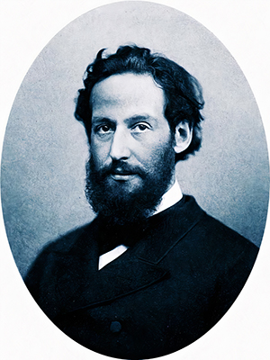
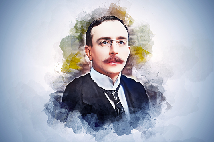
Hans Berger
Hans Berger (1873–1941), the pioneer of the human EEG, was a neuropsychiatrist, a combined specialty of neurology and psychiatry in Germany, Austria, and many other countries at that time.
These departments consisted of neurological and psychiatric wards, with training requiring rotation between the two. Pure neurology was just beginning to become its own discipline in German-speaking countries, thanks to the work of Wilhelm Erb, Max Nonne, and Otfrid Foerster, who lived during the same period as Berger.
Berger was not a leader in either neurology or psychiatry, and his name would have been lost if not for his pioneering EEG work. He was described as meticulous, conscientious, somewhat detached with patients, strict, authoritarian, and disinterested in faculty politics. He did his EEG work in a small, primitive lab, initially focused on cerebral circulation using plethysmographic methods in patients with skull defects. From 1902 to 1910, he studied the electrical activity of the dog's cerebrum using a capillary electrometer, with disappointing results. He started studying the human EEG in 1920.
While cumbersome in their original German, Berger's writings are critical for every electroencephalographer to understand, and this is made easier through Gloor’s English translation. Berger's work was titled, "On the Electroencephalogram of Man," which may not have helped its slow acceptance due to lack of appeal. His focus on linguistics is evident in his rejection of the term "electrocerebrogram" due to its mix of Greek and Latin elements, proposing instead the term "Elektrenkephalogram."
Berger's electrophysiological instrumentation evolved over time. He initially used a string galvanometer starting in 1910, later switching to the Siemens double-coil galvanometer in 1926, allowing for sensitivity up to 130 μV/cm. He recorded human EEG tracings with nonpolarizable pad electrodes, resulting in one to three minutes of data on photographic paper. His bipolar recording technique used fronto-occipital leads for his one-channel EEG tracings, along with a simultaneous ECG recording and time marker. He obtained an oscillograph in 1932 but could not acquire more amplifiers for multichannel recordings.
Berger began studying human EEG in 1924, primarily on patients with large skull defects, common after World War I in Germany. He initially believed these defects would aid in obtaining recordings, but later realized that good results could be obtained through an intact skull and scalp. Between 1926 and 1929, he successfully recorded alpha waves with a double-coil galvanometer. His initial findings were questioned, and he experienced doubts about his results.
Berger's first 1929 report highlighted the alpha rhythm and alpha blocking response, along with smaller beta waves, using various types of electrodes. His findings were only accepted after confirmation from Adrian in Cambridge in 1934. During the 1930s, Berger's research on human EEG provided valuable insights into various aspects of consciousness, sleep, the effect of hypoxia on the brain, various brain disorders, and early indications of epileptic discharges.
Despite these achievements, Berger's relationship with the Nazi regime was poor, leading to his forced retirement in 1938, which hindered his further EEG research. After a bout of illness, Berger developed severe depression, which remained undiagnosed, and committed suicide in 1941 at the age of 68. His insecurity may have been fueled by the competition from a group of EEG researchers at the Institute of Brain Research at Berlin-Buch.
Berger was complex, both as a person and as a researcher. He did not stand out clinically but was dedicated in his EEG work. His primary research motivation was to understand the nature of "mental energy", which he believed could transmit thoughts and emotions between individuals. Influenced by the Danish physiologist Alfred Lehmann, he considered this mental energy a product of metabolic energy, with EEG waves acting as messengers of mental activities.
Despite not having a formal scientific background, Berger made significant strides in EEG research, and his contribution is considered the greatest in the history of electroencephalography, despite his initial assumptions about EEG not being entirely correct (Niedermeyer & Schomer, 2011).
Berger's German Contemporaries
The Institute of Brain Research in Berlin-Bush was home to various eager neuroscientists under the directorship of Oskar Vogt, a renowned neuroanatomist and neuropathologist, until his dismissal by the Nazi government in 1936 due to his ties with a similar institute in Moscow and his refusal to expel Jewish co-workers. The institute had several departments, notably the Department of Physiology and the Department of Electrophysiology, led by M. H. Fischer and A. E. Kornmüller, respectively. They worked closely with physicist and electronic engineer J. F. Toennies, who developed the first ink-writing biological amplifier for recording brain potentials.
While in New York in 1932, Toennies designed the differential amplifier, which was crucial for EEG amplification, in parallel with Brian Matthews, Adrian's collaborator. Toennies' work provided the Berlin group with superior EEG research tools compared to Hans Berger's. Kornmüller saw the value of using more electrodes for recordings and focused on the differences between various cerebral regions. His early experimental work, particularly on epileptiform spikes, is considered more significant than his later clinical EEG studies.
Oskar Vogt (pictured below) conceived the cortex as divided into about 200 regions with clear demarcations, influencing Kornmüller's EEG work. Richard Jung joined the group in 1937 after Vogt's removal.
Vogt's replacement, Hugo Spatz, significantly altered the research goals. Toennies, in New York, constructed the first cathode follower for high-resistance electrode recording, marking the advent of microelectrode recording.
Post World War II, Kornmüller's work declined due to his fixation on an unproven theory about glia as the generator of slow brain potentials. Meanwhile, Jung became one of the leading electroneurophysiologists of his era (Niedermeyer & Schomer, 2011).
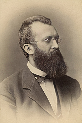
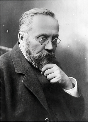
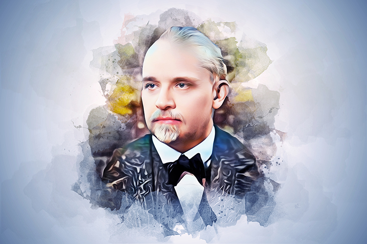
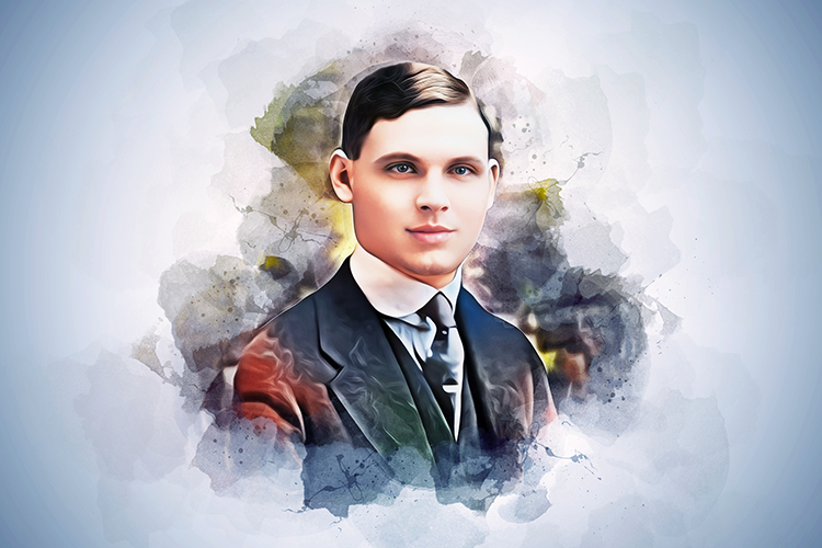
Edgar Douglas Adrian
Edgar Douglas Adrian (Baron of Cambridge, also known as Lord Adrian) (1889–1977), a preeminent electrophysiologic neurophysiologist of the 20th century, is closely linked to the discovery of the EEG, having confirmed Berger's observations. He received the 1932 Nobel Prize for Physiology or Medicine with Sir Charles Sherrington for their discoveries regarding neurons.

He demonstrated his own alpha rhythm and the blocking effect of eye opening to his peers despite his colleague Brian Matthews having a low-voltage EEG without an alpha rhythm. Interestingly, Adrian's recordings from the head ganglion of a water beetle mimicked his alpha rhythm and responded similarly to light, causing confusion among observers.
By the time Adrian corroborated Berger's findings, he was already an esteemed neurophysiologist known for showing single sensory nerve fiber potential and unit activity analysis, leading to the Adrian–Bronk law. Andrien and Matthew's demonstration of EEG recording at the 1935 Physiological Society meetings in England led to its widespread acceptance.
Adrian provided evidence supporting the all-or-none law: action potentials initiated at the axon hillock either occur or not. When they occur, they have the same amplitude and speed. He used himself as a subject and demonstrated the phenomenon of alpha blocking, where opening his eyes suppressed alpha rhythms.His co-worker, Detlef Bronk, later became president of Johns Hopkins University (Niedermeyer & Schomer, 2011).
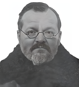
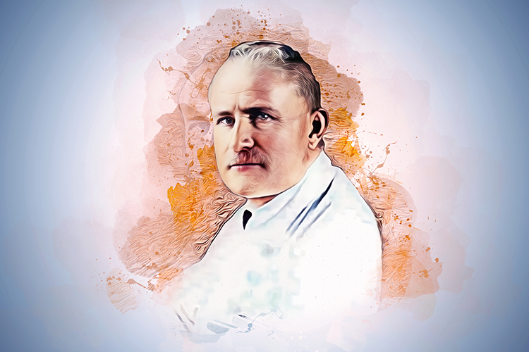
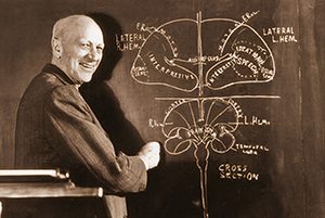
William Grey Walter
William Grey Walter, a basic scientist with a Ph.D., pioneered clinical electroencephalography in England. He discovered foci of slow activity (delta waves), sparking significant clinical interest in the EEG method. He improved Berger's electroencephalograph and pioneered EEG topography (Bladin, 2006). W. Grey Walter is pictured below.
However, Walter's academic background rather than clinical may have spurred an aversion to EEG among England’s neurologists, leaving the method mainly in the hands of lab-based Ph.D. electroencephalographers. Walter, an innovative thinker and persuasive writer, founded a highly effective school in Bristol at the Burden Institute (Niedermeyer & Schomer, 2011).
North American EEG Research
Around 1935, North America began to eclipse Europe as the focal point for the growing field of EEG research. Discoveries were emerging from the US, attracting European researchers. Hans Berger, a notable figure in this field, almost made a trip to the US in 1939, but his plans were interrupted by the outbreak of World War II.
Before Berger's involvement, the US didn't contribute significantly to the early development of EEG studies. A 1918 incident highlighted this lack of awareness. Donald McPherson, a Harvard Medical School student, identified rhythmic EEG activity in a cat's brain, but Alexander Forbes, a renowned physiologist, dismissed his findings as false.
The US's ascendancy in EEG research is often tied to the contributions of Hallowell Davis, Frederic A. Gibbs, Erna Gibbs from Harvard, and Herbert Jasper from Brown University. According to O'Leary and Goldring (1976), Hallowell Davis became aware of Berger's 1929 paper through his graduate student, A. J. Derbyshire. After initial unsuccessful attempts to replicate alpha rhythms, Davis was finally found to have a notable alpha rhythm. This marked the start of human EEG studies in the US in 1934, a precursor to the rapid increase of such research. Hallowell Davis is pictured below.
However, animal EEG experimentation in the US had begun earlier, spearheaded by researchers like Bartley and Newman in the early 1930s. Their studies, along with those by Davis and Saul, Travis and Dorsey, Travis and Herren, Bishop and Bartley, Bartley, and Gerard, laid the groundwork for future development. The work of Ralph W. Gerard in introducing a concentric needle electrode for animal brain experiments and his partnership with Franklin Offner, a pioneering electronic engineer, played a significant role in the evolution of EEG and related equipment.
The commencement of human EEG studies in the US can be traced back to Harvard, Brown University, and the University of Iowa. Key contributors included Hallowell and Pauline Davis, Frederic and Erna Gibbs, and William G. Lennox at Harvard; Herbert H. Jasper at Brown; and Lee Travis at Iowa, who established a significant school of study (Niedermeyer & Schomer, 2011).
The Gibbs–Gibbs–Lennox Period
In 1934, research on epileptic patients marked a pivotal point in clinical electroencephalography. Frederic Gibbs joined the Harvard team from Johns Hopkins University and partnered with established epileptologist William G. Lennox. Interestingly, Lennox had initially focused on cerebral circulation research.
Erna L. Gibbs, a German immigrant who began as Lennox's technical assistant, later became Frederic Gibbs's wife and a pioneering EEG technician and co-author. Their cerebral blood flow studies were milestones in the field, though EEG ultimately captivated Lennox more.
The first study of petit mal epilepsy by Gibbs and Davis in 1935, featuring twelve children, remains influential in EEG literature, primarily for its linking petit mal absences with 3/sec spike-wave complexes. This discovery eclipsed subsequent findings of grand mal and psychomotor seizure EEG patterns by the same team, even though it was later found that spike waves could occur without petit mal.
Despite their groundbreaking work, the EEG recordings produced were technically inferior. In 1935, Frederic Gibbs and his wife visited Hans Berger in Germany and studied Jan F. Toennies' "polyneurograph" instrument and Matthew's instrumentation in England. Gibbs then commissioned Albert Grass, from the Massachusetts Institute of Technology, to construct a three-channel preamplifier. By the end of 1935, the Grass Model I, featuring three channels and an ink writer, was in use.
The Gibbs–Gibbs–Lennox period in the 1930s arguably marked the most exhilarating phase in EEG history, especially regarding breakthroughs in understanding epileptic seizures. The advent of EEG partitioned epileptology into two historical periods, causing a monumental leap in understanding epilepsy. Building on Fischer's 1931 animal studies, the Gibbses and Lennox applied the knowledge to human epileptology, paving the way for future work (Niedermeyer & Schomer, 2011).
North American Sleep and Epilepsy Research
Other EEG pioneers in North America, like Hallowell and Pauline Davis, made significant contributions to understanding the normal EEG and its variants. They were among the first to investigate the human sleep EEG. In terms of sleep studies, A. L. Loomis, E. N. Harvey, and G. A. Hobart were the first to methodically research human sleep EEG patterns and stages. At Brown University, Herbert Jasper studied the EEG of children's behavior disorders, later focusing on epileptology at McGill University in collaboration with Wilder Penfield.
John R. Knott and Charles E. Henry, disciples of Lee Travis, emerged as critical figures in America's EEG work. D. Lindsley became a pioneer in researching maturational EEG aspects and directed top-tier neurophysiologic EEG research at UCLA.
Robert Schwab and Warren McCulloch also played significant roles, with McCulloch gaining recognition for his thinking that far exceeded the bounds of EEG and neurophysiology. Clinical EEG research began exploring beyond epileptology with Grey Walter’s discovery of the delta focus linked to brain tumors. By the end of the 1930s, North America had led in the field of EEG, while Europe had lagged behind.
During WWII (1939-1945), EEG research and clinical activities suffered, especially in Europe, though it was still used for localizing brain injuries and epileptogenic foci. Post-war, the gap between North America and Europe widened, with the latter's EEG research hitting a nadir.
Post-war revitalization began in England and France while Germany was struggling. W. Grey Walter, V. J. Dovey, and H. Shipton at the Burden Institute discovered the paroxysmal response to certain flickering light frequencies, a method later used by Henri Gastaut in France to establish an individual threshold for paroxysmal responses.
In 1947, the American EEG Society was founded, and the First International EEG Congress was held in London, with a follow-up in Paris in 1949. EEG activities in Germany were minimal during this time, but research efforts in Japan and Switzerland started to stand out.
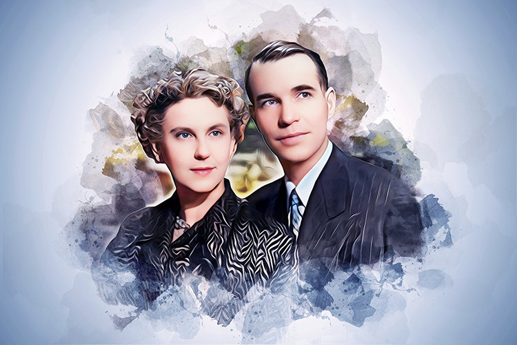
In the US, Frederic A. Gibbs, Erna Gibbs, and B. Fuster made significant strides in understanding temporal lobe epilepsy. Their findings necessitated the inclusion of sleep in EEG evaluations, causing American EEG labs to be more sophisticated than their European counterparts. Erna and Frederic Gibbs are pictured below.
Despite international acclaim, Frederic Gibbs held a modest academic rank at Harvard, reflecting a generally negative attitude towards EEG within neurologic departments. Herbert H. Jasper emerged as a key competitor to Gibbs at the Neurologic Institute of McGill University, working alongside Wilder Penfield (Niedermeyer & Schomer, 2011).
Neurophysiological Research: Thalamocortical Relationships
By the late 1940s, two new developments arose: invasive EEG techniques exploring deep brain regions and automatic frequency analysis. Also notable were neurophysiologists' studies on thalamocortical relationships, with researchers like Morison and Dempsey significantly impacting our understanding of cortical electrogenesis.
Horace W. Magoun's research on the brainstem reticular formation, carried out at Northwestern University and the University of California, had a transformative effect on neuroscience, highlighting the association between consciousness and reticular formation. However, his name is less recognized today.
The 1940s witnessed the influential role of EEG in neurophysiology, though by its end, the focus had shifted towards single neurons, and "macro-EEG" interest faded (Niedermeyer & Schomer, 2011).
The EEG Becomes Mainstream
The 1950s saw EEG becoming commonplace. Almost every university hospital had at least one EEG machine, and by the decade's end, they were in many hospitals and private practices. Specialized EEG labs began to appear, catering to children or adults. Despite clear correlations between central nervous system diseases and EEG results, many neurologists remained wary or antagonistic towards EEG. At the same time, neurosurgeons showed interest as long as EEG helped identify focal cerebral lesions.
Herbert Jasper and Wilder Penfield's work on epilepsy reached a new zenith in Montreal, establishing it as a hub for neurosurgical treatment of focal epilepsies.
 |
 |
Their collaboration led to the influential book "Epilepsy and the Functional Anatomy of the Human Brain." Controversy, however, surrounded the "centrencephalic" concept of primary generalized epilepsy, which was later proven shaky and largely dismissed.
Previously at Harvard, Frederic Gibbs moved to the University of Illinois School of Medicine, solidifying Chicago as a leader in neurologic sciences. Despite rivalries, the Chicago group under Gibbs and the Montreal group under Jasper and Gloor made significant advancements in epileptologic electroencephalography. A. Earl Walker, a master from the Chicago school, brought depth EEG, electrocorticography, epilepsy surgery, and a scientifically-oriented epileptology to Johns Hopkins in Baltimore.
In the 1950s, EEG became commonplace in hospitals and private practices. This led to the establishment of specialized EEG labs at university hospitals. While psychiatrists appreciated the neurophysiological insights EEG provided, many neurologists were hesitant or even resistant to it. However, neurosurgeons found EEG useful for identifying focal brain lesions.
During this period, Jasper and Penfield's work on epilepsy in Montreal set a new standard, culminating in their seminal book, "Epilepsy and the Functional Anatomy of the Human Brain." They also proposed the controversial "centrencephalic" concept of primary generalized epilepsy, which was heavily debated and eventually dismissed in the late 1960s.
Also in this period, Frederic Gibbs moved to the University of Illinois School of Medicine in Chicago, bolstering the city's status as a global leader in neurologic sciences. Chicago and Montreal became strong competitors in the field of epileptologic electroencephalography. A. Earl Walker, a key figure from Chicago, introduced depth EEG, electrocorticography, and epilepsy surgery to Johns Hopkins in Baltimore, solidifying his legacy in neurosurgery and epileptology.
Computational wave analysis techniques in EEG began with Hans Berger's early attempts in 1932, aided by physicist Dietsch's application of Fourier analysis. Grass, Gibbs, and Knott made further advancements. By the 1950s, automatic frequency analyzers emerged but were largely underused.
In the same decade, EEG technology advanced with the introduction of the microelectrode technique, allowing for the recording of single neurons. The invention of the cathode follower by Toennies made it technically possible to record from single cells. Extracellular microelectrode recording became more prevalent, and a decade later, intracellular microelectrode technology allowed for deeper biochemical insights.
The 1950s also saw significant progress in sleep research. Notably, Nathaniel Kleitman and his team at the University of Chicago were pioneers in studying sleep organization and rapid eye movement (REM) sleep.
However, as sleep research became more reliant on polygraphic recording, it gradually diverged from EEG research, creating a widening gap between the two fields from the 1960s onwards (Niedermeyer & Schomer, 2011).
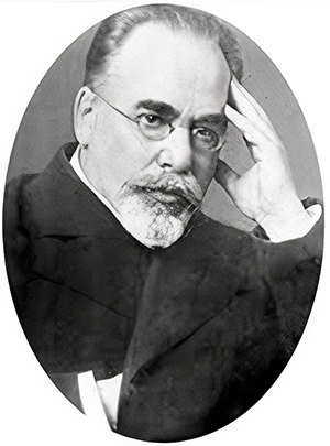
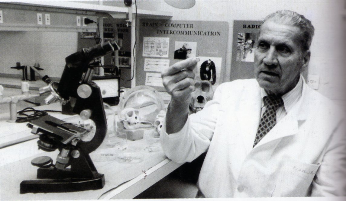
José Manuel Rodriguez Delgado's contributions to the field of electroencephalography (EEG) were significant and pioneering, albeit controversial. Delgado, a Spanish physiologist, was among the first to perform electrical brain stimulation in both animals and humans. His work in the 1960s and 1970s was groundbreaking, focusing on the electrical stimulation of specific brain areas to understand and potentially treat mental illnesses and epilepsy.
Some of his notable experiments include evoking complex behaviors in primates through brain stimulation, investigating aggressive behavior in animals, and inducing emotional responses in humans. Delgado's work with the "stimoceiver," a device he invented, allowed for two-way communication with the brain and laid the groundwork for future neuromodulation techniques.
Despite facing severe criticism and controversy, especially concerning the ethical implications and the precision of electrical stimulation effects, Delgado's contributions are acknowledged as paving the way for new modulation techniques like deep brain stimulation (Lorusso et al., 2022).
Although Delgado's work was influential in neurophysiology and the development of EEG techniques, he did not directly contribute to the fundamental development of these technologies. Instead, his work utilized these technologies to explore and manipulate brain functions in novel ways.
Samuel Sutton's work in discovering and understanding the P300 component of the event-related potential (ERP) is a major contribution to cognitive psychophysiology. Sutton and colleagues first reported the P300 slow cortical potential in 1965. They discovered the P300 in a "guessing paradigm," where participants were uncertain about the nature of the test stimulus. They observed that a large positive peak, later known as the P300 wave, occurred at about 300 ms after stimulus onset (Bashore & Molen, 1991).
The P300 (P3) wave is an event-related potential (ERP) component elicited in decision-making. It is also known as an “expectancy wave,” as it defines a degree of uncertainty in the decision-making process.
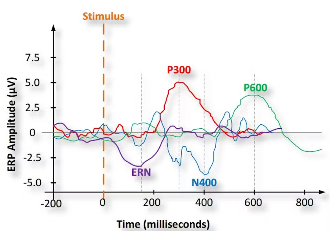
Caption: Graphic by Tamara Bonaci, CC BY-ND. Retrieved from theconversation.com.
This discovery was significant because it was one of the first demonstrations that ERPs could reflect cognitive or psychological processes, such as the subject's uncertainty or the task relevance of stimuli, rather than merely the sensory attributes of the stimuli. This revelation has profoundly impacted the field, providing a noninvasive means for studying physiological correlates of cognitive processing in humans.
Sutton's work on the P300 component has influenced a broad range of areas, including basic behavioral research, psychophysiological research, and experimental psychopathology. The P300 wave has been associated with cognitive information processing, including memory, attention, and executive function. The significance of the P300 lies in its ability to provide insights into the functional neuroanatomy of cognitive processes and its potential as a diagnostic tool in various neurological and psychiatric conditions (Bruder, 1992).
Reduced P300 amplitude is an indicator of the broad neurobiological vulnerability that underlies disorders such as alcohol dependence, drug dependence, nicotine dependence, conduct disorder, and adult antisocial behavior.
For example, P300 amplitude is reduced in patients diagnosed with alcohol use disorder (AUD) and their alcohol-naive children.
The P300 wave's discovery has also been pivotal in brain-computer interface (BCI) technology, particularly in developing P300-based BCIs for assisting individuals with motor impairments.
The Development of Fast Fourier Transforms
The peak of clinical and experimental EEG work was around 1960, but the 1960s saw a shift towards automated data analysis and computerization. James Cooley and John Tukey introduced fast Fourier transforms, foundational for power spectral analysis.
The Fast Fourier Transform (FFT) is a highly efficient algorithm for computing the Discrete Fourier Transform (DFT) and its inverse. This algorithm is widely recognized as a major breakthrough in digital signal processing, and its development is most commonly attributed to Cooley and Tukey, who published a landmark paper on the topic in 1965.
Before the development of the FFT, the calculation of the DFT was computationally intensive. This made the application of Fourier analysis unfeasible for large datasets. The FFT algorithm significantly reduced the computational burden, making it practical for larger datasets.
Cooley and Tukey's FFT algorithm leverages the symmetries and periodicities inherent in the computation of the DFT to reduce the number of necessary computations. It breaks down a DFT of any composite size N = N1*N2 into many smaller DFTs of sizes N1 and N2, recursively, to significantly reduce the number of multiplications and additions. This "divide and conquer" technique provides a dramatic speed-up for large datasets.
It is worth noting that while Cooley and Tukey are most commonly associated with the FFT, the basic principles behind the algorithm have been independently discovered and rediscovered many times throughout history. However, Cooley and Tukey's publication made the technique widely known and led to its broad adoption in the scientific and engineering communities.
The development of the FFT has had far-reaching impacts in numerous fields, including engineering, physics, mathematics, and computer science. It is extensively used in signal processing, image analysis, solving partial differential equations, and even in data compression.
Predictions of full automation in EEG interpretation proved false as EEG complexity required expert human interpretation. Despite this, computerized frequency analysis benefited psychophysiological research and neuropharmacological assessments.
The 1970s witnessed advancements in evoked potential techniques, enhancing their reliability and utility. However, during this period, the once close relationship between EEG and epileptology declined. This trend was later reversed in the 1980s due to a resurgence of interest in EEG for presurgical evaluations of seizure surgery candidates.
The advent of neuroimaging technologies like CT and MRI in the 1970s and 1980s seemed to overshadow EEG, but EEG remained crucial for identifying functional changes around CNS lesions. There's a concern that the focus on structural diagnosis may overlook functional understanding, a trend that needs to be halted (Niedermeyer & Schomer, 2011).
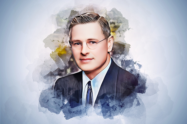
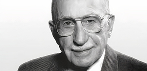
Frank Duffy's Contribution to Computerized Brain Mapping
Computerized brain mapping, associated primarily with Frank Duffy, revitalized topical EEG diagnosis. Simultaneously, from the late 1960s onwards, novel strides were made in newborn EEG exploration.
Frank H. Duffy is a prominent neurologist and a pioneer in neurophysiology, particularly noted for his contributions to pediatric neurology and computerized brain mapping.
In the 1970s and 1980s, Duffy developed techniques for computerized analysis of electroencephalography (EEG) data, leading to the emergence of the qEEG as a valuable tool in clinical and research settings. The qEEG involves using computer algorithms to analyze the electrical patterns of the brain, transforming raw EEG data into color-coded maps that provide a visual interpretation of brain activity. These brain maps can aid in the diagnosis of neurological conditions, the monitoring of disease progression, and the evaluation of treatment efficacy.
Duffy's work has been instrumental in advancing the understanding of various neurological and developmental disorders, including epilepsy, attention deficit hyperactivity disorder (ADHD), and autism (Duffy & Eksioglu, 2003). His research has shown that the qEEG can reveal distinct patterns of brain activity associated with these conditions, which may not be evident in traditional EEG readings.
Additionally, Duffy and his colleagues were among the first to use the qEEG to study brain development in infants and children (Duffy et al., 2003). This pioneering work has provided critical insights into the dynamic changes in brain function that occur throughout early development and the effects of developmental disorders on this process.
In summary, Frank Duffy's work in computerized brain mapping and quantitative EEG has significantly advanced the field of neurophysiology, particularly in pediatric neurology. His contributions have enhanced our understanding of brain function in health and disease and continue to influence current research and clinical practice.
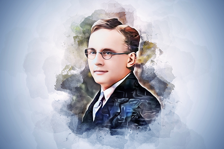
E. Roy John
E. Roy John was a leading pioneer in the field of quantitative electroencephalography. His work spanned many decades and covered a wide range of topics, including the development of novel analytical techniques, the use of the qEEG in clinical and research applications, and the application of the qEEG in neuropsychiatric disorders (John et al., 1980; John & Prichep, 2006).
John was one of the earliest advocates for using computers in EEG analysis. His work in the 1960s and 1970s led to the development of some of the first computer systems used for automated EEG analysis, a fundamental shift in how EEG data was analyzed and interpreted.
John also developed the concept of neurometrics, which involves using qEEG measures to quantify deviations from normal brain function (John et al., 1988). Neurometrics is based on the idea that brain dysfunction can be quantified in terms of deviations from normative data in qEEG measures. This concept has had a significant impact on the clinical use of QEEG, including its use in diagnosing and treating neuropsychiatric disorders (John et al., 2007).
John's work has led to the use of the qEEG in a variety of clinical applications. For example, he and his colleagues have shown that the qEEG can predict a coma's outcome, helping clinicians make more informed decisions about treatment and prognosis.
John has also been instrumental in applying the qEEG to the study of various neuropsychiatric disorders, including ADHD, schizophrenia, and Alzheimer's disease. His research has shown that QEEG can reveal abnormalities in brain function that are not evident from standard clinical assessments.
Overall, E. Roy John's contributions to the qEEG have been instrumental in its development and widespread use in clinical and research settings. His work has transformed how we understand and assess brain function and dysfunction.
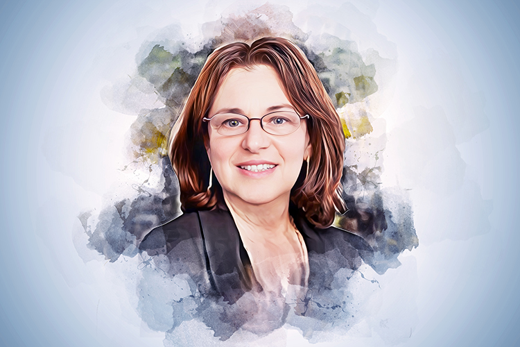
Leslie S. Prichep
Leslie S. Prichep is a renowned figure in the qEEG field and has made considerable contributions, particularly concerning clinical applications. Prichep, in collaboration with E. Roy John, was instrumental in developing neurometrics, a quantitative approach to understanding neurological and psychiatric disorders. This approach uses normative databases to identify deviations in QEEG measures, which can aid in diagnosing a range of disorders (Prichep, 2005).
Prichep has played a major role in demonstrating the qEEG's clinical utility. She has conducted extensive research using the qEEG in various contexts, including assessing brain function in psychiatric disorders, predicting treatment outcomes, and identifying biomarkers for various neurological conditions (Prichep et al., 2006).
Prichep's research has been fundamental in identifying qEEG-based biomarkers for a range of neuropsychiatric disorders. For example, she has identified qEEG patterns that can assist in the diagnosis of conditions such as ADHD and depression.
She has also worked extensively on the use of the qEEG to assess traumatic brain injury, demonstrating that the qEEG can reveal brain abnormalities even when conventional imaging methods such as MRI or CT are normal (Prichep et al., 2013).
Prichep's work has greatly expanded our understanding of how the qEEG can be used to assess brain function and dysfunction. Her research has demonstrated the clinical utility of the qEEG and its potential for improving the diagnosis and treatment of various neuropsychiatric conditions.
Roberto Pascual-Marquis
Roberto Pascual-Marquis has made significant contributions to the field of qEEG, particularly in developing and applying techniques to analyze brain electrical activity. His research has helped enhance our understanding of how the brain functions in various physiological and pathological states.
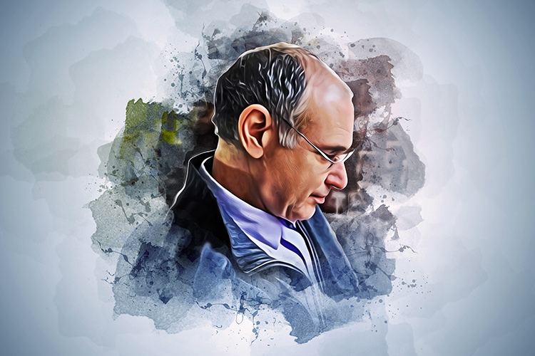
Pascual-Marquis is perhaps best known for developing LORETA, a method used for localizing sources of brain activity based on EEG recordings (Pascual-Marquis et al., 1994). This technique uses scalp-recorded electrical activities to generate a 3D image of electrical activity within the brain. It is considered a significant advancement in EEG analysis because it helps identify the source of electrical activity with a high degree of precision.
Later, Pascual-Marquis developed a standardized version of LORETA, known as sLORETA, which improved the resolution of the original algorithm and removed the localization bias present in LORETA (Pascual-Marquis, 2002).
Pascual-Marquis also contributed to developing methods to assess functional and effective connectivity using qEEG data (Pascual-Marquis et al., 2011). His work included the use of linear and non-linear methods to understand how different brain regions interact, which is fundamental to understanding many neurological and psychiatric conditions.
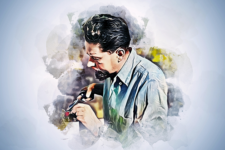
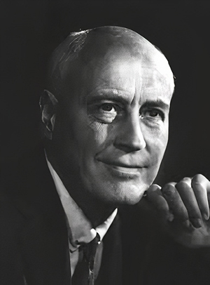
Hershel Toomim
Hershel Toomim made significant contributions to biofeedback, particularly in the development of hemoencephalography (HEG).
His work focused on noninvasive monitoring of brain functioning using light in the wavelength region of 650 to 1000 nanometers, which can penetrate human tissue, including bone. In 1994, Toomim discovered that it was possible to measure and teach individuals to control the amount of oxygenated blood flowing in the prefrontal regions of the brain using an optical device. This method, termed hemoencephalography or HEG, represents a form of neurofeedback based on blood flow biofeedback.
HEG, as developed by Toomim, allows for the training of brain functions by providing feedback on cortical circulatory changes. This technique uses a biofeedback device that emits red light into the skull and detects changes in the returning refracted light, representing variations in cortical circulation. Toomim's discovery and subsequent research have been particularly influential in treating conditions like attention deficit hyperactivity disorder (ADHD) and other cognitive deficits, showing promise in improving attention, executive control, and other cognitive functions through neurofeedback training.
Toomim also developed the first standardized and calibrated systems spanning the measures of electromyography (EMG), temperature, galvanic skin response (GSR), and EEG.
J. Peter Rosenfeld and Elsa Baehr
J. Peter Rosenfeld and Elsa Baehr have made significant contributions to the field of neurofeedback and alpha asymmetry protocols, particularly in the context of treating mood disorders such as depression.
Using operant conditioning programs, Rosenfeld and colleagues designed experiments demonstrating that cortical asymmetry (A-score) could be modified in subjects. This work laid the foundation for neurofeedback protocols to treat mood disorders (Hammond & Baehr, 2009).

Baehr, Rosenfeld, and colleagues conducted studies where depressed patients were trained to change their frontal alpha asymmetry to resemble non-depressed persons. This clinical application showed that alpha asymmetry neurofeedback training could be an effective adjunct to psychotherapy in treating certain mood disorders (Baehr et al., 1997).
Their research also includes follow-up studies assessing the long-term effectiveness of alpha asymmetry neurofeedback in treating mood disorders. These studies reported that patients who underwent neurofeedback maintained normal scores over time and showed reduced depression symptoms (Baehr, Rosenfeld, & Baehr, 2001).
Further extending their work, Baehr and colleagues explored the use of alpha asymmetry protocols in conditions like premenstrual dysphoric disorder (PMDD), showing the versatility of this approach in different clinical scenarios (Baehr et al., 2004).
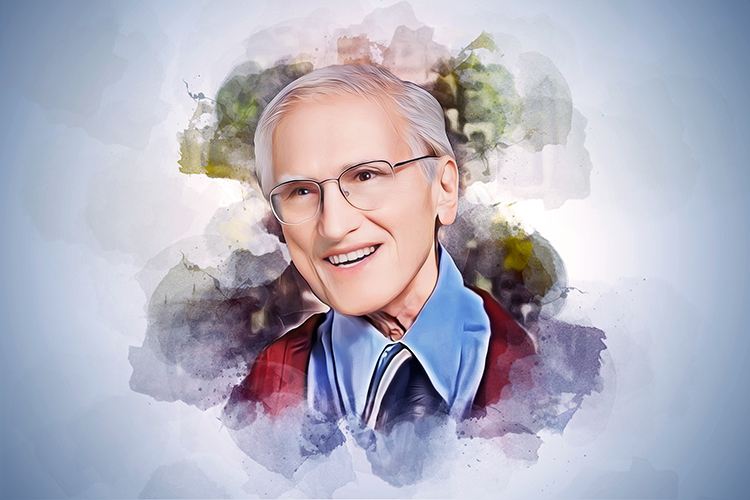
Joel F. Lubar
Joel F. Lubar was best known for his pioneering work in using neurofeedback to treat attention deficit hyperactivity disorder (ADHD; Lubar, 1989; 1991; Lubar & Shouse, 1977).
He developed a treatment protocol that uses the qEEG to identify abnormal brainwave patterns and then trains individuals to modify these patterns through neurofeedback.
Lubar documented the importance of theta-to-beta ratios (the ratio of power in the theta and beta bands) in ADHD and developed theta suppression-beta enhancement protocols to decrease these ratios and improve student performance. His research has demonstrated the effectiveness of this approach in reducing symptoms of ADHD (Lubar, 2020).
Lubar contributed significantly to developing and refining qEEG techniques used in neurofeedback (Lubar, 1997). His work helped establish the scientific foundation for using neurofeedback in treating various neurological and psychiatric conditions (Monastra, Lubar, & Linden, 2001).
Jay Gunkelman
Jay Gunkelman is known for his deep EEG knowledge and ability to distill complex concepts into understandable terms. He has spent considerable time educating others about the qEEG, giving lectures, conducting workshops, and mentoring new professionals. Jay has contributed to the understanding and application of the qEEG in clinical and research settings.
Gunkelman has vast experience in interpreting qEEG patterns and has shared his insights and approaches to analysis on various platforms. His pragmatic approach has assisted countless clinicians and researchers in interpreting qEEG data and using it effectively (Gunkelman, 2020).
Gunkelman has applied his expertise to neurofeedback. He has played a critical role in demonstrating the application of the qEEG in this field, which has contributed to the understanding and effective use of neurofeedback techniques.
Robert Thatcher
Robert Thatcher is a key figure in the qEEG, with many significant contributions to its development and its practical applications.
Thatcher has been instrumental in creating normative databases for the qEEG, which provide reference standards for different age groups and conditions. These databases are used to compare individual patient data with normative values, which can assist in diagnosing a wide variety of neurological and psychiatric disorders (Thatcher et al., 2014).
Thatcher developed the NeuroGuide software, one of the most widely used tools for qEEG analysis. NeuroGuide incorporates Thatcher's normative database and provides user-friendly tools for qEEG analysis, including various measures of brain connectivity and functionality (Thatcher, 2010).
Thatcher has conducted extensive research on the application of the qEEG in assessing traumatic brain injury. His work has shown that the qEEG can reveal subtle abnormalities that are not visible in conventional imaging studies, which can contribute to diagnosing and treating TBI (Thatcher et al., 1989).
Thatcher has also contributed to understanding brain connectivity (Thatcher, North, & Biver, 2005). His work has shown that the qEEG can be used to assess the functional connections between different brain regions, which can aid in understanding various brain disorders.
Robert Thatcher's work has significantly advanced our understanding of the qEEG and its clinical applications. His normative databases and analytical software development have played a critical role in enabling clinicians and researchers to apply the qEEG in a practical setting. His work on traumatic brain injury and brain connectivity has opened up new areas of research and has contributed to improved patient care.
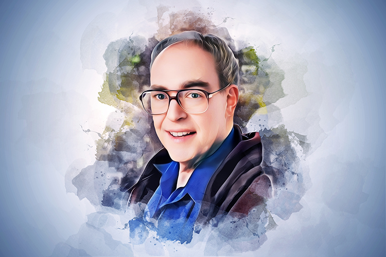
Thomas F. Collura
Collura has significantly contributed to developing neurofeedback technologies, particularly in creating accessible and practical systems for the qEEG and neurofeedback. He founded BrainMaster Technologies, a company that produces hardware and software for neurofeedback, biofeedback, and qEEG analysis.
Collura has been influential in advancing z-score neurofeedback, a form of neurofeedback training that involves comparing a patient's brainwave patterns to a normative database and providing feedback based on the statistical difference, or z-score. His work has supported the practical application of this approach in a clinical setting (Collura, 2008; Collura, 2020; Collura et al., 2010).
Collura has contributed significantly to the field as an educator and mentor (Collura, 2013). He has delivered numerous workshops and lectures on the qEEG and neurofeedback and has also written extensively on these subjects. This has helped promote understanding and use of these methods among clinicians and researchers.
In summary, Collura's contributions have substantially impacted the field of qEEG and neurofeedback, both through his practical work in developing technologies and his theoretical work in promoting understanding of these techniques. His work in z-score neurofeedback, in particular, has greatly expanded the potential applications of neurofeedback in treating a wide range of conditions.
Juri Kropotov
Juri Kropotov has been instrumental in the development of qEEG normative databases. His work in this area has provided important reference data that can be used to compare individual patient data, contributing to the diagnosis and assessment of various neurological and psychiatric conditions (Kropotov, 2020).

Kropotov has significantly contributed to our understanding of ERPs (Kropotov et al., 2005). His work has demonstrated how ERP components can be used to gain insight into various cognitive processes and how deviations from normal ERP patterns can indicate the presence of certain disorders.
Kropotov has also conducted extensive research on the application of the qEEG in psychiatry, demonstrating its utility in understanding and diagnosing a wide range of psychiatric conditions, including ADHD, schizophrenia, and post-traumatic stress disorder.
Kropotov has significantly contributed to neurofeedback, demonstrating the efficacy of qEEG-guided neurofeedback in treating various conditions. His work has helped establish the scientific basis for using neurofeedback in clinical practice.
Overall, Kropotov's work has greatly advanced our understanding of the qEEG and its applications in clinical practice and research. His work in developing normative databases, understanding ERPs, applying the qEEG in psychiatry, and advancing neurofeedback has significantly impacted the field.
Glossary
absence seizure: a seizure usually lasting less than 15 s in which a client blanks out; also called petit mal seizures.
Adrian–Bronk law: the law that states the strength of a nerve impulse is related to the size of the nerve cell.
all-or-none law: the principle that the strength by which a nerve or muscle fiber responds to a stimulus is not dependent on the strength of the stimulus. If the stimulus is any strength above threshold, the nerve or muscle fiber will either give a complete response or no response at all.
alpha blocking: the replacement of the alpha rhythm by low-amplitude desynchronized beta activity during movement, attention, mental effort like complex problem-solving, and visual processing.
alpha asymmetry protocols: neurofeedback techniques used in the treatment of mood disorders, based on the principle that frontal alpha brainwave asymmetry (differences in alpha wave activity between the left and right frontal lobes) correlates with emotional states. The protocols aim to modify this asymmetry to resemble patterns observed in non-depressed individuals, thereby potentially alleviating symptoms of depression and anxiety.
alpha rhythm: the first EEG rhythm discovered by Berger that ranges from 8 to 12 Hz which appears in three-quarters of adults when they are calm, awake, and not actively processing information.
alpha-theta training: training to progressively slow the EEG, increasing alpha and then theta abundance, developed by Brown and the Menninger Foundation and then adopted by Peniston and Kulkovsky in treating alcoholics.
beta rhythm: the second EEG rhythm discovered by Berger, 12-16 Hz.
brain connectivity: the patterns of links, or connections, between different areas of the brain. This can be measured structurally (anatomical connectivity) or functionally (functional connectivity).
capillary electrometer: a device used for the detection and measurement of small electric currents using the movement of a liquid in a thin tube.
complex partial seizure: a seizure in which awareness is impaired and repetitive behaviors called automatisms (e.g., lip smacking) may occur.
computerized brain mapping: a technology used to create images of brain activity or other measures, often used in research or clinical settings.
concentric needle electrode: an electrode used in electroencephalography (EEG) and electromyography (EMG) for detecting electrical activity.
contingent negative variation (CNV): a steady, negative shift in potential (15 microvolts in young adults) detected at the vertex discovered by Walter. This slow cortical potential may reflect expectancy, motivation, intention to act, or attention. The CNV appears 200-400 ms after a warning signal (S1), peaks within 400-900 ms, and sharply declines after a second stimulus that requires the performance of a response (S2).
cortical gradient: the difference in electrical charge between cortical neurons and scalp electrodes. When cortical networks are activated, their neurons are positively charged, and scalp electrodes are negatively charged.
d’Arsonval galvanometer: a type of galvanometer that uses a moving coil of wire in a fixed magnetic field to measure electric current.
delta focus in epilepsy: a brain region where abnormal delta wave (slow wave) activity is seen, often associated with seizure disorders.
delta rhythm: an EEG rhythm named by Walter that ranges from 0.5 to 3.5 Hz and is increased in adult Stage 3 sleep, brain injury, brain tumor, and developmental disability.
EEG topography: the display that maps EEG activity across the brain surface in color.
event-related potentials (ERPs): responses that are directly related to a specific sensory, cognitive, or motor event.
evoked potentials (EPs): electrical signals generated by the nervous system in response to stimuli, which are measured via electrodes and analyzed to assess sensory and motor pathways.
Fast Fourier Transform (FFT): an algorithm to compute the discrete Fourier transform and its inverse. In the context of neuroscience, it is often used to analyze the frequencies present within EEG signals.
galvanometer: a device for measuring current.
generalized seizures: seizures characterized by a peculiar cry, loss of consciousness, falling, tonic-clonic convulsions of all extremities, incontinence, and amnesia for the episode. These were previously called grand mal seizures.
Golgi's network theory: an early theory of brain structure, proposed by Camillo Golgi, that suggested the brain was a single, interconnected network.
hemoencephalography (HEG): a neurofeedback technique that involves monitoring and providing feedback on the brain's blood flow or oxygenation levels, typically using near-infrared (NIR) technology.
independent component analysis (ICA): a computational method for separating a multivariate signal into additive subcomponents. In the context of neuroscience, it can be used to separate out different sources of signal within EEG data.
interictal activity: EEG potentials between seizures.
live z-score neurofeedback: a type of neurofeedback where real-time EEG data is statistically compared to a normative database, and feedback is provided based on how much the individual's data deviates from the norm.
LORETA (Low-Resolution Electromagnetic Tomography): a technique used in neuroscience to localize the source of electrical activity within the brain, such as the source of an EEG signal.
machine learning: a type of artificial intelligence (AI) that allows computers to learn from and make decisions or predictions based on data.
neurometrics: the quantitative measurement of brain function, often using techniques such as EEG.
neuron: an excitable nervous system cell that processes and distributes information, chemically and electrically, and usually contains a soma (cell body), dendrites, and axon.
normative database: means and standard deviations for EEG variables such as amplitude, power, coherence, and phase that are calculated for single hertz bins, frequency bands, or band ratios based on the EEG data collected from healthy normal subjects who are grouped by age, eyes open or eyes closed conditions, and sometimes gender and task, which also allows for the specification of z-scores with a mean of 0 and standard deviation of 1 for the various combinations of EEG variables, frequency ranges, subject ages, eyes-open or eyes-closed conditions and other variables.
P300 (P3) wave: a component of the event-related potential (ERP) measured in electroencephalography (EEG). It is characterized by a positive deflection in voltage peaking around 300 milliseconds after the presentation of a stimulus, particularly when the stimulus is unexpected or significant to the task at hand. The P300 wave is widely used in cognitive neuroscience and clinical neurology for studying attention, information processing, and for detecting cognitive impairment and mental disorders.
photographic galvanometer: a type of galvanometer that uses a beam of light to produce a photographic record of electric current.
quantitative electroencephalography (qEEG): a method of analyzing the electrical activity of the brain to provide quantitative measures, often including comparison to normative databases.
quantitative electrophysiology: the use of quantitative methods to analyze and measure electrical activity within the nervous system.
Ramón y Cajal’s neuron theory: the theory that the nervous system is made up of discrete individual cells, a foundational concept in neuroscience.
rapid eye movement (REM) sleep: a sleep cycle stage characterized by random/saccadic rapid movements of the eyes, low muscle tone throughout the body, and the propensity of the sleeper to dream vividly. This stage comprises about 20-25% of total sleep in adults. It exhibits a mixed frequency pattern on EEG, similar to wakefulness, hence also termed as paradoxical sleep.
standardized low-resolution brain electromagnetic tomography (sLORETA): a method used to identify sources of brain electrical activity using an inverse solution.
sensorimotor rhythm (SMR): an EEG rhythm that ranges from 12-15 Hz and appears when you inhibit movement and relax your muscles.
sleep spindles: short bursts of 12-15 Hz activity during stage 2 sleep.
slow cortical potentials (SCPs): gradual changes in the membrane potentials of cortical dendrites that last from 300 ms to several seconds. These potentials include the contingent negative variation (CNV), readiness potential, movement-related potentials (MRPs), and P300 and N400 potentials. SCPs modulate the firing rate of cortical pyramidal neurons by exciting or inhibiting their apical dendrites. They group the classical EEG rhythms using these synchronizing mechanisms.
spike: a sharp EEG transient with a duration of less than 70 ms.
string galvanometer: a type of galvanometer that uses a very fine string of conductive wire to measure current. It was used in early electrocardiography and electroencephalography.
synapse: specialized chemical and electrical junctions across which neurons communicate with each other and non-neural cells.
thalamus: a forebrain structure above the hypothalamus that receives, filters, and distributes most sensory information. The thalamus contains neurons that can block or relay ascending sensory information. When these thalamic neurons rhythmically fire, this blocks the transmission of information to the cortex. When they depolarize in response to sensory information, this integrates and transmits this information to the cortex. Inputs to the thalamus determine whether these neurons block or relay sensory information.
theta rhythm: the EEG rhythm discovered by Walter that ranges from 4 to 7 Hz and is associated with drowsiness, the transition from wakefulness to sleep, rapid eye movement (REM) sleep, and the processing of information.
theta/beta training: a protocol that decreases theta amplitude and increases beta amplitude.
theta-to-beta ratio: the ratio of power in the theta and beta bands.
topographic mapping: the creation of a visual representation of the distribution of brain activity on the surface of the head, often using color to represent different levels of activity.
toposcope: a device for locating the direction and distance of a source of sound, it has also been used to describe tools used for localizing activity in the brain.
wavelet transformation: a mathematical function useful in signal processing for analyzing different frequency components with different resolutions. In neuroscience, it can be used for time-frequency analysis of brain signals.
z-score training: a strategy that attempts to normalize brain function with respect to mean values in a clinical database. EEG amplitudes that are 2 or more standard deviations above or below the database means are down-trained or uptrained to treat symptoms and improve performance.
TEST YOURSELF ON CLASSMARKER
Click on the ClassMarker logo below to take a 10-question exam over this entire unit.

REVIEW FLASHCARDS ON QUIZLET
Click on the Quizlet logo to review our chapter flashcards.

Visit the BioSource Software Website
BioSource Software offers Physiological Psychology, which satisfies BCIA's Physiological Psychology requirement, and Neurofeedback100, which provides extensive multiple-choice testing over the Biofeedback Blueprint.

Assignment
From your perspective, which EEG researcher contributed most to qEEG development? Why?
References
Baehr, E., Miller, L., Rosenfeld, J., & Baehr, R. (2004). Changes in frontal brain asymmetry associated with Premenstrual Dysphoric Disorder: A single case study. Journal of Neurotherapy, 8, 29-42. https://doi.org/10.1300/J184V08N01_03.
Baehr, E., Rosenfeld, J., & Baehr, R. (1997). The clinical use of an alpha asymmetry protocol in the neurofeedback treatment of depression: Two case studies. Journal of Neurotherapy, 2, 10-23. https://doi.org/10.1300/J184V02N03_02.
Baehr, E., Rosenfeld, J., & Baehr, R. (2001). Clinical use of an alpha asymmetry neurofeedback protocol in the treatment of mood disorders: Follow-up study one to five years post therapy. Journal of Neurotherapy, 4, 11-18. https://doi.org/10.1300/J184V04N04_03.
Bashore, T., & Molen, M. (1991). Discovery of the P300: A tribute. Biological Psychology, 32, 155-171. https://doi.org/10.1016/0301-0511(91)90007-4.
Berger, H. (1929). Über das elektrenkephalogramm des menschen. Archiv für Psychiatrie und Nervenkrankheiten, 87(1), 527-570.
Bladin, P. F. (2006). W. Grey Walter, pioneer in the electroencephalogram, robotics, cybernetics, artificial intelligence. Journal of Clinical Neuroscience, 13(2), 170-177. https://doi.org/10.1016/j.jocn.2005.04.010
Bruder, G. (1992). P300 Findings for Depressive and Anxiety Disorders. Annals of the New York Academy of Sciences, 658. https://doi.org/10.1111/j.1749-6632.1992.tb22846.x.
Burrus, C. S., Gopinath, R. A., & Guo, H. (1998). Introduction to wavelets and wavelet transforms: A primer. Prentice Hall.
Caton, R. (1875). The electric currents of the brain. British Medical Journal, 2, 278.
Collura, T. F. (2008). Neuronal dynamics in relation to normative electroencephalography assessment and training. Biofeedback, 36(4), 134-139.
Collura, T. F. (2013). Technical foundations of neurofeedback. Routledge.
Collura, T. F. (2020). My place in neurofeedback -- The last 50 years. In J. R. Evans, M. B. Dellinger, & H. LR. Russell (Eds.). Neurofeedback: The first fifty years. Academic Press.
Collura, T. F., Guan, J., Tarrant, J., Bailey, J., & Starr, F. (2010). EEG biofeedback case studies using live Z-score training and a normative database. Journal of Neurotherapy, 14(1), 22-46. https://doi.org/10.1080/10874200903543963
Congedo, M., Lubar, J. F., & Joffe, D. (2004). Low-resolution electromagnetic tomography neurofeedback. IEEE Transactions on Neural Systems and Rehabilitation Engineering: A Publication of the IEEE Engineering in Medicine and Biology Society, 12(4), 387–397. https://doi.org/10.1109/TNSRE.2004.840492
Cooley, J. W., & Tukey, J. W. (1965). An algorithm for the machine calculation of complex Fourier series. Mathematics of Computation, 19(90), 297-301.
Duffy, F. H., Als, H., & McAnulty, G. B. (2003). Infant EEG spectral coherence data during quiet sleep: Unrestricted principal components analysis--Relation of factors to gestational age, medical risk, and neurobehavioral status. Clinical EEG (Electroencephalography), 34(2), 54–69. https://doi.org/10.1177/155005940303400204
Duffy, F. H., Bartels, P. H., & Burchfiel, J. L. (1981). Significance probability mapping: An aid in the topographic analysis of brain electrical activity. Electroencephalography and Clinical Neurophysiology, 51(5), 455–462. https://doi.org/10.1016/0013-4694(81)90221-2
Duffy, F. H., & Eksioglu, Y. Z. (2003). Neurophysiological assessment of autistic spectrum disorders. Pediatric neurology: Principles & practice (4th ed.), 1361-1370. Elsevier.
Duffy, F. H., Hughes, J. R., Miranda, F., Bernad, P., & Cook, P. (1994). Status of quantitative EEG (QEEG) in clinical practice, 1994. Clinical EEG (Electroencephalography), 25(4), VI–XXII. https://doi.org/10.1177/155005949402500403
Gibbs, F. A., & Davis, H. (1935) Changes in the human electroencephalogram associated with loss of consciousness. Am J Physiol, 113, 49–50.
Gunkelman, J. (2020). My neurofeedback-related adventures. In J. R. Evans, M. B. Dellinger, & H. LR. Russell (Eds.). Neurofeedback: The first fifty years. Academic Press.
Hammond, D., & Baehr, E. (2009). Neurofeedback for the treatment of depression: Current status of theoretical issues and clinical research, 295-313. https://doi.org/10.1016/B978-0-12-374534-7.00012-5.
John, E. R., Ahn, H., Prichep, L., Trepetin, M., Brown, D., & Kaye, H. (1980). Developmental equations for the electroencephalogram. Science, 210(4475), 1255–1258. https://doi.org/10.1126/science.7434026
John, E. R., & Prichep, L. S. (2006). The relevance of QEEG to the evaluation of behavioral disorders and pharmacological interventions. Clinical EEG and Neuroscience, 37(2), 135–143. https://doi.org/10.1177/155005940603700210
John, E. R., Prichep, L. S., Fridman, J., & Easton, P. (1988). Neurometrics: computer-assisted differential diagnosis of brain dysfunctions. Science, 239(4836), 162–169. https://doi.org/10.1126/science.3336779
John, E. R., Prichep, L. S., Winterer, G., Herrmann, W. M., diMichele, F., Halper, J., Bolwig, T. G., & Cancro, R. (2007). Electrophysiological subtypes of psychotic states. Acta Psychiatrica Scandinavica, 116(1), 17–35. https://doi.org/10.1111/j.1600-0447.2006.00983.x
Kropotov, D. D. (2020), My neurofeedback narrative. In J. R. Evans, M. B. Dellinger, & H. LR. Russell (Eds.). Neurofeedback: The first fifty years. Academic Press.
Kropotov, J. D., Grin-Yatsenko, V. A., Ponomarev, V. A., Chutko, L. S., Yakovenko, E. A., & Nikishena, I. S. (2005). ERPs correlates of EEG relative beta training in ADHD children. International Journal of Psychophysiology: Official Journal of the International Organization of Psychophysiology, 55(1), 23–34. https://doi.org/10.1016/j.ijpsycho.2004.05.011
Lorusso, N., Mohan, U., & Jacobs, J. (2022). Jose Delgado: A controversial trailblazer in neuromodulation. Artificial Organs. https://doi.org/10.1111/aor.14200.
Lotte, F., Congedo, M., Lécuyer, A., Lamarche, F., & Arnaldi, B. (2007). A review of classification algorithms for EEG-based brain-computer interfaces. Journal of Neural Engineering, 4(2), R1–R13. https://doi.org/10.1088/1741-2560/4/2/R01
Lubar, J. F. (1989). Electroencephalographic biofeedback and neurological applications. In J. V. Basmajian (Ed.), Biofeedback: Principles and practice for clinicians (3rd ed.), pp. 67-90. Williams and Wilkins.
Lubar, J. F. (1991). Discourse on the development of EEG diagnostics and biofeedback treatment for attention-deficit/hyperactivity disorders. Biofeedback and Self-regulation, 16, 201-225. https://doi.org/10.1007/bf01000016
Lubar J. F. (1997). Neocortical dynamics: implications for understanding the role of neurofeedback and related techniques for the enhancement of attention. Applied psychophysiology and biofeedback, 22(2), 111–126. https://doi.org/10.1023/a:1026276228832
Lubar, J. F. (2020). 50 years of neurofeedback. In J. R. Evans, M. B. Dellinger, & H. LR. Russell (Eds.). Neurofeedback: The first fifty years. Academic Press.
Lubar, J. F., & Shouse, M. N. (1977). Use of biofeedback in the treatment of seizure disorders and hyperactivity. In B. B. Lahey & A. E. Kazdin (Eds.), Advances in clinical child psychology (pp. 203-265). Plenum Press.
Monastra, V. J., Lubar, J. F., & Linden, M. (2001). The development of a quantitative electroencephalographic scanning process for attention deficit-hyperactivity disorder: Reliability and validity studies. Neuropsychology, 15(1), 136–144. https://doi.org/10.1037//0894-4105.15.1.136
Niedermeyer, R., & Schomer, D. L. (2011). Historical aspects of EEG. In D. L. Schomer & F. Lopes da Silva (Eds.). Niedermeyer's Electroencephalography: Basic principles, clinical applications, and related fields (6th ed.). Lipincott Williams & Wilkins.
O’Leary, J. L., & Goldring, S. (1976). Science and epilepsy. Raven Press.
Pascual-Marqui R. D. (2002). Standardized low-resolution brain electromagnetic tomography (sLORETA): Technical details. Methods and Findings in Experimental and Clinical Pharmacology, 24 (Suppl D), 5–12. PMID: 12575463
Pascual-Marqui, R. D., Lehmann, D., Koukkou, M., Kochi, K., Anderer, P., Saletu, B., Tanaka, H., Hirata, K., John, E. R., Prichep, L., Biscay-Lirio, R., & Kinoshita, T. (2011). Assessing interactions in the brain with exact low-resolution electromagnetic tomography. Philosophical Transactions. Series A, Mathematical, Physical, and Engineering Sciences, 369(1952), 3768–3784. https://doi.org/10.1098/rsta.2011.0081
Pascual-Marqui, R. D., Michel, C. M., & Lehmann, D. (1994). Low resolution electromagnetic tomography: A new method for localizing electrical activity in the brain. International Journal of Psychophysiology: Official Journal of the International Organization of Psychophysiology, 18(1), 49–65. https://doi.org/10.1016/0167-8760(84)90014-x
Prichep L. S. (2005). Use of normative databases and statistical methods in demonstrating clinical utility of QEEG: Importance and cautions. Clinical EEG and Neuroscience, 36(2), 82–87. https://doi.org/10.1177/155005940503600207
Prichep, L. S., John, E. R., Ferris, S. H., Rausch, L., Fang, Z., Cancro, R., Torossian, C., & Reisberg, B. (2006). Prediction of longitudinal cognitive decline in normal elderly with subjective complaints using electrophysiological imaging. Neurobiology of Aging, 27(3), 471–481. https://doi.org/10.1016/j.neurobiolaging.2005.07.021
Prichep, L. S., McCrea, M., Barr, W., Powell, M., & Chabot, R. J. (2013). Time course of clinical and electrophysiological recovery after sport-related concussion. The Journal of Head Trauma Rehabilitation, 28(4), 266–273. https://doi.org/10.1097/HTR.0b013e318247b54e
Sherrington, C.S. (1942). Man on his nature. Cambridge University Press.
Thatcher, R. W. (2010). Validity and reliability of quantitative electroencephalography (qEEG). Journal of Neurotherapy, 14(2), 122-152. https://doi.org/10.1080/10874201003773500
Thatcher, R. W., North, D., & Biver, C. J. (2005). Evaluation and validity of a LORETA normative EEG database. Clinical EEG and Neuroscience, 36(2), 116–122. https://doi.org/10.1177/155005940503600211
Thatcher, R. W., North, D. M., & Biver, C. J. (2014). LORETA EEG phase reset of the default mode network. Frontiers in Human Neuroscience, 8, 529. https://doi.org/10.3389/fnhum.2014.00529
Thatcher, R. W., Walker, R. A., Gerson, I., & Geisler, F. H. (1989). EEG discriminant analyses of mild head trauma. Electroencephalography and Clinical Neurophysiology, 73(2), 94–106. https://doi.org/10.1016/0013-4694(89)90188-0
Walter, W. G. (1937). Electroencephalogram in cases of cerebral tumour. Proceedings of the Royal Society of Medicine, 30, 579-578. PMID: 19991061