- Assessment:
- Home
- Intake
- EEG
- Ongoing
- Demonstration
- Treatment Protocols:
- Early Protocols
- Steps
- Demonstration
- Trends
- Ethics
- Treatment Implementation:
- Client Preparation
- Relationship
- Procedures
- Alpha-Theta
- Remote
- Demonstration
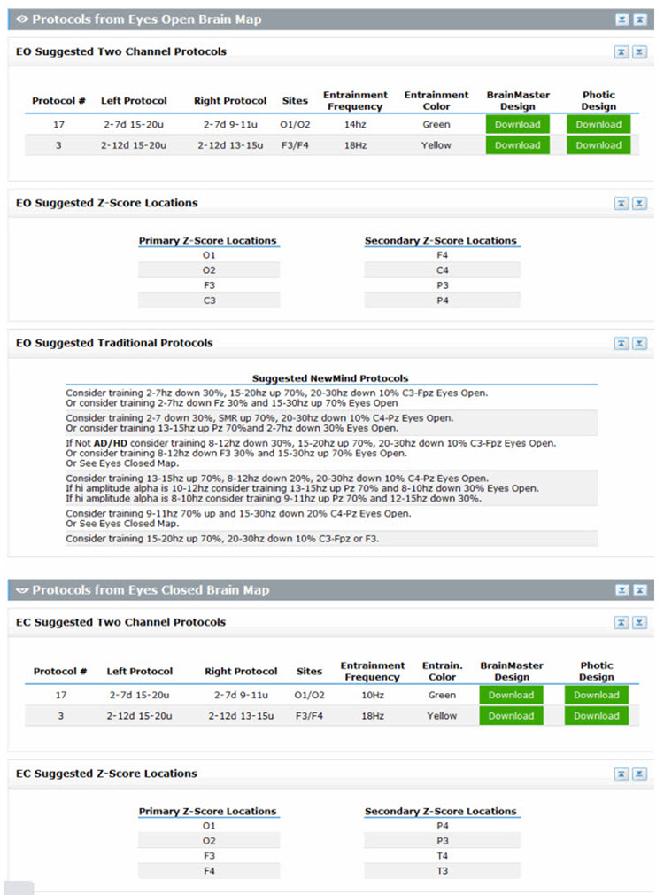
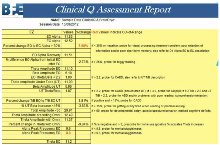
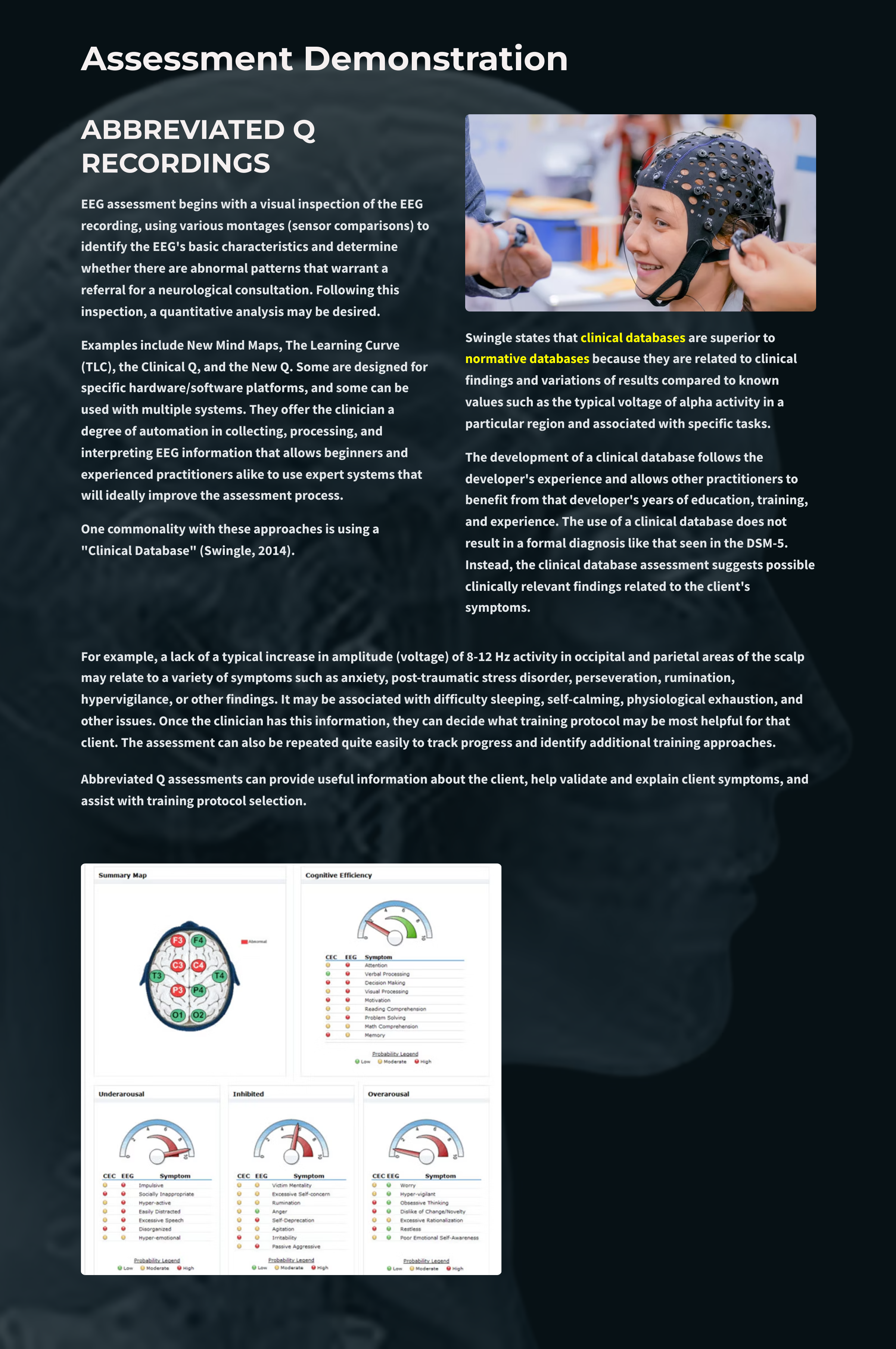
Assessment Demonstration
Overview
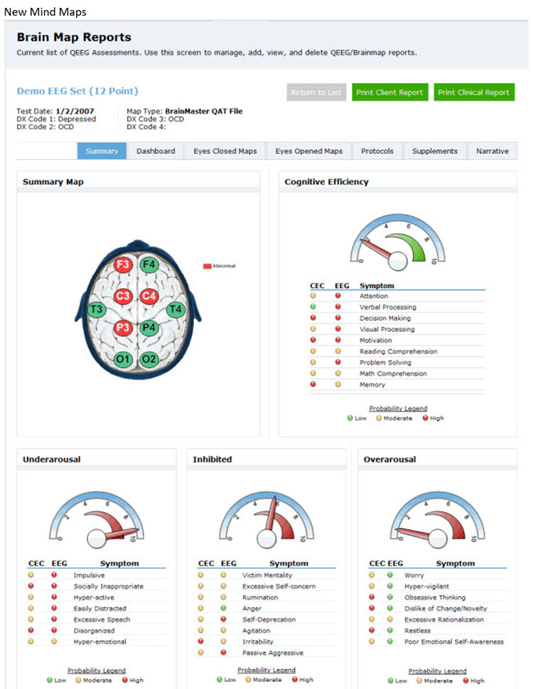
We have discussed the assessment of the EEG from a variety of perspectives in other sections of Neurofeedback Tutor. This section will provide examples of relatively simple and more complex EEG assessment approaches.
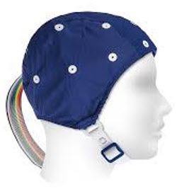
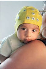
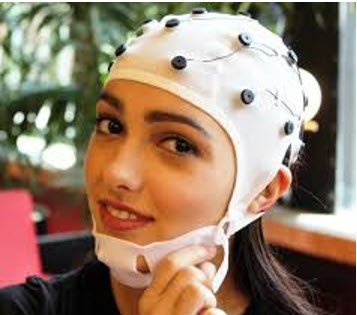
Dr. Ronald Swatzyna has generously permitted the authors to share the Houston Neuroscience Brain Center's client qEEG cap orientation video.