Current Trends in Neurofeedback
What You Will Learn in This Chapter
This unit explores the evolving landscape of neurofeedback, from qEEG-based approaches and z-score training to infra-low frequency methods and functional connectivity training. You will also learn how clinicians combine neurofeedback with complementary modalities—including heart rate variability biofeedback, respiration training, hemoencephalography, and neuromodulation—to improve training outcomes and accelerate the acquisition of self-regulation skills.
We have divided this unit into two major sections. The first covers current neurofeedback trends, including z-score training, LORETA z-score training, infra-low and infra-slow frequency training, connectivity training, and fMRI neurofeedback. The second covers combining neurofeedback with other modalities, including heart rate variability (HRV), respiration, hemoencephalography (HEG), and neuromodulation systems.
BCIA Blueprint Coverage: This unit addresses X. Current Trends in Neurofeedback.
Learning Objectives
After completing this section, you will be able to:
Describe the development and rationale of qEEG-based neurofeedback training and z-score training.
Compare infra-low frequency training and infra-slow frequency training approaches.
Explain how functional connectivity training and fMRI neurofeedback extend traditional EEG-based methods.
Describe how heart rate variability biofeedback, respiration training, and hemoencephalography can complement neurofeedback.
Distinguish neuromodulation approaches (AVE, tACS, rTMS, PEMF, photobiomodulation) from neurofeedback.
Overview
This section covers the role of ongoing assessment in neurofeedback practice, including its ethical foundations and its contribution to clinical decision-making. Intake and initial EEG assessment establish a baseline against which you can gauge subsequent progress. This matters because, despite strong research evidence supporting neurofeedback efficacy in group studies (Schwartz & Andrasik, 2016; Tan, Shaffer, Lyle, & Teo, 2016), benefits are never guaranteed for an individual client. Ongoing assessment allows you to verify that expected outcomes are occurring and, when they are not, to use continuous data to adjust training protocols before time and resources are wasted.
The ethical principles guiding ongoing assessment are both practical and profound. Benevolence asks whether training is helping, while nonmalevolence asks whether it is causing harm (Beauchamp, 2003). Even the ongoing cost of time and money for an ineffective treatment constitutes a form of harm. The principle of autonomy requires that clients remain well-informed about their training outcomes so they can provide meaningful consent to continue. Finally, the principle of justice challenges you to deliver training efficiently enough that you do not unreasonably deny access to others.
Beyond ethics, ongoing assessment strengthens the therapeutic alliance by allowing you and your client to examine outcomes collaboratively. Seeing measurable progress motivates clients to persevere with training and to practice self-regulation skills outside the clinic. Clients may also begin to recognize links between the subjective states they experience during neurofeedback and real-world situations, empowering them to reproduce those states intentionally when it matters most.
Ultimately, neurofeedback trains EEG activity, but the goal extends well beyond the EEG itself. The client and practitioner want to know whether changes in brain activity generalize to emotional experience, cognition, physiology, and real-life behavior. For this reason, ongoing assessment should periodically include non-EEG measures—repeating the same instruments used during intake—to evaluate whether brain-based changes translate into the improvements that originally motivated the client to seek neurofeedback.
Ongoing assessment serves ethical, clinical, and motivational purposes. It verifies training effectiveness, informs collaborative decision-making between client and practitioner, and helps clients develop self-awareness of the relationship between their subjective states and real-world behavior.
BCIA Blueprint Coverage
This unit covers X. Current Trends in Neurofeedback.

We have divided this unit into A. Current trends (e.g., z-score training, LORETA z-score training) and B. Combining neurofeedback with other modalities, including heart rate variability (HRV), respiration, HEG, and neuromodulation systems.
Neurofeedback Trends
This section surveys the major neurofeedback approaches that have emerged over recent decades. We will cover qEEG-based neurofeedback training, the development of z-score training, infra-low training, infra-slow training, and connectivity training—including fMRI neurofeedback.
qEEG-Based Neurofeedback Training
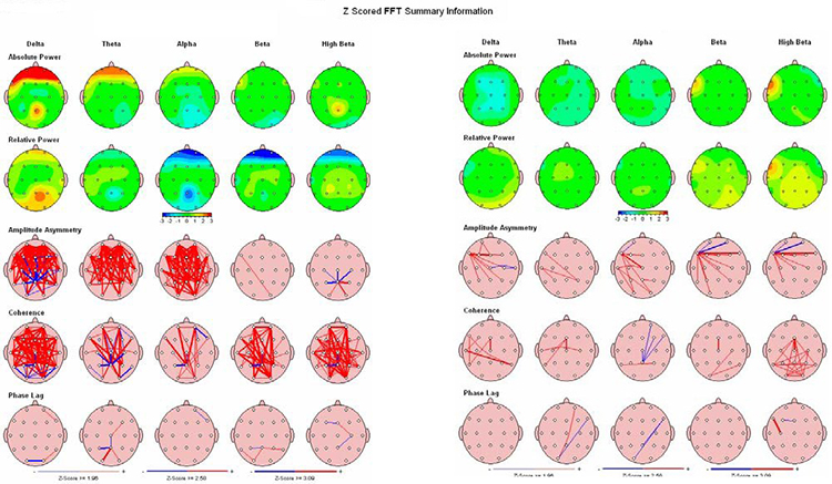
This subsection traces how quantitative EEG evolved from a research curiosity into a cornerstone of individualized neurofeedback protocol design. The story begins with hardware and mathematics. The development of high-quality EEG recording devices capable of capturing 19 scalp electrode locations simultaneously, combined with software that could generate topographic images from recorded data, opened the door to a quantitative approach to the EEG. The Fast Fourier Transformation (FFT) algorithm, introduced in 1965 (Cooley & Tukey, 1965; Dumermuth & Fluhler, 1967), made it possible to deconstruct the complex, multi-frequency EEG signal into frequency and power spectral displays—first as tables and later as topographic head maps, commonly known as brain maps.
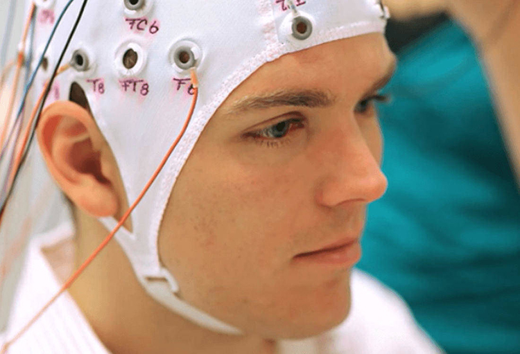
As affordable personal computers became widely available in the 1990s, clinicians and researchers gained access to these computational methods and began identifying EEG patterns associated with various conditions and disorders. This progress was propelled by the development of normative EEG databases—collections of EEG recordings from healthy, screened individuals that serve as benchmarks for comparison.
Pioneers included E. Roy John at New York University, Robert Thatcher at the University of Maryland, and Frank Duffy of Harvard University. The EEG has demonstrated strong stability and specificity across multiple ethnic and cultural groups (John, Ahn, & Prichep, 1980) and high consistency in test-retest reliability evaluations (Fein et al., 1983; Oken & Chiappa, 1988), making these databases trustworthy reference points.


Because of this consistency and reliability, the quantitative EEG (qEEG) became a powerful clinical tool for individualized protocol development. Each client could be assessed using multiple techniques, including a qEEG with normative database comparison (see the Assessment unit).
As Thatcher (1998) described, normative database comparison allowed clinicians to assess a client's neuropsychological status, identify strengths and weaknesses in their neurophysiology, design an optimal training regimen, and evaluate results following intervention. Many clinicians adopted this approach: if a brain region showed excess or deficient activity in one or more frequency bands, that area would be targeted with uptraining or down-training to address the specific dysregulation, often resolving the client's presenting complaints.
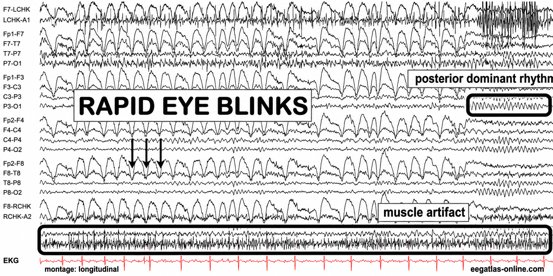
The benefits of database-guided training were compelling: clinicians no longer needed to guess at the training location, target frequency, or direction of training, and follow-up qEEG assessment could verify results. However, several challenges emerged. Neurologists (Nuwer & Coutin-Churchman, 2014) highlighted the artifact problem—inexperienced practitioners often made errors in data selection and rejection when preparing EEG recordings for database comparison.
Eye artifacts such as blinks, lateral movements, and flutter introduce excessive delta-frequency activity (1–4 Hz) in frontal electrode locations, producing false-positive findings. EMG (muscle) artifact artificially elevates beta frequencies, cable sway contaminates the delta range, and electromagnetic frequency (EMF) artifact inflates fast beta values (please see the section on EEG artifacts). Graphic © eegatlas-online.com.
A second major pitfall was the assumption that every out-of-range finding on a qEEG should be trained. In some cases, training an apparently abnormal finding worsened the client's symptoms, revived previously resolved complaints, or introduced entirely new problems. This painful experience taught clinicians that not all deviations from normative values represent pathology—some are compensatory, reflecting the brain's attempts to restore balance or homeostasis. Removing these compensatory patterns often produced the very negative effects practitioners sought to avoid.
From this understanding emerged the idea that training should target only the specific symptom or network most problematic for the client and should include built-in safeguards against moving EEG variables in undesirable directions—an idea that led directly to z-score training.
The development of the FFT algorithm, normative EEG databases, and affordable computers enabled qEEG-based neurofeedback. While these tools allowed individualized training protocol development, challenges emerged with artifact contamination and the discovery that not all abnormal findings represent pathology; some EEG deviations are compensatory.
The Development of Z-Score Training
This subsection explains how z-score training addressed the limitations of earlier qEEG-guided approaches. To solve the problems that arose from training every out-of-range finding, Robert Thatcher, developer of the NeuroGuide database, and Thomas Collura of BrainMaster Technologies created Live Z-Score Training (LZT). Beginning in 1996 (Thatcher et al., 2019), they started with a simple 1- or 2-channel system and quickly developed a 4-channel approach that provided clients with real-time feedback showing how closely their EEG matched a database of age-matched typical controls. The Thatcher database initially offered approximately 72 variables—including power, relative power, phase, and coherence values across standard EEG frequencies—and now contains over 600 qEEG variables (Thatcher, 2005).
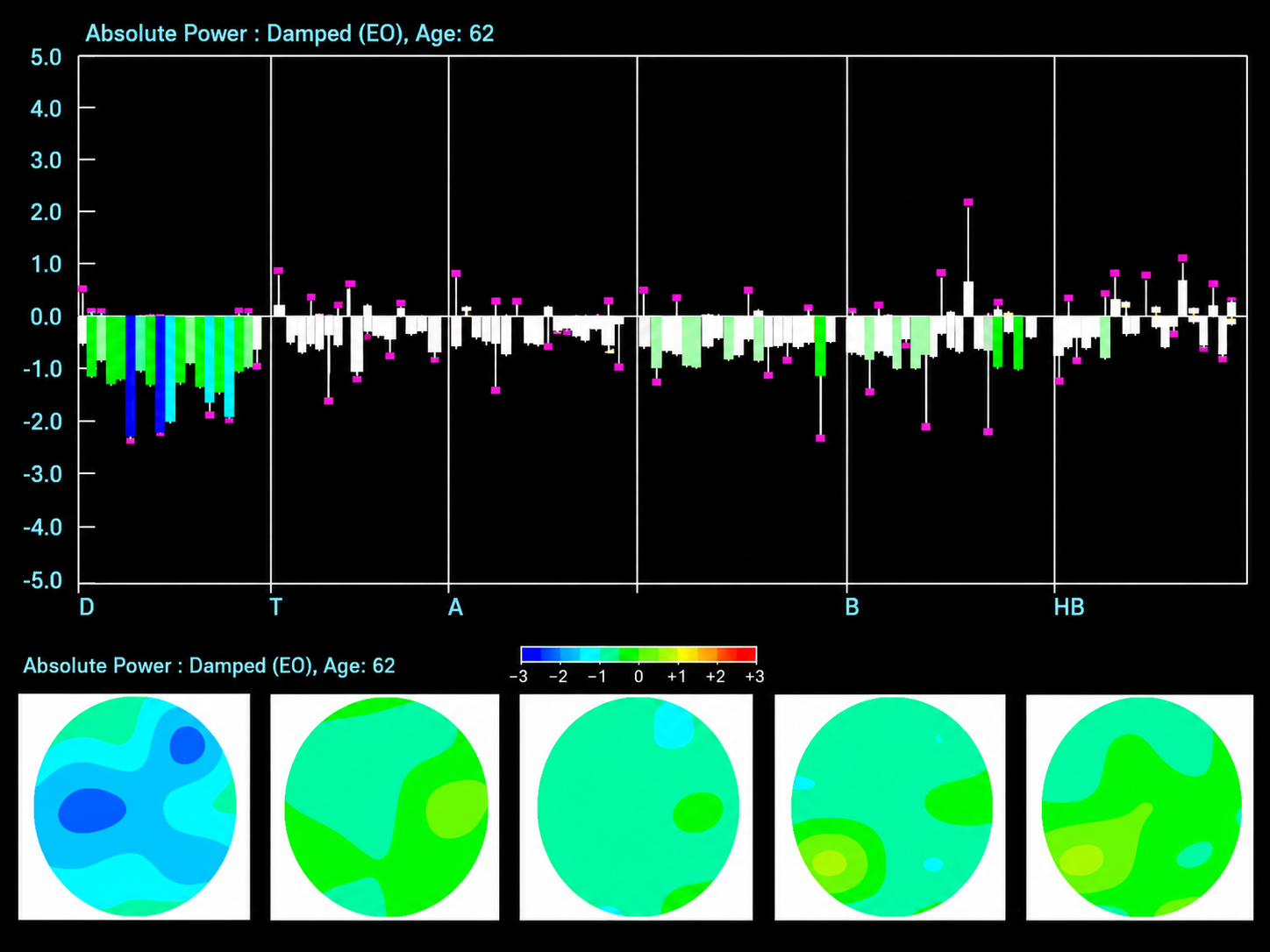
The client's EEG values are compared to normative values, and differences are expressed in standard deviations (z-scores). The training goal is to move these z-scores toward zero—the statistical norm. Subsequent advances extended the system from 4 channels to 19-channel real-time surface z-score training, and then to 19-channel LORETA z-score training, which uses the LORETA source localization method to train brain activity in three dimensions. Collura and colleagues initially worked with the Thatcher database, then transitioned to the E. Roy John database (now known as BrainDX), and more recently shifted to qEEGPro, a database built by screening client EEG recordings using a clinical questionnaire (qEEG.pro/database/). This approach rests on assumptions that many qEEG researchers have viewed with skepticism (personal communication, John Anderson, 2019–2021).

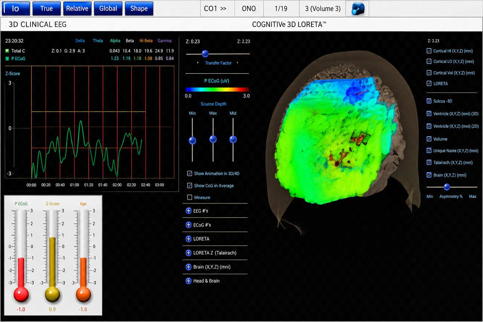
Meanwhile, Thatcher and his team continued refining the NeuroGuide database and expanding the capabilities of 19-channel z-score training. Their most recent innovation is swLORETA, a more precise and accurate iteration of the LORETA source localization method, which has attracted a large base of clinicians. More than 50 publications have presented evidence supporting the efficacy of z-score neurofeedback training, suggesting widespread scientific acceptance (https://www.appliedneuroscience.com/PDFs/Z_Score_NFB_Publications.pdf).
Live Z-Score Training (LZT) was developed by Thatcher and Collura to provide real-time feedback comparing client EEG to normative databases. The approach has progressed from 1-2 channel to 19-channel LORETA and swLORETA systems, with more than 50 publications supporting its efficacy.
Infra-Low Training
This subsection introduces infra-low frequency training and the ongoing debate surrounding its mechanisms. Susan and Siegfried Othmer are among the most prominent figures to contribute to this branch of neurofeedback. They initially studied with Margaret Ayers when she worked with their son, Brian, who experienced temporal lobe epilepsy. After several professional transitions, they developed a training institute and began working with clients while training new practitioners.

The evolution of the Othmer method led to training in infra-low frequencies (ILF)—EEG activity below 0.1 Hz. This approach stirred considerable controversy because this region of the EEG spectrum is poorly understood, and the mechanisms governing these very slow electrical fluctuations remain unclear. Compounding the debate, the infra-low frequency range overlaps with common artifacts, including eye blinks, eye movements, cable sway, electrodermal responses (GSR), and electrode drift. Siegfried Othmer, who holds a doctorate in physics, has stated that proprietary signal processing methods address these contamination issues and that training proceeds effectively despite them (personal communication, 2021).
Multiple published reports describe clinical applications of infra-low frequency training (Legarda et al., 2011; Othmer & Legarda, 2011; Sasu & Othmer, 2020), and other clinicians have independently developed their own approaches to training brain activity below 1 Hz. The Othmers have continued to expand their methodology, incorporating alpha-theta (A-T) training with a two-channel sum training approach and more recently adding two-channel synchrony training in the alpha and gamma frequency bands. They have a large following, and many clinics and individual practitioners employ these methods, reporting positive client outcomes. However, a clear explanation of what exactly is being trained in the ILF protocol remains elusive and awaits future investigation.
Infra-Slow Training
Another prominent clinician, Mark Smith, has pursued a related approach called infra-slow frequency (ISF) training, utilizing different amplifiers and software. Smith initially trained with the Othmers but then began developing his own method for providing feedback on EEG activity below 0.1 Hz. Over time, he incorporated z-score training using the NeuroGuide database developed by Robert Thatcher, and he learned quantitative EEG techniques from Jonathan Walker, MD, and others.
Smith's explanation of ISF training, developed in collaboration with Thomas Collura (Smith et al., 2014), centers on a key technical distinction: rather than training typical amplitude fluctuations of an alternating-current EEG signal, ISF training provides clients with information about the phase of the infra-slow signal. Because the time constant is very long, the filters require significant time—approximately 3 minutes in the published example—to return to baseline following a substantial signal shift.
As noted in the discussion of infra-low training, significant voltage shifts also occur with typical eye movement, eye blink, electrodermal, and cable sway artifacts. There is no published discussion of how these artifacts are managed in the ISF approach, leaving questions about the validity of the training signal. There may, however, be additional signal processing methods beyond what the published literature describes.
Despite these open questions, clinical results using a combination of ISF, z-score, synchrony, and referential enhancement training have been positive, and clinical examples and book chapters (Smith, 2014, 2017, 2018) support continued investigation.
Infra-low frequency (ILF) training by the Othmers and infra-slow frequency (ISF) training by Mark Smith both operate below 0.1 Hz. While both approaches have produced positive clinical results, questions remain about artifact contamination in this frequency range and the mechanisms underlying training effects.
Functional Connectivity Training
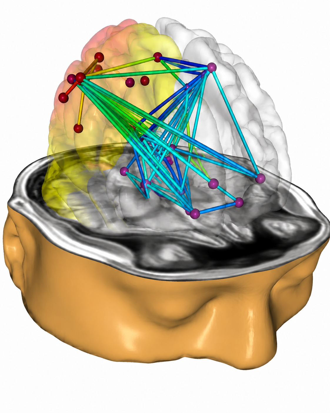
This subsection examines how training communication between brain regions has become a major frontier in neurofeedback. Studying how different brain areas are interconnected is now a central focus in both the EEG/qEEG community and among researchers using functional magnetic resonance imaging (fMRI) to track the blood oxygen level-dependent (BOLD) signal—a measure of metabolic activity that indicates which brain regions are most active. One major advantage of EEG is its faster time scale: blood flow changes occur over seconds to minutes, while EEG changes happen within milliseconds. Evoked potential (EP) and event-related potential (ERP) studies using EEG can detect brain responses to stimuli in as little as 50 milliseconds (ms). Furthermore, the cost of fMRI-based training is orders of magnitude greater than EEG neurofeedback, making EEG the more practical choice for most clinical settings.
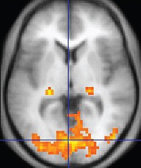
Connectivity changes following even a single neurofeedback session have been demonstrated. Kluetsch and colleagues (2014) used pre- and post-fMRI measurements to identify changes in network communication and found a rebound effect on the EEG following alpha desynchronization training, along with increased connectivity within the salience network (SN) and default mode network (DMN)—two large-scale brain networks involved in detecting relevant stimuli and in self-referential thought, respectively. Graphic courtesy of BrainMaster Technologies.
Coben and colleagues (2018) studied 174 participants from 11 US clinical sites who received an average of 21.49 sessions of coherence feedback training—a form of connectivity training that targets the degree to which EEG signals at two or more electrode sites are synchronized. They found significant changes in EEG power values and showed 50% greater change in participants randomly assigned to four-channel vs. two-channel coherence training (113 vs. 61 participants). The improved results in the four-channel group may reflect more sophisticated approaches to measuring coherence. Separately, Walker and Horvath (2010) found no clear advantage for either power or coherence training alone but observed that power training often introduced new coherence abnormalities requiring subsequent coherence-focused intervention.
An important technical consideration in EEG coherence training is reference contamination from linked-ears or linked-mastoid montages. Nunez and Srinivasan (2006) demonstrated that using either a single mastoid or averaged linked-mastoid reference artificially inflates coherence values across all electrode combinations, because the reference signal contributes to every electrode. They reported that common average reference montages yield more accurate coherence estimates, particularly when using dense electrode arrays, although values remain elevated by volume conduction for electrode pairs closer than about 8–10 cm.
These findings suggest that future coherence training should account for reference effects, and the work of Coben and colleagues points to the need for continued refinement of the mathematical analysis and display of coherence measures. Importantly, many neurofeedback approaches appear to improve functional connectivity whether or not they are explicitly designed to target it, consistent with the principle that nothing occurs in the central nervous system (CNS) without activating and exercising network structures.
fMRI Neurofeedback
Real-time neurofeedback based on functional magnetic resonance imaging (rt-fMRI) has been applied to conditions including pain, anxiety, depression, schizophrenia, cognitive dysfunction, and post-stroke motor impairment (Linhartova et al., 2019; Watanabe et al., 2017). fMRI provides superior spatial resolution compared to EEG and can image deep cerebral structures that EEG cannot reach. However, because fMRI relies on blood oxygen level responses, its temporal resolution is considerably slower than that of EEG. Recent work has begun exploring the integration of rt-fMRI neurofeedback with EEG neurofeedback (Bezmaternykh et al., 2021), potentially combining the spatial precision of fMRI with the temporal sensitivity of EEG.
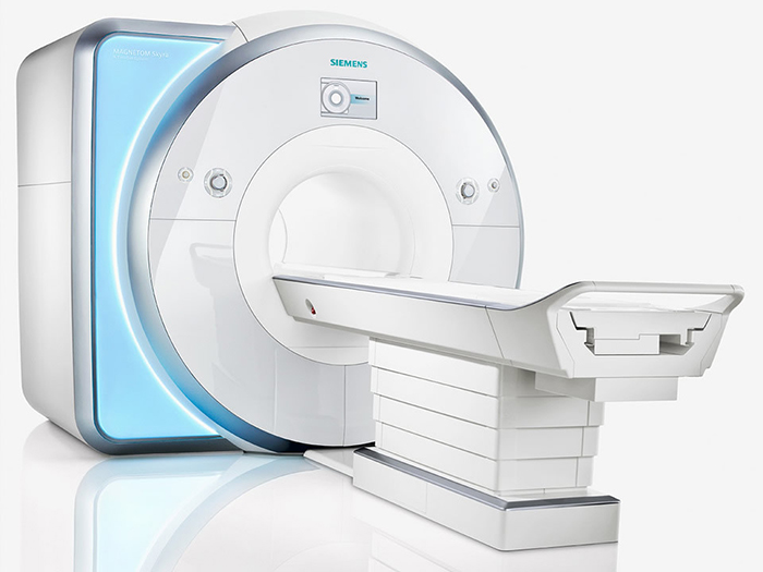
Functional connectivity training targets communication between brain regions, with both EEG and fMRI approaches available. EEG offers superior temporal resolution and lower cost, while fMRI provides better spatial resolution. Research suggests many neurofeedback approaches improve connectivity, and reference contamination remains an important consideration for EEG coherence training.
Combining Neurofeedback with Other Modalities
This section explores how clinicians integrate complementary modalities with neurofeedback to accelerate the acquisition of self-regulation skills and improve training outcomes. We will cover heart rate variability (HRV), respiration, hemoencephalography (HEG), and neuromodulation.
Heart Rate Variability
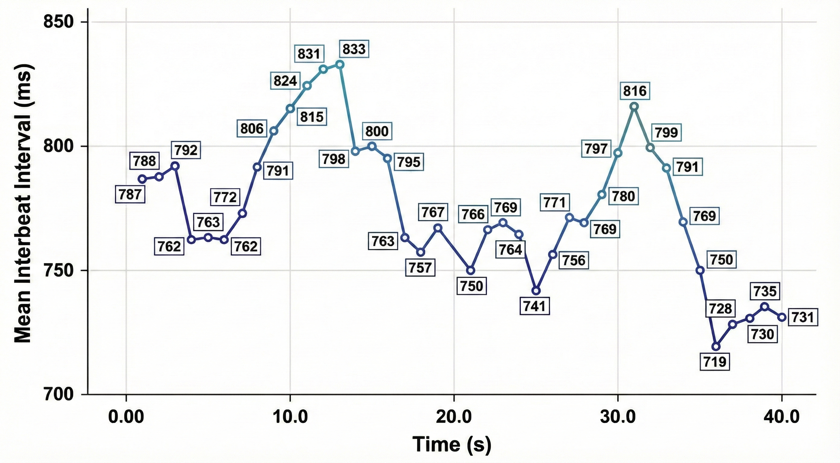
Heart rate variability (HRV) refers to the organized fluctuation of time intervals between successive heartbeats, defined as interbeat intervals (IBIs) (Shaffer, Meehan, & Zerr, 2020). The oscillations of a healthy heart are complex, not random. HRV indexes how efficiently we mobilize and utilize limited self-regulatory resources to maintain homeostasis—the body's dynamic equilibrium. Because a healthy heart can rapidly adjust to sudden challenges through interlocking and well-calibrated control systems, HRV plays a vital role in regulatory capacity, executive functions, health, and performance.
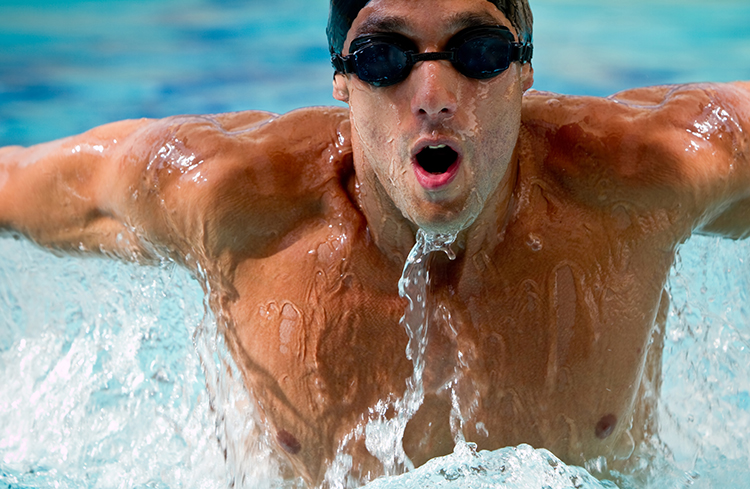
Behavioral interventions like aerobic exercise, healthy breathing, compassion, and mindfulness meditation are powerful strategies for increasing HRV. Graphic © Maridav/Shutterstock.com.

We have divided this section into The Meaning of HRV, A Healthy Heart is Not a Metronome, How HRV Biofeedback Can Support Neurofeedback, and Heart-Brain Interactions.
The Meaning of HRV
This section introduces the concept of heart rate variability and explains why it matters for clinical and optimal performance practice. You will learn how heart rate constrains HRV, why a healthy heart is not a metronome, and how key theoretical frameworks connect HRV to self-regulation and health. Understanding these fundamentals will shape how you assess clients, set training goals, and explain biofeedback rationale.
Heart Rate and Its Consequences
Heart rate is the number of heartbeats per minute. In clinical practice, a client's resting heart rate is often the first physiological metric you observe, and it carries more prognostic weight than many clinicians realize.

Elevated HR Is Associated with Dementia and Cognitive Decline
Imahori et al. (2021) conducted a cohort study of 2,147 adults aged 60 and older who were free of dementia at enrollment. Resting heart rates of 80 bpm or higher, compared with 60 to 69 bpm, were associated with a greater risk of dementia and more rapid cognitive decline, independent of cardiovascular disease. For practitioners working in VA settings or geriatric care, this finding suggests that elevated resting heart rate may be a modifiable risk factor worth monitoring alongside traditional cognitive screening.
Elevated HR Limits HRV
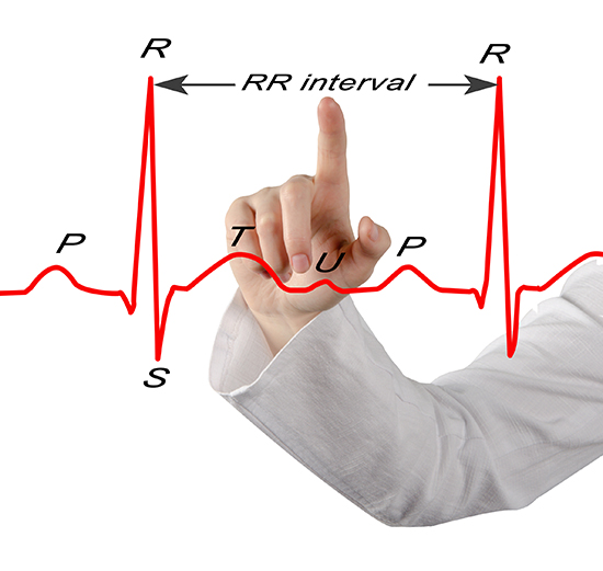
Heart rate matters for biofeedback practitioners because a high rate can reduce heart rate variability (HRV)—the changes in the time intervals between consecutive heartbeats (Task Force of the European Society of Cardiology and the North American Society of Pacing and Electrophysiology, 1996). We measure these interbeat intervals (IBIs) in milliseconds, and the degree to which they fluctuate reflects the heart's capacity to adapt moment by moment.
Mini-Lecture: Heart Rate Variability Overview
Faster heart rates reduce the time between successive beats, leaving less opportunity for IBIs to vary. This is a straightforward but clinically important point: an anxious client presenting with a resting heart rate of 90+ bpm has limited room for beat-to-beat fluctuation, which constrains HRV before training even begins. Resting heart rates that exceed 90 bpm are also associated with an elevated risk of mortality (Zhang et al., 2016).
The next three scatterplots illustrate this inverse relationship between HR and three widely used HRV metrics: RMSSD, SDNN, and low-frequency power.
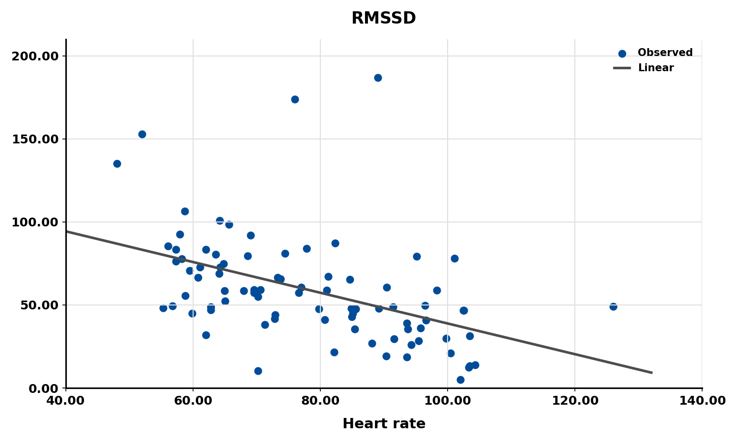
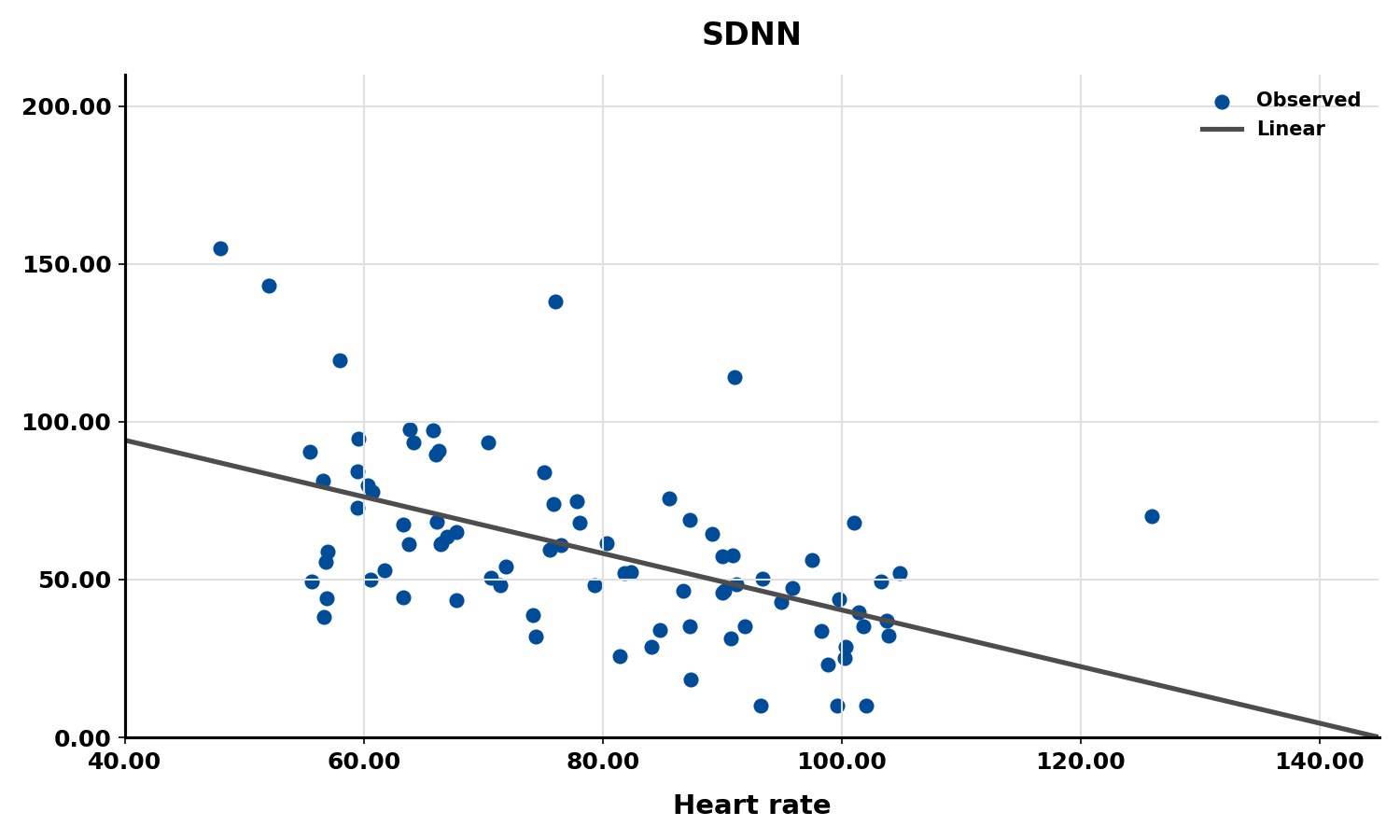
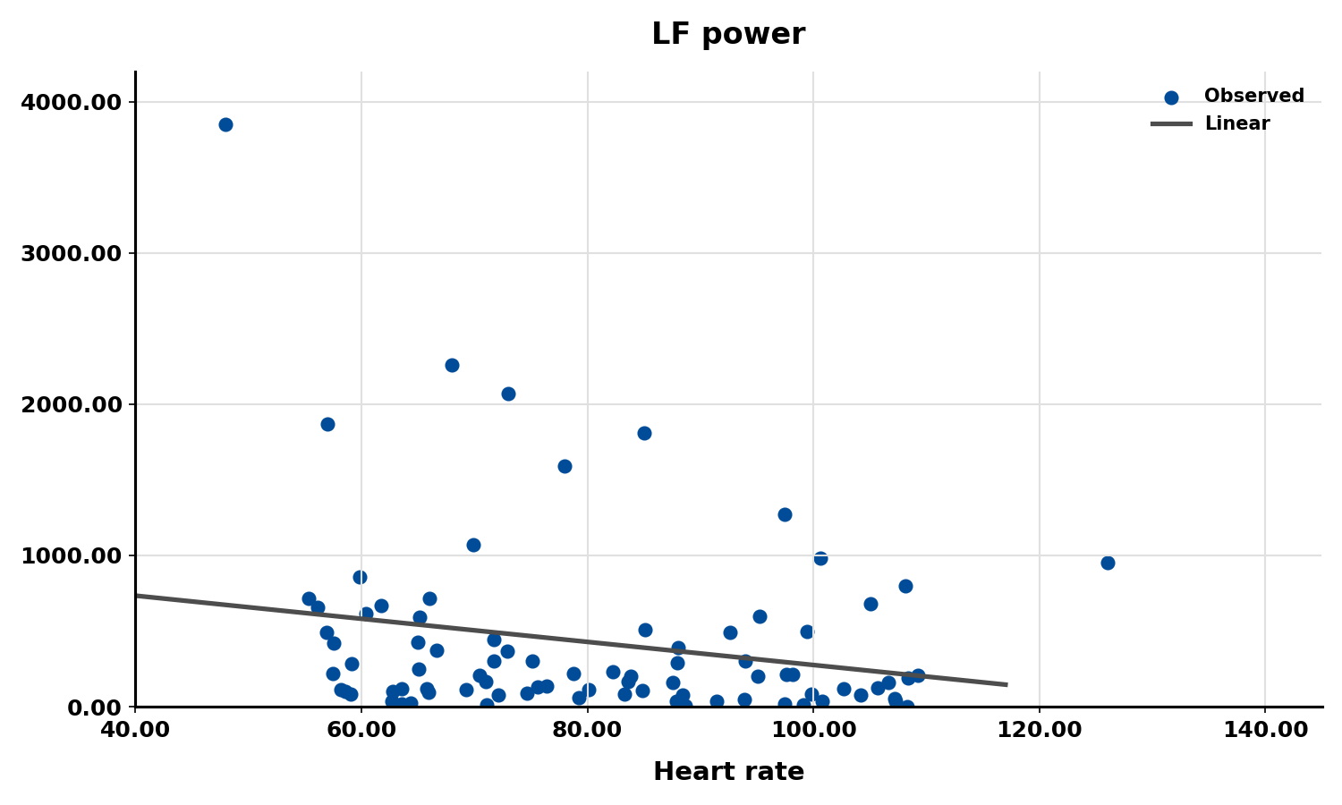
Conversely, the slower heart rates seen in endurance athletes—such as trail runners—increase the time between adjacent heartbeats, creating more opportunity for IBIs to vary and raising HRV. This phenomenon is called cycle length dependence (McCraty & Shaffer, 2015), and it explains why aerobic conditioning is one of the most reliable ways to improve HRV outside the clinic.

Typical non-athlete resting heart rates range from 60 to 80 bpm, while athletes may have resting rates between 40 and 60 bpm (Khazan, 2019). When you encounter an athlete with unusually low resting heart rate, expect correspondingly higher HRV values—an important consideration when establishing normative baselines for optimal performance training.
A Healthy Heart Is Not a Metronome
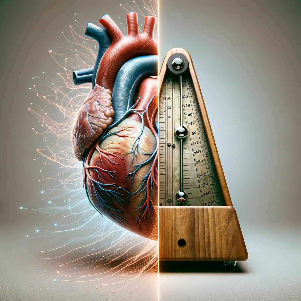
A healthy heart is not a metronome. This simple idea is one of the most important concepts in HRV biofeedback. When the time intervals between heartbeats change substantially across successive breathing cycles, it demonstrates that the cardiovascular center can effectively modulate vagal tone—the parasympathetic "brake" on the heart.
Mini-Lecture: Why Is Heart Rate Variability Important?
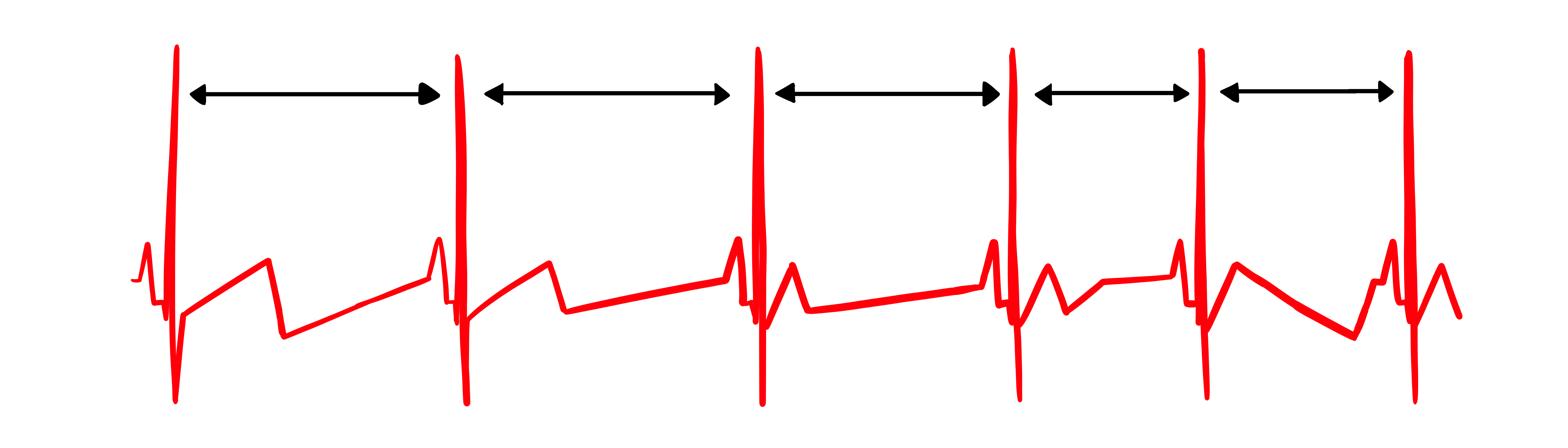
The record below shows healthy variability—the time intervals between successive heartbeats clearly differ. For your clients, this is the pattern you want to see and help them develop.
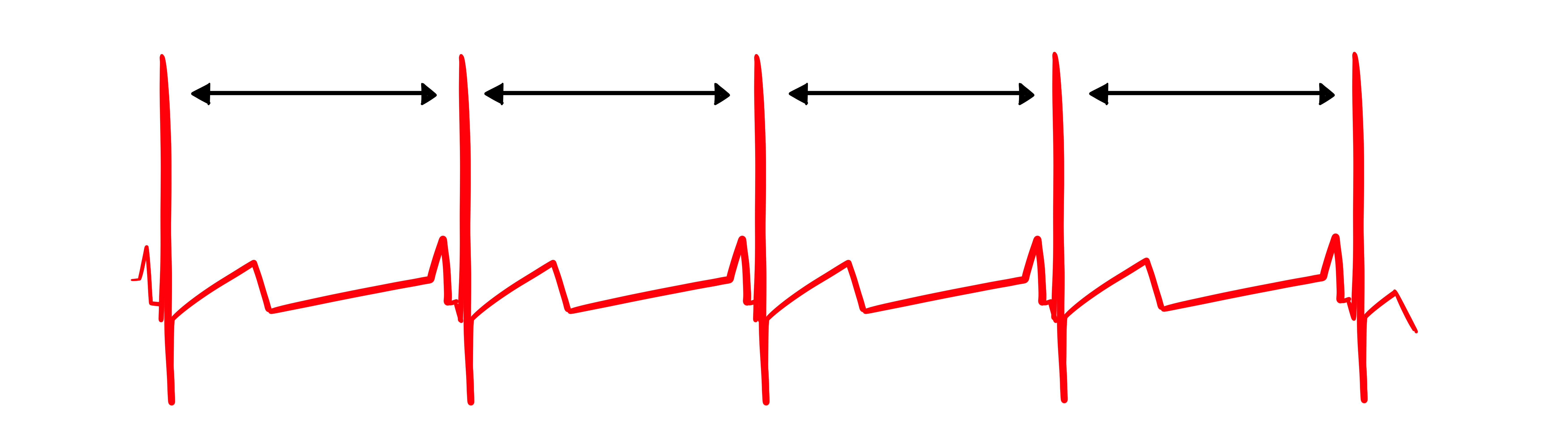
In contrast, this record shows no variability: the IBIs are identical. This display could represent a heart driven by a pacemaker or one that needs one. When you see this pattern in a client who is not pacemaker-dependent, it signals significantly compromised autonomic regulation.
"The complexity of a healthy heart rhythm is critical to the maintenance of homeostasis because it provides the flexibility to cope with an uncertain and changing environment. HRV metrics are important because they are associated with regulatory capacity, health, and performance and can predict morbidity and mortality" (Shaffer et al., 2020).
Check out the YouTube video HRV Training and its Importance.
"HRV is associated with executive function, regulatory capacity, and health. Cardiac vagal control indexes how efficiently we mobilize and utilize limited self-regulatory resources during resting, reactivity, and recovery conditions" (Shaffer et al., 2020).
HRV as a Multisystem Biomarker
Vagal tone modulation helps maintain the dynamic autonomic balance critical to cardiovascular health. Autonomic imbalance due to deficient vagal inhibition is implicated in increased morbidity and all-cause mortality (Thayer, Yamamoto, & Brosschot, 2010). For the clinician, this means that HRV is not merely a research metric—it is a practical indicator of how well a client's autonomic nervous system is functioning day to day.
HRV appears to index autonomic functioning, blood pressure, neurocardiac functioning, digestion, oxygen and carbon dioxide exchange, vascular tone (the diameter of resistance vessels), and possibly facial muscle regulation (Gevirtz et al., 2016). It also reflects the vagal contribution to executive functions, affective control, and social self-regulation (Byrd et al., 2015; Laborde et al., 2017; Mather & Thayer, 2018). This breadth of associations is why HRV biofeedback has applications across such diverse clinical populations—from cardiac rehabilitation patients to service members managing stress.
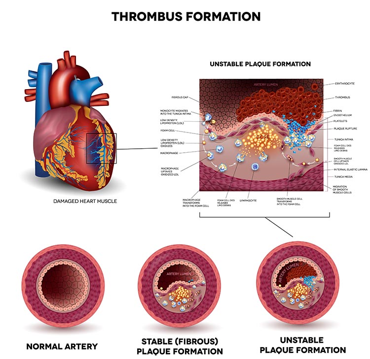
HRV is a multisystem biomarker that indexes autonomic functioning, blood pressure, digestion, gas exchange, vascular tone, and the vagal contribution to executive function and emotion regulation. Deficient vagal inhibition and autonomic imbalance predict greater morbidity and all-cause mortality, which is why HRV biofeedback applies across diverse clinical populations.
Influential HRV Theories
Two theoretical frameworks help clinicians understand why HRV matters and how to interpret it. The Vagal Tank Theory frames cardiac vagal control as a dynamic, depletable resource for self-regulation, while the Autonomic Space Theory reveals that sympathetic and parasympathetic activity interact more flexibly than older models assumed. Together, they provide a comprehensive framework for understanding HRV as a marker of physiological adaptability, emotional regulation, and cognitive flexibility.
Vagal Tank Theory
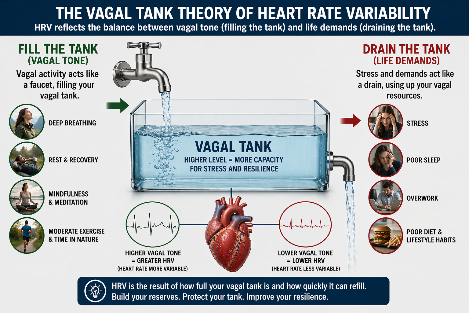
The Vagal Tank Theory (Laborde et al., 2018) conceptualizes cardiac vagal control as a dynamic resource that can be depleted or replenished—much like a fuel tank. It consists of three Rs: (1) Resting vmHRV, reflecting baseline self-regulation capacity; (2) Reactivity, the vagal withdrawal response to stressors; and (3) Recovery, the ability to restore autonomic balance post-stressor. A higher vagal "tank" is associated with better stress resilience, emotional regulation, and health outcomes. In practice, this means you need to assess clients at rest, during challenge, and during recovery to get a complete picture of their autonomic resources. A single resting vmHRV measurement tells only part of the story.
From Laborde and colleagues' perspective, vagal traffic to the heart indicates how efficiently we mobilize and use scarce self-regulatory resources.
Autonomic Space Theory
The Autonomic Space Theory (Berntson et al., 1994) challenges the simplistic reciprocal model of autonomic control by proposing three modes: (1) Reciprocal Activation, where one branch is active and the other is suppressed; (2) Coactivation, where both sympathetic and parasympathetic systems are active simultaneously; and (3) Coinhibition, where both are suppressed. This framework matters clinically because it explains why some clients show paradoxical physiological patterns—such as elevated heart rate alongside high HRV—that a simple "seesaw" model of autonomic balance cannot explain.
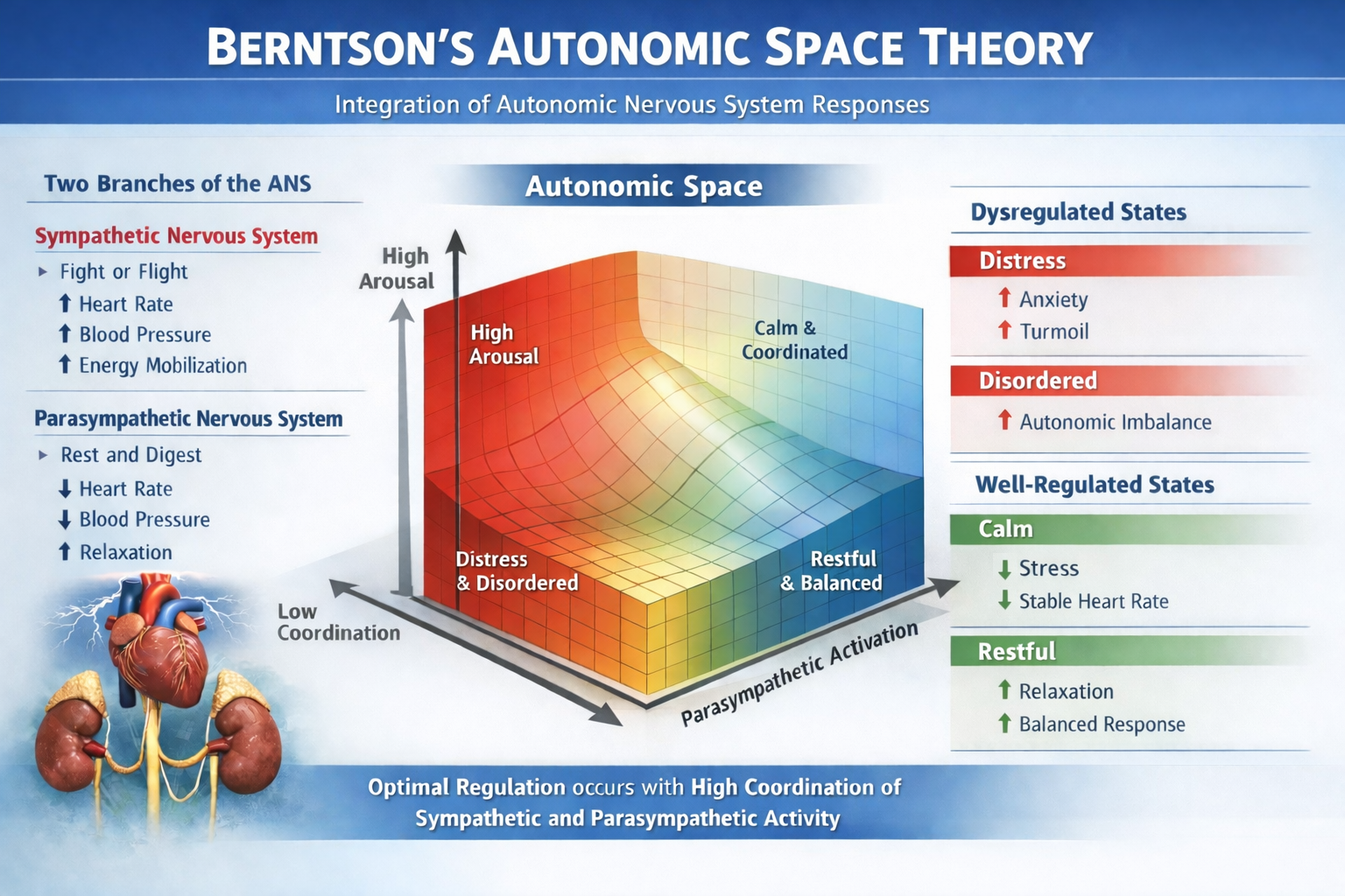
The Vagal Tank Theory frames cardiac vagal control as a depletable resource assessed across resting, reactivity, and recovery conditions. The Autonomic Space Theory replaces the simple reciprocal "seesaw" model with reciprocal activation, coactivation, and coinhibition, explaining paradoxical patterns such as elevated heart rate alongside high HRV.
Vagally-Mediated HRV (vmHRV)
This subsection covers vagally-mediated HRV (vmHRV) as a clinical biomarker, its companion concept of sympathetically-mediated HRV (smHRV), and the health implications of vmHRV across multiple body systems. Understanding vmHRV is essential because it provides the clearest autonomic signal you can track with standard biofeedback equipment.
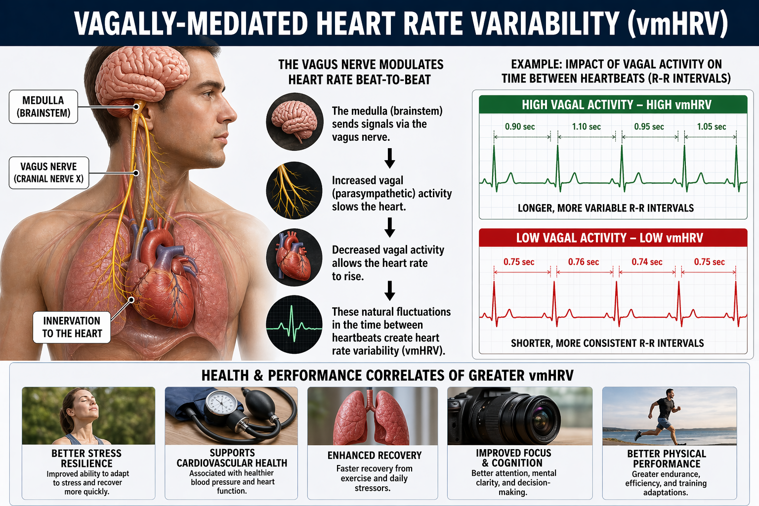
Vagally-mediated heart rate variability (vmHRV) has emerged as a critical biomarker for self-regulation and health, offering insights into psychological and physiological adaptation. As a noninvasive and cost-effective tool, vmHRV serves as an actionable measure in physical and mental health, social interactions, stress regulation, and performance optimization (Laborde et al., 2023). For clinicians and performance coaches alike, vmHRV is one of the most practical psychophysiological metrics available.
The sympathetic nervous system is another source of HRV. Sympathetically-mediated HRV (smHRV) refers to HRV components influenced by the sympathetic nervous system, typically assessed through measures such as low-frequency (LF) power in HRV analysis. However, the interpretation of LF power as a direct marker of sympathetic activity remains debated—a nuance that matters when you interpret spectral analysis results for clients.
How the Autonomic Nervous System Shapes Heart Rate Variability
HRV captures the moment-to-moment push and pull of the autonomic nervous system on the heart's pacemaker, the sinoatrial node. The two branches of this system operate on very different timelines. Parasympathetic (vagal) signals act almost instantaneously, modulating heart rate on a beat-to-beat basis through acetylcholine release. Sympathetic signals, by contrast, work more slowly, taking several seconds to exert their effects through norepinephrine. This speed difference is why high-frequency HRV (roughly 0.15–0.40 Hz), and especially the familiar breathing-linked rhythm known as respiratory sinus arrhythmia, is understood to be primarily a vagal phenomenon in healthy adults (Berntson et al., 1997; Task Force of the European Society of Cardiology and the North American Society of Pacing and Electrophysiology, 1996).
Slower Rhythms Tell a More Complex Story
The question of whether sympathetic activity leaves a detectable signature in HRV has been explored for decades. Foundational spectral analysis work by Akselrod and colleagues (1981) first demonstrated that HRV contains separable frequency components tied to distinct autonomic control processes. Later, Pomeranz et al. (1985) used pharmacologic blockade combined with postural changes to show that low-frequency oscillations (roughly 0.04–0.15 Hz) shift from predominantly parasympathetic mediation when a person is lying down to a mixed sympathetic-parasympathetic pattern when standing. Perhaps the most compelling evidence comes from microneurography studies, in which researchers directly record sympathetic nerve traffic in peripheral nerves. Pagani et al. (1997) showed that as sympathetic drive increases, LF oscillations become more prominent and track closely with sympathetic nerve firing patterns and arterial pressure fluctuations, pointing to a shared sympathetic rhythm during activation.
When Sympathetic Signatures Emerge Most Clearly
A sympathetic contribution to HRV appears most consistently under conditions that ramp up sympathetic drive and strongly engage the baroreflex, the negative-feedback loop in which blood pressure changes trigger reflex autonomic adjustments. Classic examples include standing up (orthostatic stress), head-up tilt testing, and experimentally induced blood pressure changes using vasoactive drugs (Pomeranz et al., 1985; Pagani et al., 1997). In these situations, vagal withdrawal typically reduces HF power and short-term time-domain measures like RMSSD, while spectral energy shifts toward the LF band. Tilt experiments illustrate this dramatically: LF power becomes dominant, and the LF/HF ratio rises markedly; when beta-adrenergic blockers are administered, this LF predominance is blunted, supporting the case for sympathetic involvement (Pagani et al., 1986). For longer recordings, broader measures like SDNN capture total variance across many time scales, including slower sympathetically linked and circadian influences, so they may include sympathetic contributions without being specific to sympathetic cardiac control (Berntson et al., 1997; Task Force of the European Society of Cardiology and the North American Society of Pacing and Electrophysiology, 1996).
Why "Sympathovagal Balance" Remains Contentious
A heated debate persists over whether standard HRV metrics can validly quantify cardiac sympathetic tone or so-called sympathovagal balance in any straightforward, context-independent way. Critics point out that LF power is heavily shaped by baroreflex modulation of both autonomic branches and often contains substantial parasympathetic contributions, making it an ambiguous stand-in for sympathetic control (Eckberg, 1997; Goldstein et al., 2011). Adding to the complexity, LF power and the LF/HF ratio can shift simply because vagal tone drops, even when direct sympathetic measures show no parallel increase. Reviews synthesizing blockade studies and cross-method comparisons have concluded that LF and LF/HF do not reliably track sympathetic cardiac activity across different conditions and should not be treated as direct sympathetic measures (Billman, 2013; Reyes del Paso et al., 2013). Instead, clinicians and researchers who need to draw inferences about sympathetic function are best served by triangulating HRV data with independent measures such as blood pressure variability, pre-ejection period, microneurography, or catecholamine levels, while carefully controlling for respiration and signal nonstationarity.
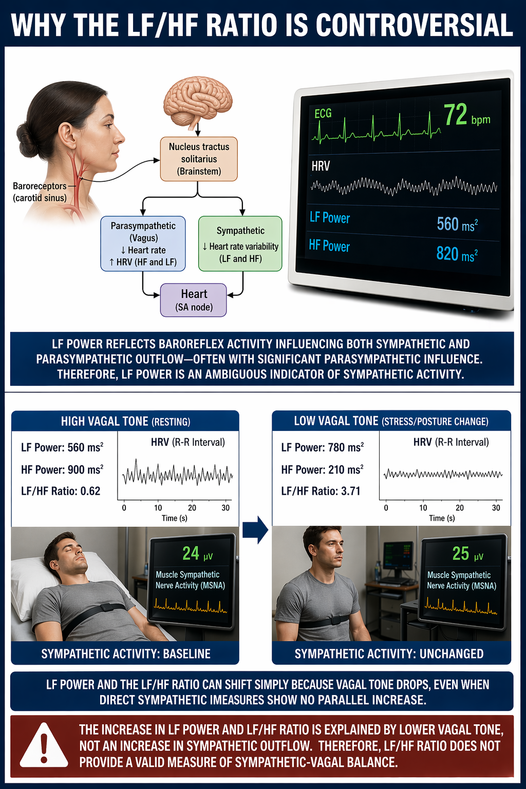
vmHRV and Self-Regulation
Heart rate variability, particularly its vagally mediated component, reflects the dynamic interplay between the autonomic nervous system and cognitive-emotional regulation. The neurovisceral integration model (Thayer et al., 2009) posits that vmHRV is linked to self-regulation processes via prefrontal cortical control over the vagus nerve, facilitating adaptive responses to environmental demands. In plain terms, the prefrontal cortex acts as a top-down regulator of the heart through vagal pathways, and vmHRV is the measurable signal of that regulation.
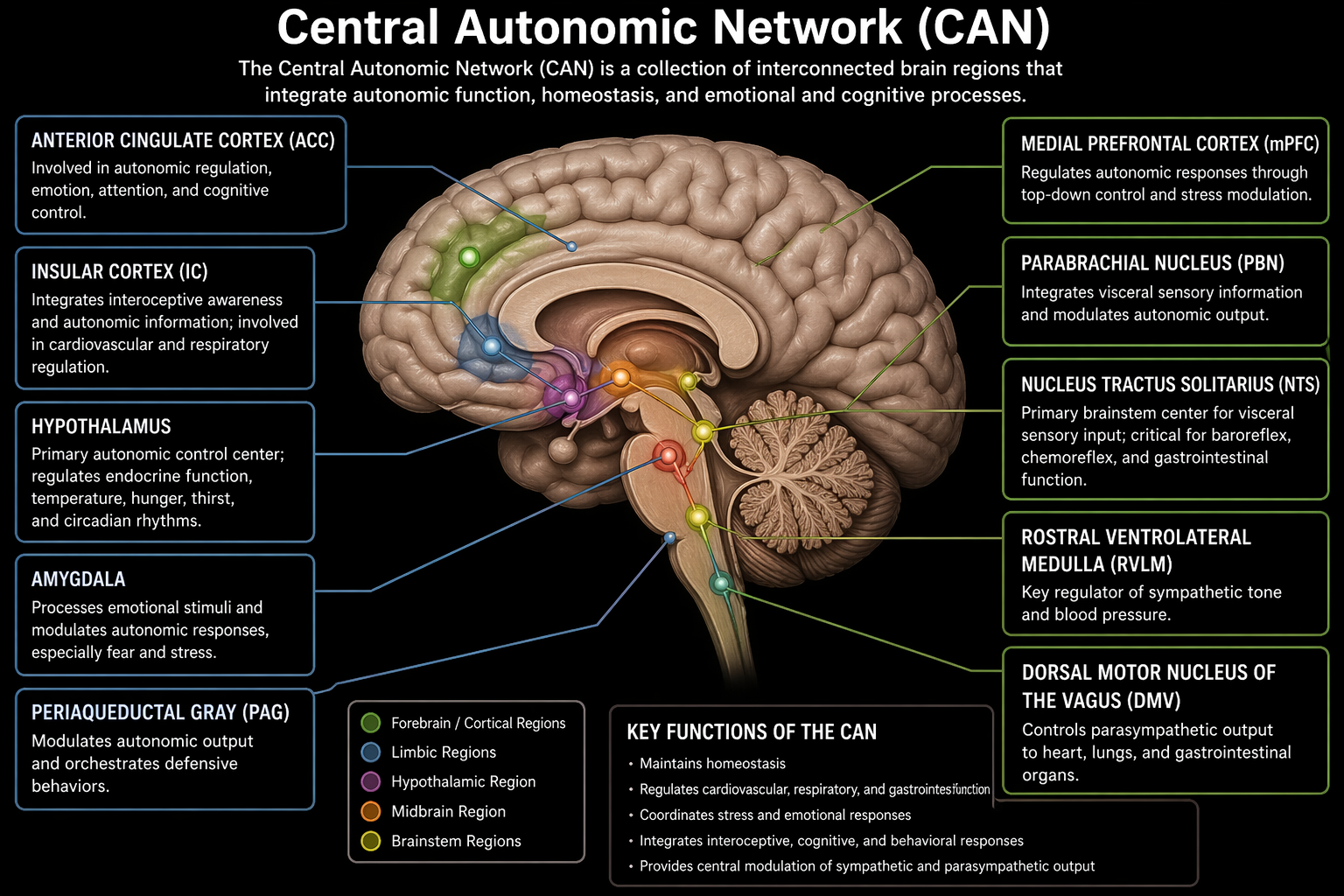
Vagally-mediated HRV (vmHRV) is a noninvasive, actionable biomarker of self-regulation and health, indexed by RMSSD and HF-HRV. Because vagal signals act almost instantly while sympathetic signals are slow, high-frequency HRV is primarily vagal. Whether LF power and the LF/HF ratio index sympathetic tone remains contentious, so sympathetic inferences require triangulation with independent measures. The neurovisceral integration model links vmHRV to top-down prefrontal control of the heart.
Health Implications of vmHRV
Cardiovascular Health
The autonomic nervous system (ANS) plays a critical role in maintaining physiological stability, and vmHRV is an essential biomarker of its function. Higher vmHRV indicates robust parasympathetic activity, which supports greater adaptability to environmental and internal stressors. Lower vmHRV, on the other hand, signals autonomic dysregulation and increased health risks.
Numerous studies have demonstrated that lower vmHRV predicts cardiovascular disease (Hillebrand et al., 2013), including hypertension, atherosclerosis, and heart failure. Individuals with higher vmHRV tend to exhibit greater cardiovascular efficiency, characterized by better baroreceptor sensitivity, improved endothelial function, and enhanced myocardial perfusion. In contrast, diminished vmHRV is often accompanied by increased arterial stiffness, reduced heart rate recovery following exertion, and an overall heightened risk for adverse cardiac events, including myocardial infarction and stroke.
Metabolic Regulation
Beyond cardiovascular health, vmHRV has significant implications for metabolic regulation. Research has consistently linked low vmHRV to metabolic disorders such as diabetes mellitus (Benichou et al., 2018), insulin resistance, and obesity. The autonomic imbalance observed in individuals with lower vmHRV contributes to dysregulated glucose metabolism, increased systemic inflammation, and greater susceptibility to metabolic syndrome.
High vmHRV, conversely, is associated with better glycemic control and improved pancreatic beta-cell function. The modulation of insulin sensitivity via vagal pathways suggests that vmHRV-enhancing interventions—such as slow-paced breathing and vagus nerve stimulation—could serve as valuable therapeutic strategies for individuals with metabolic disorders. For clinicians treating diabetic veterans or hospital patients with metabolic syndrome, HRV biofeedback offers a complementary approach that targets an upstream regulatory mechanism.
Immune Function
Another critical dimension of vmHRV's health implications is its connection to immune function. The cholinergic anti-inflammatory pathway, mediated by vagal activity, plays a pivotal role in regulating inflammatory responses (Williams et al., 2019).
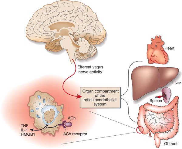
Lower vmHRV is associated with increased levels of pro-inflammatory cytokines such as interleukin-6 (IL-6), tumor necrosis factor-alpha (TNF-α), and C-reactive protein (CRP), which are implicated in the pathogenesis of numerous chronic diseases, including autoimmune conditions, neurodegenerative diseases, and cancer.
Conversely, higher vmHRV correlates with an anti-inflammatory state that facilitates faster recovery from infections and injuries while mitigating chronic inflammation-related pathologies. This has led to growing interest in using vmHRV as a biomarker for immune resilience, particularly in conditions such as rheumatoid arthritis, lupus, and inflammatory bowel disease. The clinical implication is clear: by training clients to improve vagal tone, you may help modulate their inflammatory burden as well.
Stress and Sleep Regulation
The relationship between vmHRV and stress-related health outcomes is particularly compelling. Chronic stress and prolonged exposure to psychological stressors contribute to dysregulation of the hypothalamic-pituitary-adrenal (HPA) axis—the body's central stress response system—and sustained autonomic imbalance. Individuals with lower vmHRV exhibit heightened sympathetic activity and reduced parasympathetic regulation, leading to persistently elevated cortisol levels, increased oxidative stress, and greater allostatic load (the cumulative wear and tear from chronic stress).
This autonomic dysfunction is linked to conditions including irritable bowel syndrome, chronic fatigue syndrome, and fibromyalgia—conditions frequently seen in VA, hospital, and clinic settings. Individuals with higher vmHRV demonstrate more efficient stress recovery mechanisms, allowing for better emotional and physiological resilience.
The significance of vmHRV in sleep regulation further highlights its broad health implications. Sleep disturbances, including insomnia, sleep apnea, and restless leg syndrome, have been associated with reduced vmHRV, indicating impaired autonomic balance during sleep (Chouchou & Desseilles, 2014). Poor sleep quality triggers a cascade of negative health outcomes, including impaired cognitive function, weakened immune defense, and increased cardiovascular and metabolic risk.
Individuals with higher vmHRV tend to experience more stable sleep patterns, enhanced slow-wave sleep, and improved nocturnal autonomic regulation, which collectively support overall well-being and cognitive performance. For clinicians, this connection between vmHRV and sleep quality provides a strong rationale for incorporating HRV biofeedback into treatment plans for clients with insomnia or other sleep disorders.
Higher vmHRV supports cardiovascular efficiency, glycemic control, an anti-inflammatory state through the cholinergic anti-inflammatory pathway, efficient stress recovery, and more stable sleep. Lower vmHRV predicts cardiovascular disease, metabolic disorders, elevated inflammatory cytokines, HPA-axis dysregulation with greater allostatic load, and disturbed sleep. These broad associations provide a strong rationale for HRV biofeedback across many clinical populations.
Clinical and Psychological Relevance
Mental Health Disorders
In clinical psychology and psychiatry, vmHRV is increasingly recognized as a transdiagnostic biomarker—a marker that cuts across traditional diagnostic categories rather than being specific to any single disorder. Individuals with low vmHRV consistently exhibit greater vulnerability to psychiatric disorders, including anxiety disorders, major depressive disorder (MDD), posttraumatic stress disorder (PTSD), and bipolar disorder (Beauchaine & Thayer, 2015). Reduced vmHRV in these populations reflects diminished vagal regulation of emotional responses, heightened sympathetic arousal, and impaired top-down control from prefrontal cortical regions.
For anxiety disorders, lower vmHRV is associated with excessive autonomic arousal, persistent worry, and hypervigilance (Wang et al., 2023). Individuals with generalized anxiety disorder (GAD) often show reduced parasympathetic tone, leading to increased heart rate, respiratory irregularities, and prolonged physiological recovery from stressors. This is precisely the pattern you will observe on biofeedback screens during intake assessments.
Similarly, individuals with PTSD exhibit pronounced autonomic dysregulation, with lower vmHRV reflecting impaired fear extinction and heightened amygdala reactivity to trauma-related cues. Targeted interventions such as HRV biofeedback and vagal nerve stimulation have shown promise in improving autonomic regulation and reducing symptoms of hyperarousal in these populations. For practitioners working with military service members and veterans, HRV biofeedback offers an evidence-based approach to addressing the autonomic roots of trauma-related symptoms.
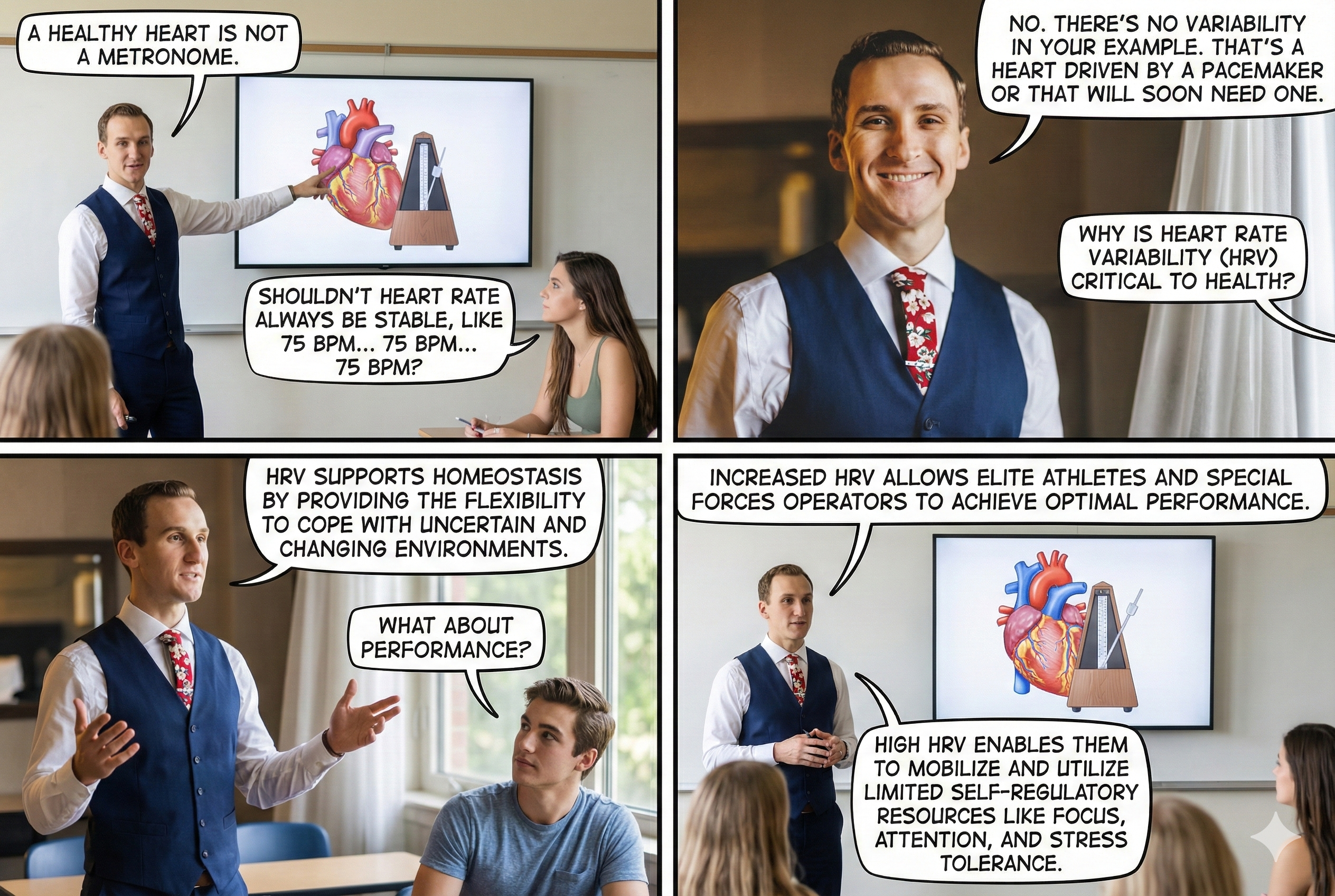
HRV is the organized fluctuation of time intervals between successive heartbeats. A healthy heart is not a metronome; greater beat-to-beat variability reflects better regulatory capacity, executive function, and health. Elevated heart rate limits HRV through cycle length dependence. Vagally-mediated HRV (vmHRV) serves as a critical biomarker for cardiovascular health, metabolic regulation, immune function, stress resilience, and sleep quality. Key theoretical frameworks include the Vagal Tank Theory and Autonomic Space Theory. Low vmHRV is a transdiagnostic marker for psychiatric disorders including anxiety, depression, and PTSD.
How HRV Biofeedback Can Support Neurofeedback
HRV biofeedback (HRVB) is the real-time display of HRV back to the individual. When HRVB training uses a paced-breathing protocol, clinicians can assess and correct dysfunctional breathing behaviors that might otherwise interfere with both HRVB and neurofeedback. Increased HRV is associated with improved executive function and may strengthen descending medial prefrontal cortex regulation of emotion (Mather & Thayer, 2018; McCraty & Shaffer, 2015)—benefits that directly support neurofeedback goals. These gains are made possible by the reciprocal interactions between the heart and the brain.
Dr. Gevirtz discusses Evgeny Vaschillo's findings regarding HRVB effects on the BOLD signal peak in multiple brain areas © Association for Applied Psychophysiology and Biofeedback.
Heart-Brain Interactions
This section explores the bidirectional communication between the heart and brain. Understanding these pathways explains why HRV is more than just a cardiac metric—it is a window into how the brain regulates emotion, attention, and threat processing.
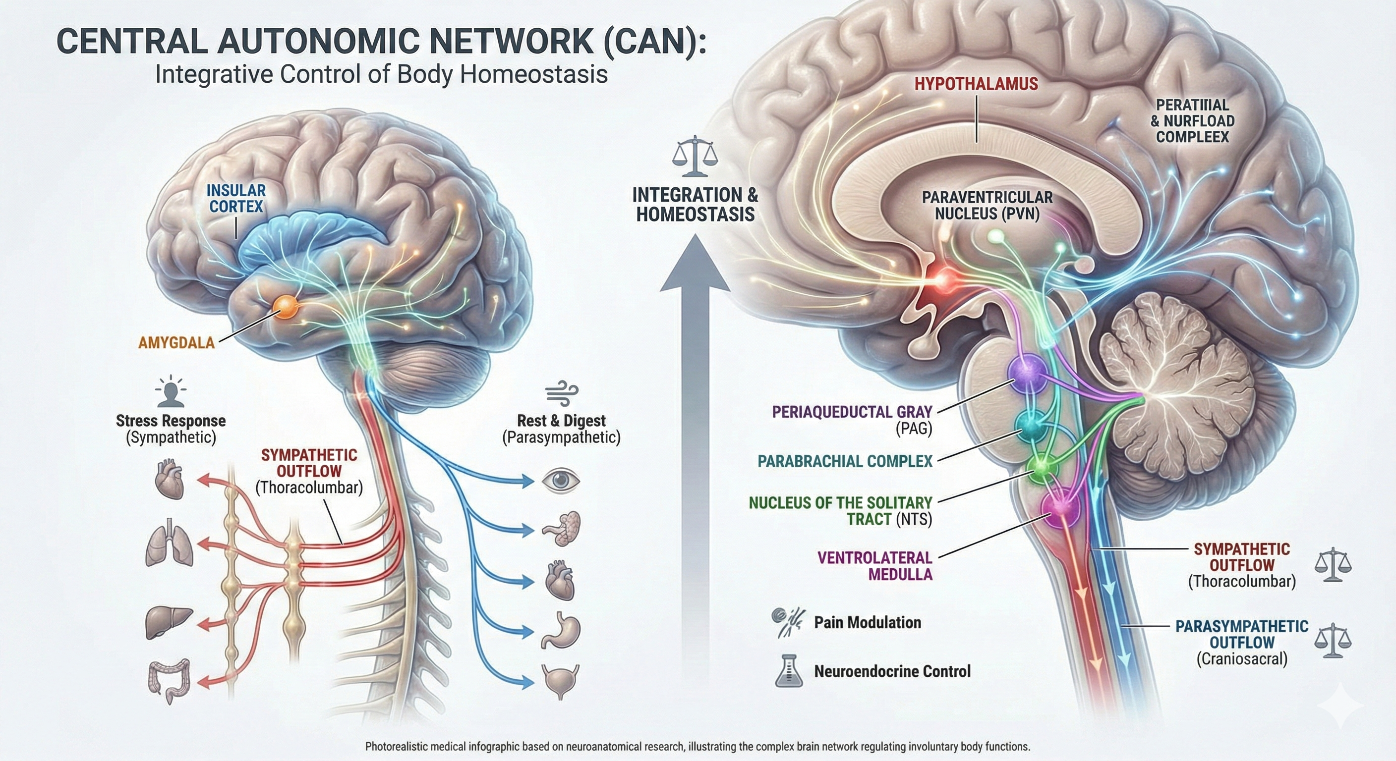
Thayer and Lane (2000) proposed a neurovisceral integration model that describes how a central autonomic network (CAN)—a set of interconnected brain structures including the anterior cingulate, insula, ventromedial prefrontal cortex, amygdala, and hypothalamus—links with the brainstem's nucleus of the solitary tract (NST) through feedback and feed-forward loops. They speculated that a breakdown in negative feedback within this network may produce the increased SNS arousal that characterizes anxiety disorders. Thayer et al. (2012, p. 754) further contended that regions including the amygdala and medial prefrontal cortex, which evaluate "threat and safety," help regulate HRV through their connections with the NST. For clinicians treating anxiety, PTSD, or panic disorder, this model explains why enhancing HRV through biofeedback may improve emotional regulation at the brain level.
The Heart's "Little Brain"
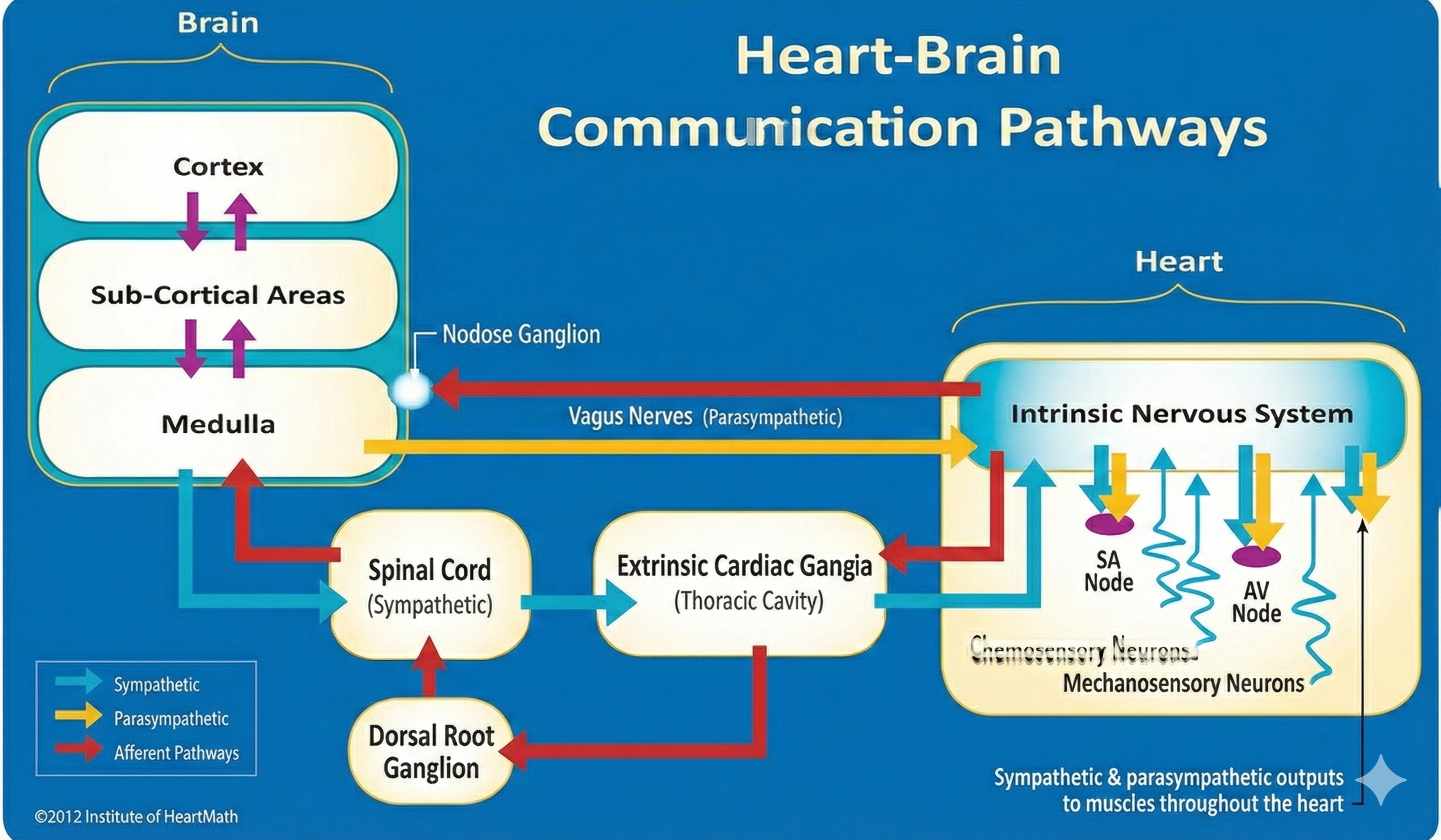
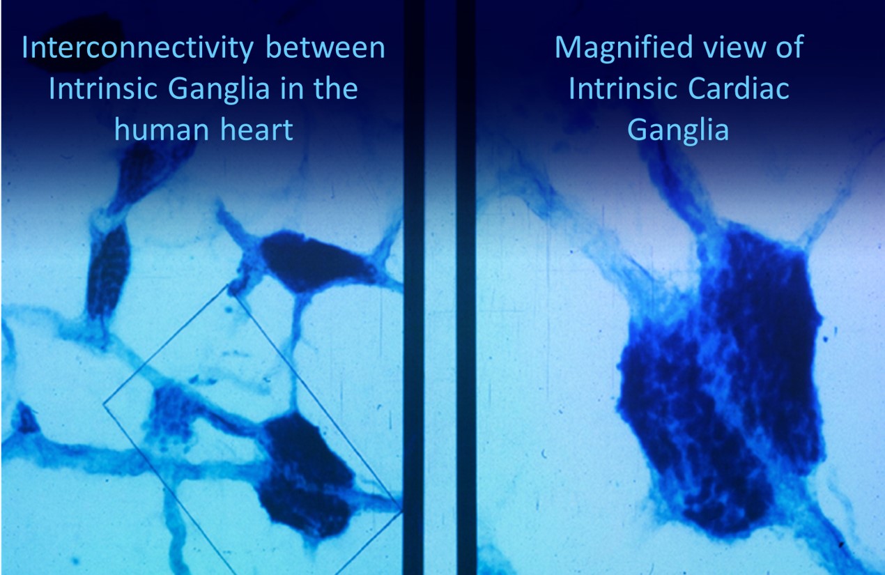
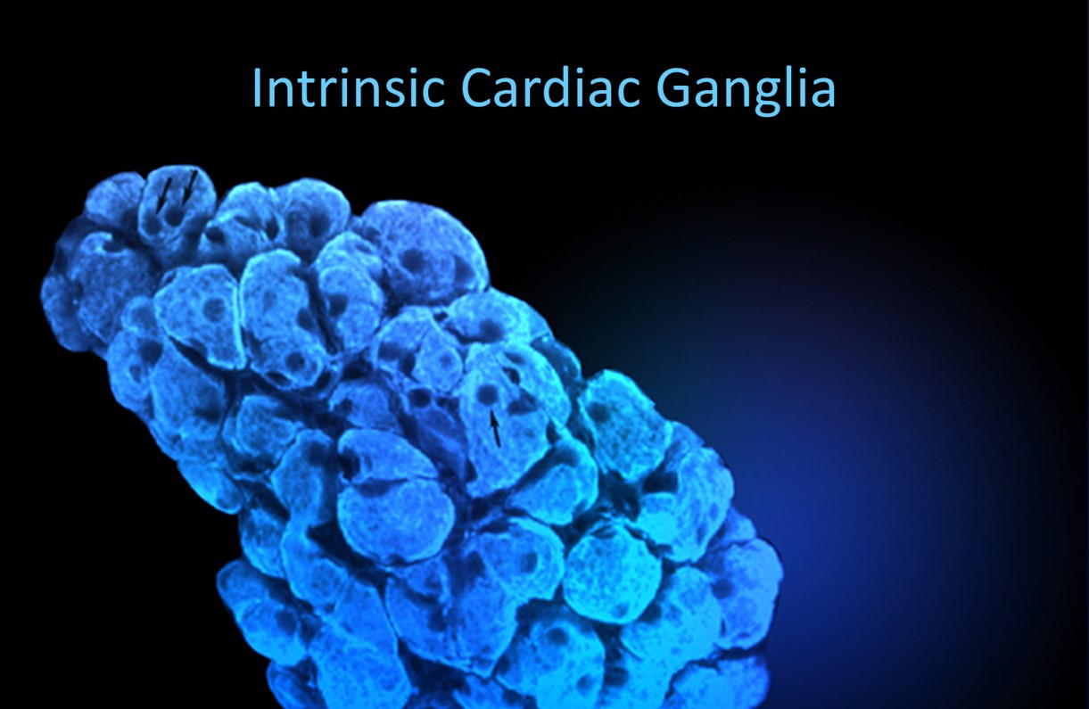
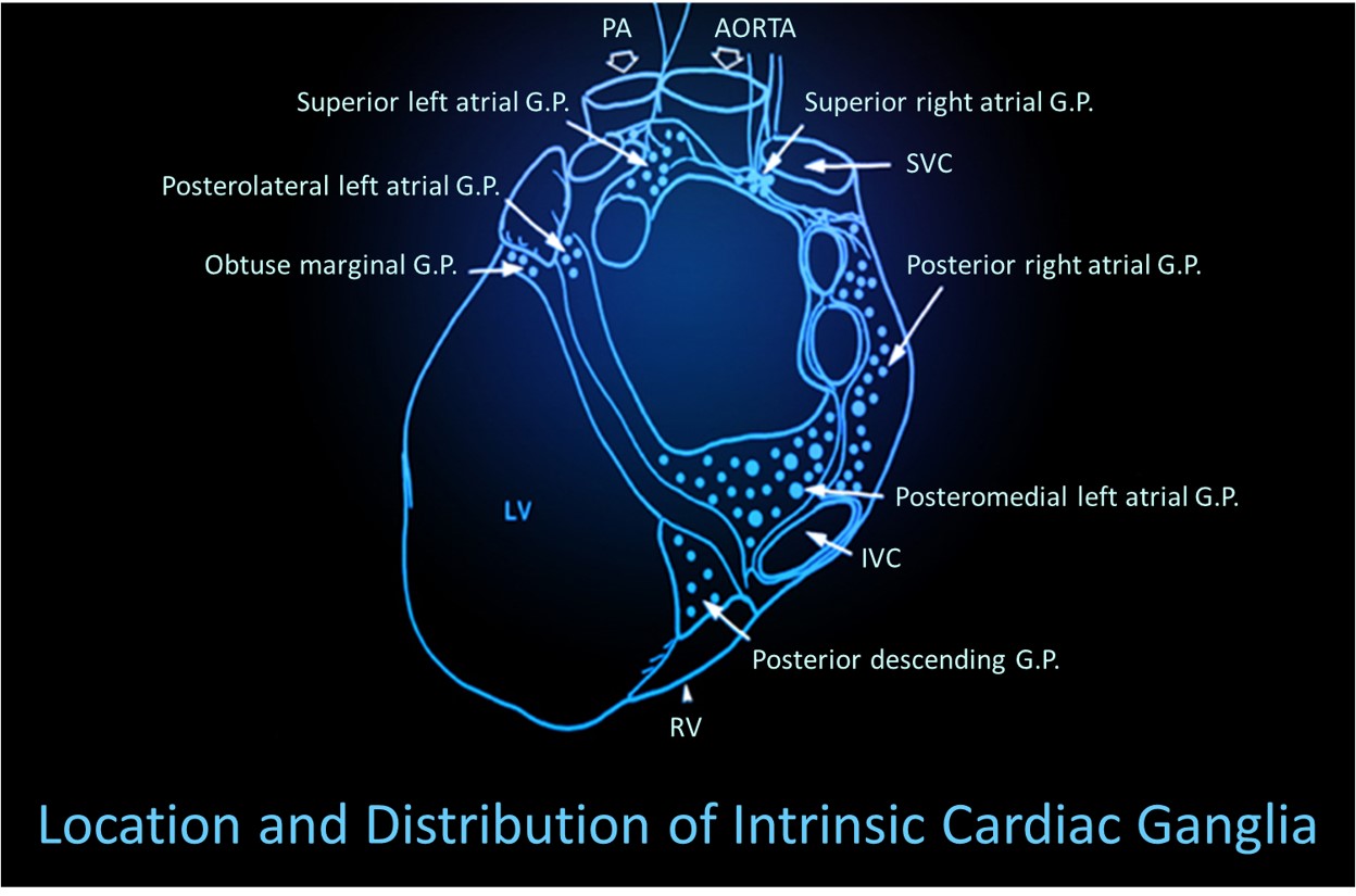
Shaffer et al. (2014) proposed that interconnected cardiac ganglia create an intrinsic nervous system within the heart that influences the SA and AV node pacemakers and forms reciprocal connections with the extrinsic cardiac ganglia found in the chest cavity and the medulla. The sensory, interconnecting, afferent, and motor neurons within the heart can function independently, constituting what researchers call a "little brain" on the mammalian heart.
Mini-Lecture: Heart-Brain Interactions
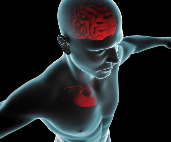
The ascending afferent nerves from the heart help regulate cardiac rhythms and influence efferent SNS and PNS activity. A remarkable 85-90% of vagus nerve fibers are afferents (carrying information toward the brain), and more afferent projections from the heart target the brain than from any other major organ. These afferent signals from the intrinsic cardiac nervous system appear to affect attention, motivation, perceptual sensitivity, and emotional processing (Shaffer et al., 2014). This finding has profound implications for performance practitioners: optimizing cardiac coherence through HRV biofeedback may enhance cognitive function by improving the quality of signals the heart sends to the brain.
Resonance Frequency Breathing and Cardiac-Brain Communication
MacKinnon et al. (2013) provided compelling evidence for how HRV biofeedback strengthens heart-brain communication. They found that HRV influences the amplitude of heartbeat event-related potentials (HERPs)—negative EEG potentials that appear about 200-300 milliseconds after each R-spike and index cardiac afferent communication with the brain. Both negative and positive emotion conditions reduced HRV and HERP amplitude, suggesting that emotional states can disrupt heart-to-brain signaling. In contrast, resonance frequency breathing increased HRV above baseline and increased HERP amplitude.
The authors speculated that resonance frequency breathing reduces interference with vagal afferent signal transmission from the heart to the cerebral cortex. For practitioners, this finding offers a compelling mechanism to share with clients: resonance frequency breathing does not just calm the body—it may actually enhance the clarity of the information your heart sends to your brain, improving emotional regulation and cognitive performance.
Respiration
This section covers why healthy breathing is critical to the success of both HRV biofeedback and neurofeedback training. Unless clinicians identify and correct dysfunctional breathing patterns like overbreathing, these interventions may be less effective or fail altogether. You will learn respiratory anatomy and physiology, the physiological mechanisms that regulate blood gases, disordered breathing patterns, healthy breathing principles, and how overbreathing affects the EEG.
We encourage you to review breathing misconceptions in our A Comprehensive Breathing Myths Guide.
Respiratory Physiology
Listen to a mini-lecture on Respiratory PhysiologyUnderstanding pH: The Foundation of Blood Chemistry
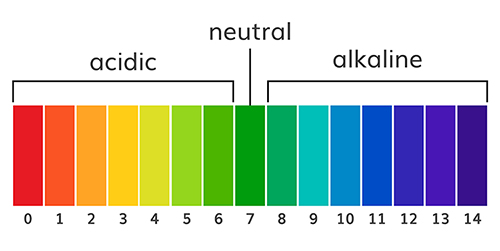
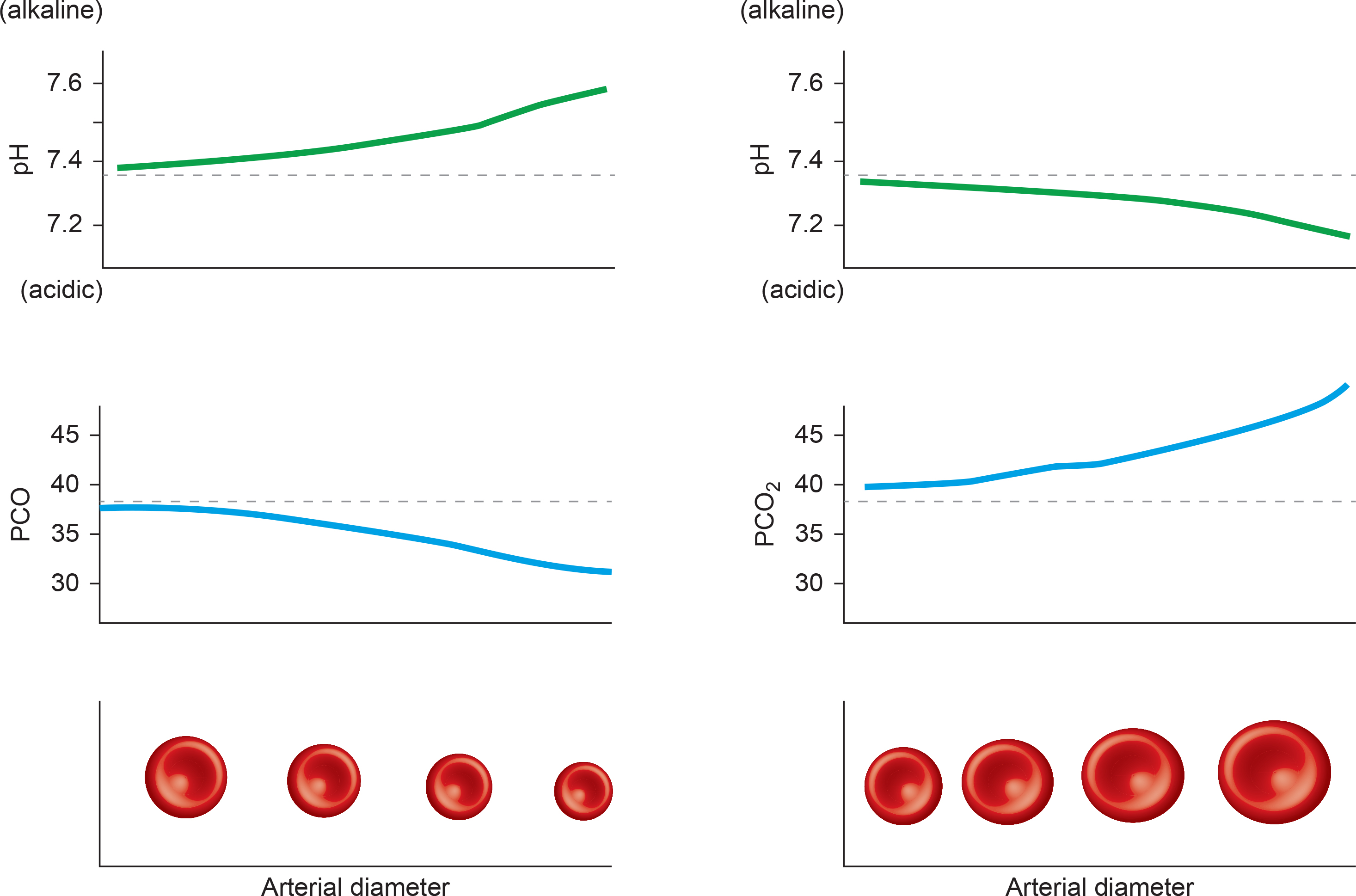
The abbreviation pH refers to the power of hydrogen, which is the concentration of hydrogen ions. Acidic solutions have a low pH (less than 7) due to a high concentration of hydrogen ions. A neutral solution of distilled water has a pH of 7. Alkaline or basic solutions have a high pH (greater than 7) due to a low concentration of hydrogen ions. The pH level regulates oxygen and nitric oxide release.
Here is a surprising fact that challenges our intuition about breathing: the "waste gas" we exhale with every breath is actually a master regulator that keeps us alive. As CO2 travels through our bloodstream, it transforms into carbonic acid, a weak acid that serves as the primary buffer for maintaining blood pH at precisely 7.35 to 7.45 (Gilbert, 2005). This delicate balance is so critical that even small deviations can trigger cascading symptoms throughout the body. When we breathe normally, our bodies maintain about 5% CO2 in arterial blood, creating perfect harmony between production and elimination.
The consequences of pH imbalance extend far beyond discomfort. Blood pH below 7.0 leads to severe acidosis, causing disorientation, coma, and potentially death, while pH above 7.7 creates dangerous alkalosis with similar life-threatening outcomes (Gilbert, 2005). Medical professionals sometimes use controlled hyperventilation therapeutically, such as reducing brain swelling after head injuries, demonstrating both the power and the danger of manipulating this fundamental physiological process (Tanaka, Sato, & Kasai, 2020).
How Breathing Maintains Healthy CO2 Levels
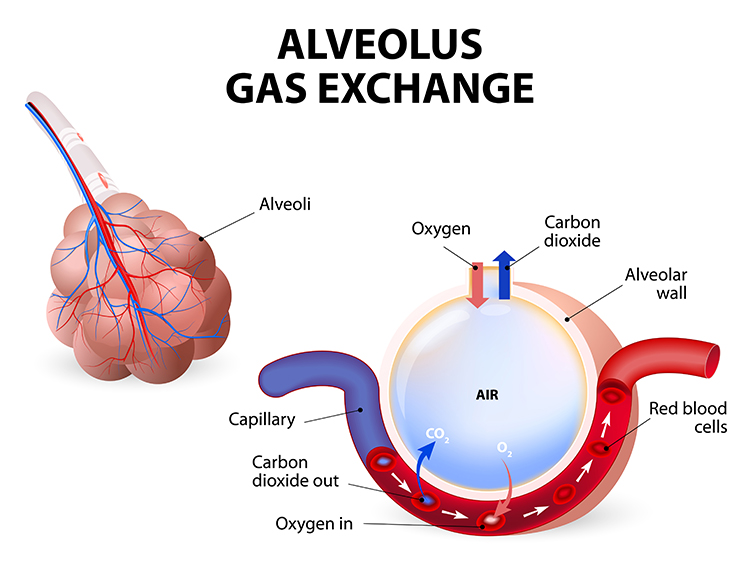
The main functions of breathing are gas exchange and acid-base (pH) regulation. Respiratory system alveoli exchange oxygen for carbon dioxide (CO2) released by cells during metabolism. Alveoli are tiny, thin-walled gas exchange sacs in the lung (Fox & Rompolski, 2022).
CO2: The Unsung Hero of Blood Chemistry
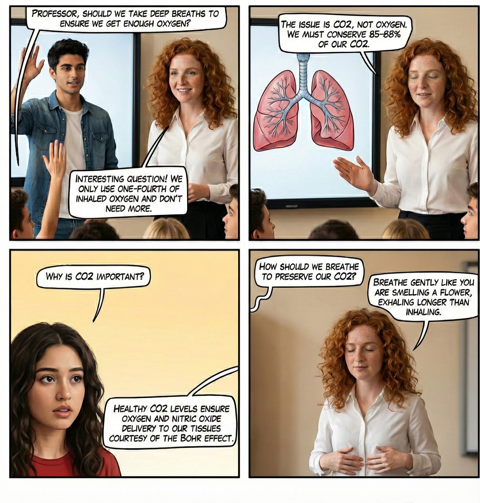
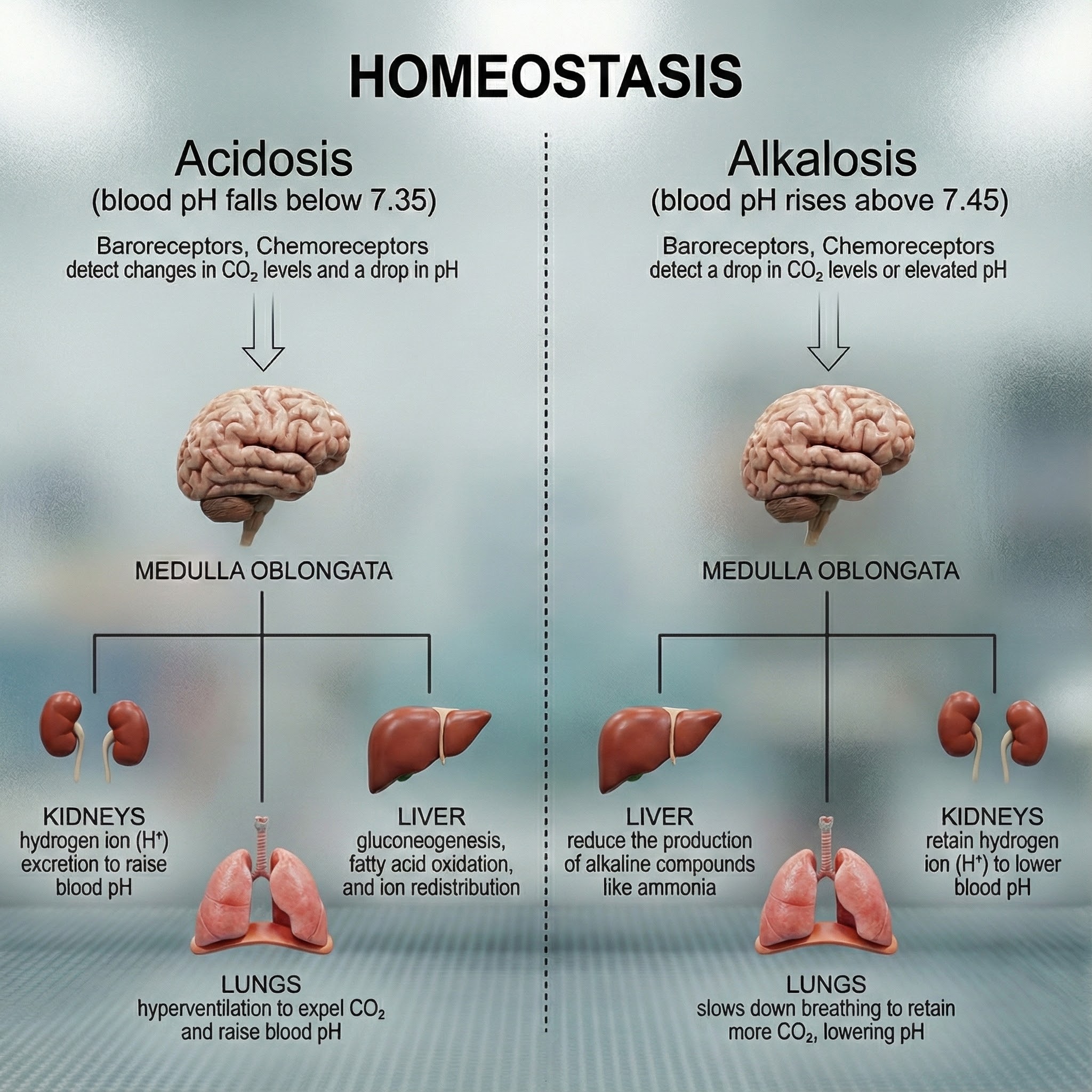
Here is a fact that surprises many people: our body uses 85-88% of blood CO2 to ensure a healthy acid-base balance to prevent our blood from becoming too acidic (acidosis) or basic (alkalosis).
Breathing allows the respiratory system to maintain a blood pH level between 7.35 and 7.45 (Hopkins, Sanvictores, & Sharma, 2022).
Hemoglobin: The Oxygen Delivery System
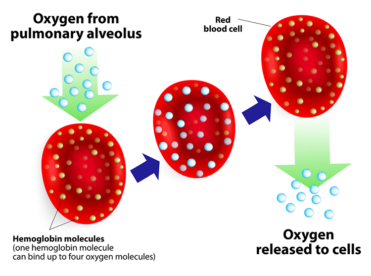
Hemoglobin molecules on red blood cells transport oxygen and nitric oxide through the bloodstream. Each human red blood cell contains about 270 million hemoglobin molecules. One hemoglobin molecule can carry four oxygen or nitric oxide molecules. This allows a single red blood cell to carry over 1 billion oxygen molecules under full saturation. Oxygen and nitric oxide compete for attachment to hemoglobin's binding sites.
.jpg)
How CO2 Controls pH Levels
CO2 regulates pH levels to distribute oxygen and nitric oxide. When cells are active, they produce CO2.
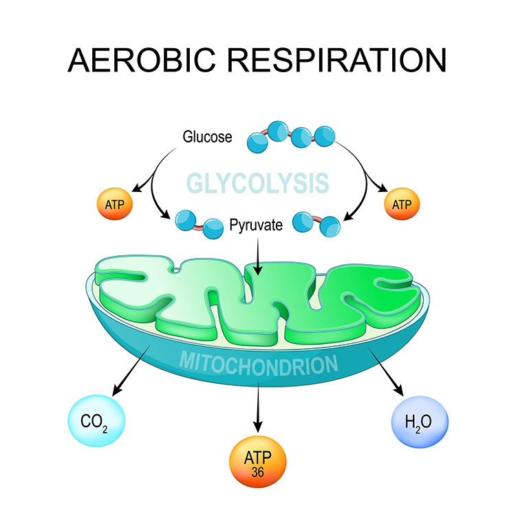
Inspiratory muscle activity (such as diaphragm and the external intercostals) increases metabolism. Muscles break down glucose and fatty acids to power contraction, which produces CO2 (Milic-Emili & Tyler, 1963; Swenson, 2017).
Slow-paced breathing with low tidal volumes retains more CO2 in arterial blood than when overbreathing. Tidal volume (TV) is the amount of air inhaled or exhaled during a normal breath (Fox & Rompolski, 2022).
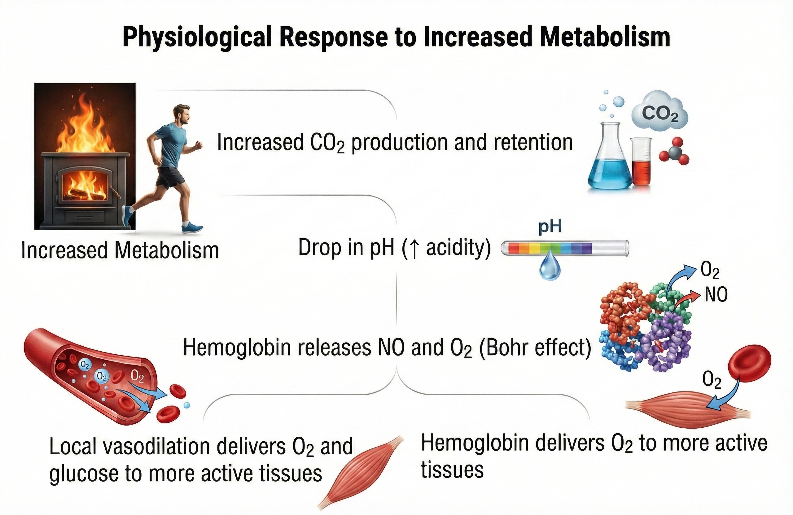
In both cases, rising CO2 lowers blood pH (more acidic), weakening the bond between hemoglobin and oxygen. Oxygen loss changes hemoglobin's shape, destabilizing its bond with NO. Oxygen and NO release support physical activity.
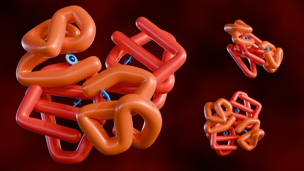
Breathing plays a pivotal role in regulating the pH level of the blood. By adjusting the rate and depth of breathing, the body can control the amount of CO2 expelled, directly influencing the blood's acidity. This regulation is vital for the proper functioning of enzymes and metabolic processes (Guyenet & Bayliss, 2015).
The infographic below shows how CO2 regulates blood pH and is redrawn from Gilbert (2005).
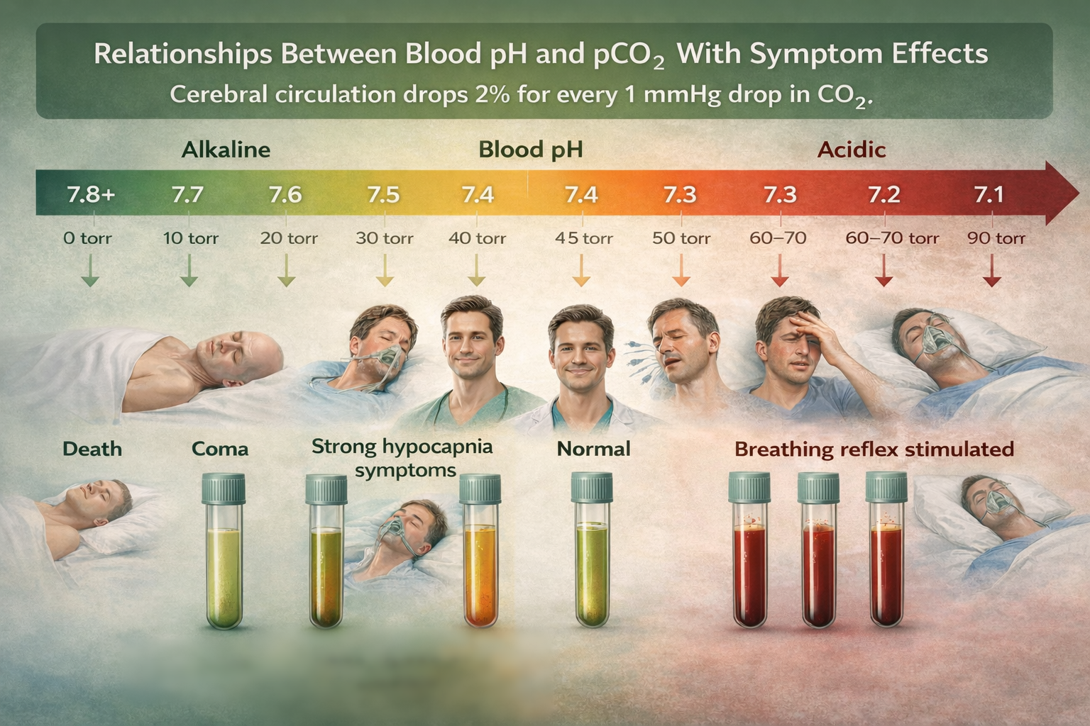
Breathing supports the efficient exchange of gases in the lungs, where oxygen is absorbed into the bloodstream, and CO2 is released for exhalation. This exchange is crucial for metabolic activities and energy production within cells (Hsia, 2023).
The Bohr Effect: Why CO2 Matters for Oxygen Delivery
The Bohr effect enables oxygen to leave its hemoglobin carrier to enter blood vessels and cells (Riggs, 1988). Think of it this way: CO2 acts like a key that unlocks oxygen from hemoglobin. Without adequate CO2, oxygen stays bound to hemoglobin and cannot reach the tissues that need it.
Recent research has demonstrated that slow-paced breathing at approximately 6 breaths per minute, often called resonance breathing, optimizes the balance between oxygen delivery and CO2 retention (Laborde et al., 2022). A comprehensive meta-analysis of 223 studies found that voluntary slow breathing consistently increases vagally-mediated heart rate variability both during breathing practice and immediately afterward, with effects persisting after multi-session interventions (Laborde et al., 2022). This finding has profound clinical implications: slow-paced breathing appears to enhance parasympathetic nervous system activity, which in turn supports better oxygen utilization through the Bohr effect.
Jung and colleagues (2024) demonstrated that slow breathing at 6 breaths per minute not only increases heart rate variability but also enhances connectivity between the amygdala and medial prefrontal cortex, improving emotional regulation. Interestingly, larger effects were found in older adults and women. These findings suggest that breathing rate influences far more than gas exchange; it appears to modulate the brain circuits involved in stress responses and emotional processing.
Oxygen's Role in Cellular Energy Production
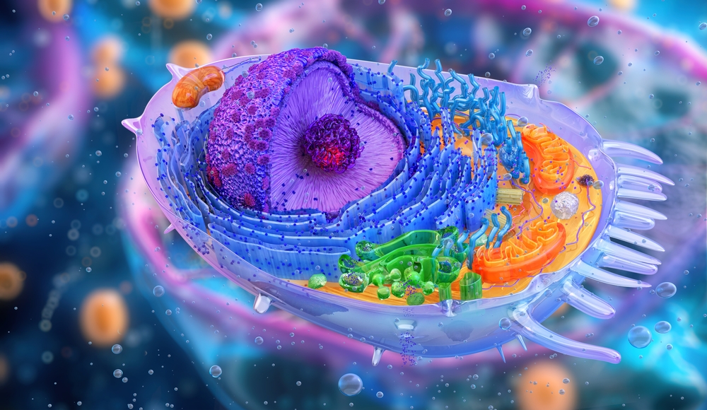
In cells, oxygen supports mitochondrial ATP production and metabolism to support activity.
Nitric Oxide: The Vasodilator
Slow-paced breathing releases more nitric oxide as blood CO2 rises and pH falls, resulting in vasodilation.
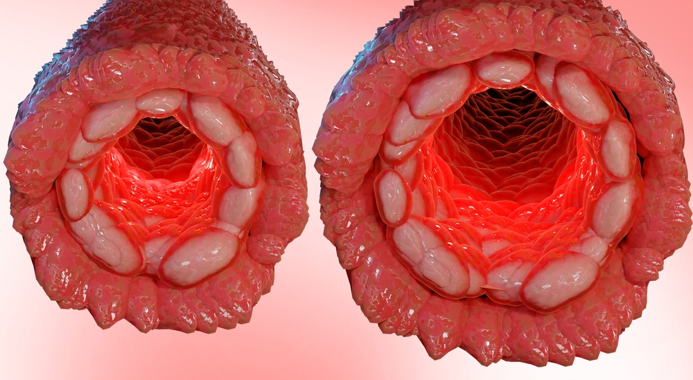
Nitric oxide dilates capillaries and arterioles, increasing oxygen, nitric oxide, and nutrient delivery via blood flow.
Healthy Breathing Roadmap
Listen to a mini-lecture on Healthy Breathing RoadmapDr. Khazan explains internal respiration.
Why We Need to Conserve CO2, Not Get More Oxygen
Here is a counterintuitive truth that surprises many clients: we do not need more oxygen! (Khazan, 2021). Near sea level, the air healthy clients inhale contains 21% oxygen, while the air they exhale has 15%. We only use 25% of inhaled oxygen and do not need more. We need to conserve blood CO2 by retaining 85-88% of it.
Breathing Serves More Than Gas Exchange
The respiratory system also delivers odorants to the olfactory epithelium, produces the airway pressure required for speech, anticipates cognitive and skeletal muscle metabolic demands, and helps to modulate systems regulated by the autonomic nervous system (ANS), especially the cardiovascular system. Respiration is an important regulator of heart rate variability, consisting of beat-to-beat changes in the heart rhythm (Lorig, 2007). Check out the YouTube video The Respiratory System.
The Respiratory Cycle
We breathe about 20,000 times a day. Typical adult resting breathing rates are 12-20 breaths per minute (bpm; Saatchi et al., 2025). Disorders that affect respiration may raise rates to 18-28 bpm (Fried, 1987; Fried & Grimaldi, 1993).
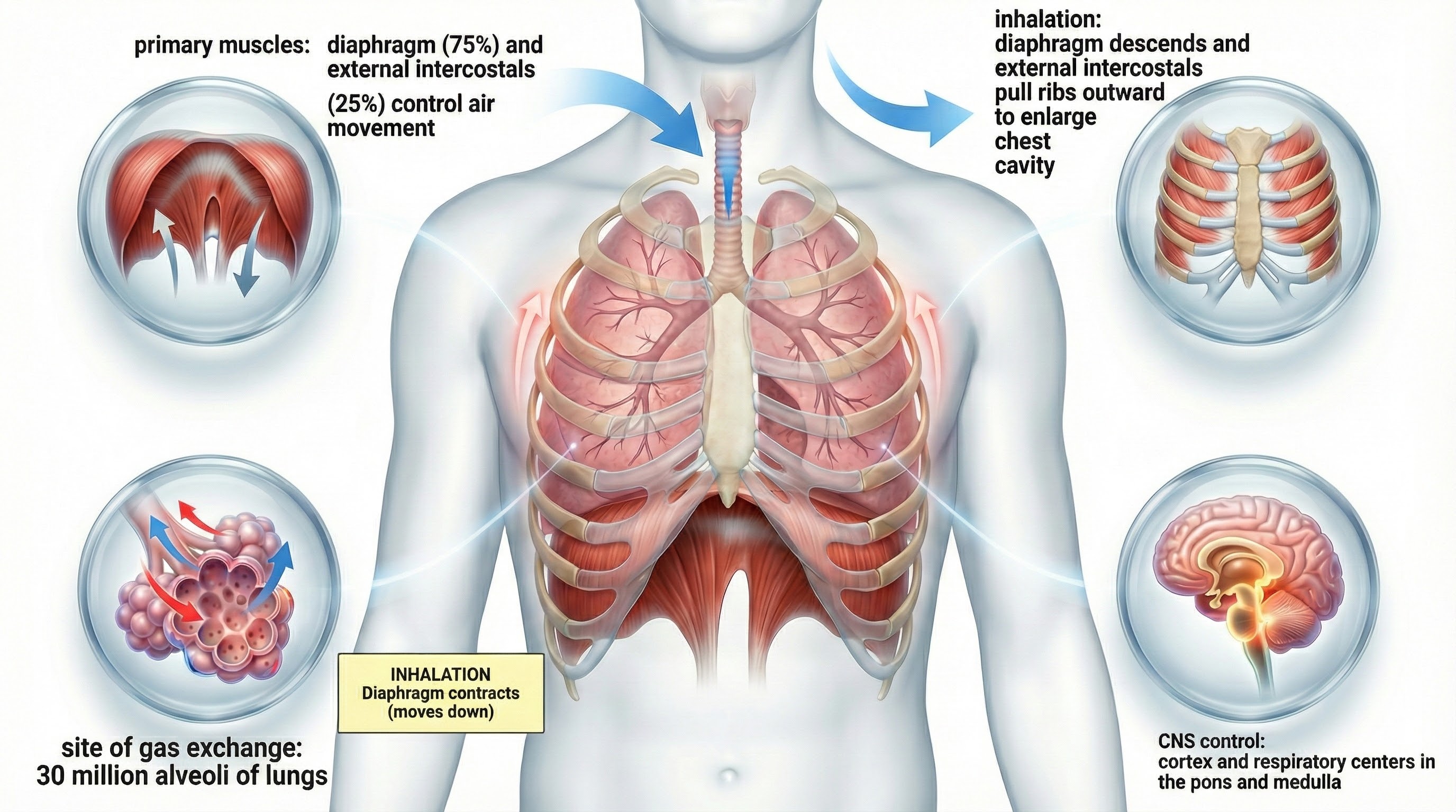
The respiratory cycle consists of inhalation (breathing in) and exhalation (breathing out), controlled by separate mechanisms.
The lungs cannot inflate themselves since they lack skeletal muscles. Instead, they passively inflate by creating a partial vacuum by the diaphragm and external intercostal muscles (Gevirtz, Schwartz, & Lehrer, 2016).
During inhalation, contraction by the diaphragm and external intercostal muscles ventilate the lungs.
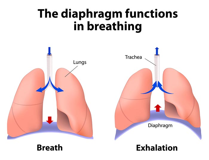
The dome-shaped diaphragm muscle plays the lead role during inhalation. The diaphragm comprises the floor of the thoracic cavity. When the diaphragm contracts, it flattens, and its dome drops, increasing thoracic cavity volume. Contraction of the diaphragm pushes the rectus abdominis muscle of the stomach down and out.
In the animation below, watch the lungs inflate as the diaphragm descends.
In relaxed breathing, a 1-cm descent creates a 1-3 mmHg pressure difference and moves 500 milliliters of air. In labored breathing, a 10-cm descent produces a 100-mmHg pressure difference and transports 2-3 liters of air. The diaphragm accounts for about 75% of air movement into the lungs during relaxed breathing.
A systematic review and meta-analysis by Abdullahi, Wong, and Ng (2024) found that diaphragmatic breathing exercise significantly improves respiratory function in stroke patients, with marked improvements in forced vital capacity, forced expiratory volume, and peak expiratory flow. These findings extend beyond stroke rehabilitation to suggest that diaphragmatic breathing training can benefit anyone seeking to optimize respiratory mechanics and CO2 retention.
Research by Vranich and colleagues (2025) introduced the Breathing IQ, an anthropometric index that measures the efficiency of diaphragmatic breathing by assessing abdominothoracic expansion during inhalation and exhalation. In a study of 384 participants, a single 90-minute intervention improved breathing mechanics dramatically, with the percentage of participants demonstrating efficient diaphragmatic breathing increasing from just 3.7% to 26.6%. This finding suggests that dysfunctional breathing patterns are common but highly correctable with proper instruction.
The external intercostals play a supporting role during inhalation. External intercostal muscle contraction pulls the ribs upward and enlarges the thoracic cavity. The external intercostals account for about 25% of air movement into the lungs during relaxed breathing.
The contraction of the diaphragm and the external intercostals expands the thoracic cavity, increases lung volume, and decreases the pressure within the lungs below atmospheric pressure. This pressure difference causes air to inflate the lungs until the alveolar pressure returns to atmospheric pressure.
During forceful inhalation, accessory muscles of inhalation (sternocleidomastoid, scalene, pectoralis major and minor, serratus anterior, and latissimus dorsi) also contract (Khazan, 2021).
How the Dome-Shaped Diaphragm Produces Exhalation
The relaxation of the diaphragm and external intercostal muscles, contraction of the internal intercostals, the elastic recoil of the chest wall and lungs, and surface tension produce exhalation during relaxed breathing. When the diaphragm relaxes, its dome moves upward. When the external intercostals relax, the ribs move downward. These changes reduce the thoracic cavity volume and the lungs and increase the pressure within the lungs above atmospheric pressure. This pressure difference causes air to deflate the lungs until the alveolar pressure returns to atmospheric pressure.
Forceful exhalation during exercise recruits the rectus abdominis, external and internal obliques, and transversus abdominis abdominal muscles (Khazan, 2021; Lorig, 2007; Tortora & Derrickson, 2021).
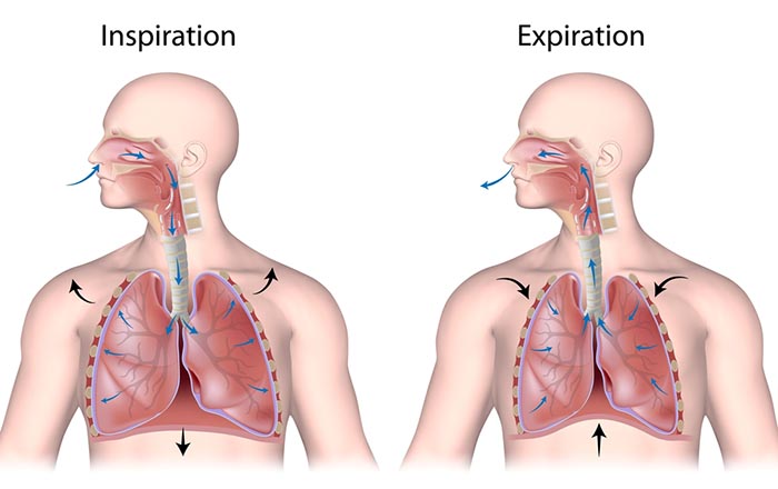
The BioGraph® Infiniti display below shows healthy inhalation and exhalation in which the abdomen gradually expands and then contracts.
Types of Respiration
The term respiration refers to external, internal, and cellular processes. External respiration transports gases in and out of our lungs. Internal respiration transports oxygen from the air we inhale, delivers it to our cells, and returns metabolic CO2 to the lungs for 12-15% to be exhaled and 85-88% retained to regulate pH (Khazan, 2021).
Respiratory gases are exchanged (between the lungs and blood) across the respiratory membrane, comprised of the alveolar and capillary walls.
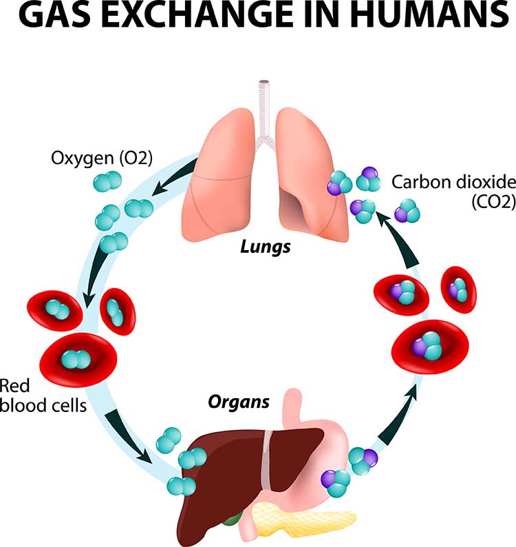
The lungs contain about 300 million pulmonary alveoli (air sacs) that create an incredible 760 ft2 surface for gas exchange (Fox & Rompolski, 2022).
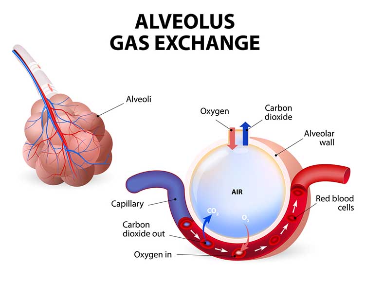
The alveoli collapse like "wet balloons" during normal breathing. Since deflated alveoli cannot absorb normal oxygen levels, the brain triggers sighs to reopen these air sacs. Humans initiate sighs every 5 minutes to increase oxygen delivery and activate the brain through double inhalation (Long, 2016).
The respiratory cycle consists of an inspiratory phase, inspiratory pause, expiratory phase, and expiratory pause. Abdominal respirometer excursion, which indexes respiratory amplitude (the peak-to-trough difference), is often greatest during the inspiratory pause. The diagram below was adapted from Stern, Ray, and Quigley (2001).
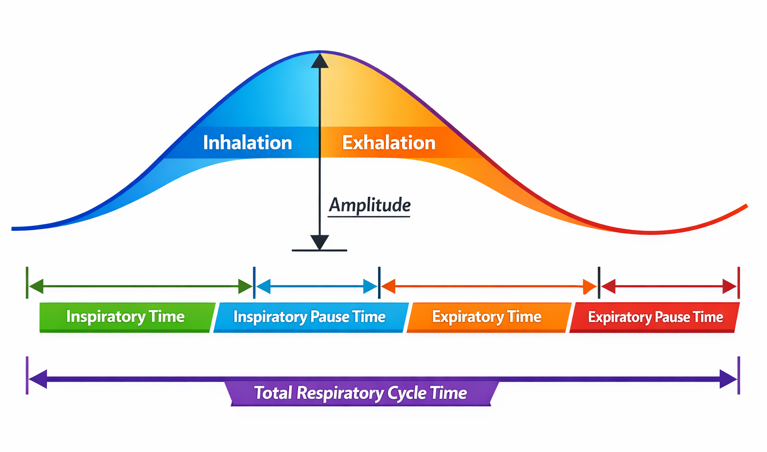
Clinicians should examine all components of the respiratory cycle, not just respiration rate, to understand their clients' respiratory mechanics. Everyday activities like speaking and writing checks may affect individual components differently. Apnea, breath suspension, lowers respiration rate. Clinicians teaching effortless breathing training may instruct their clients to lengthen the expiratory pause with respect to the inspiratory pause. Simple inspection of their respiration rates will not show whether they have successfully changed the relative durations of these two pauses. Finally, in heart rate variability (HRV) biofeedback, clinicians encourage slow (5-7 bpm) and rhythmic breathing.
Neural Control of Respiration
Respiration is controlled by a respiratory center in the medulla and pontine respiratory group. The dorsal respiratory group (DRG) and ventral respiratory group (VRG) are neuron clusters in two medulla regions. Pacemaker cells located in the VRG (analogous to the heart's sinoatrial node) organize the basic breathing rhythm. Check out the Khan Academy YouTube video The Respiratory Center.
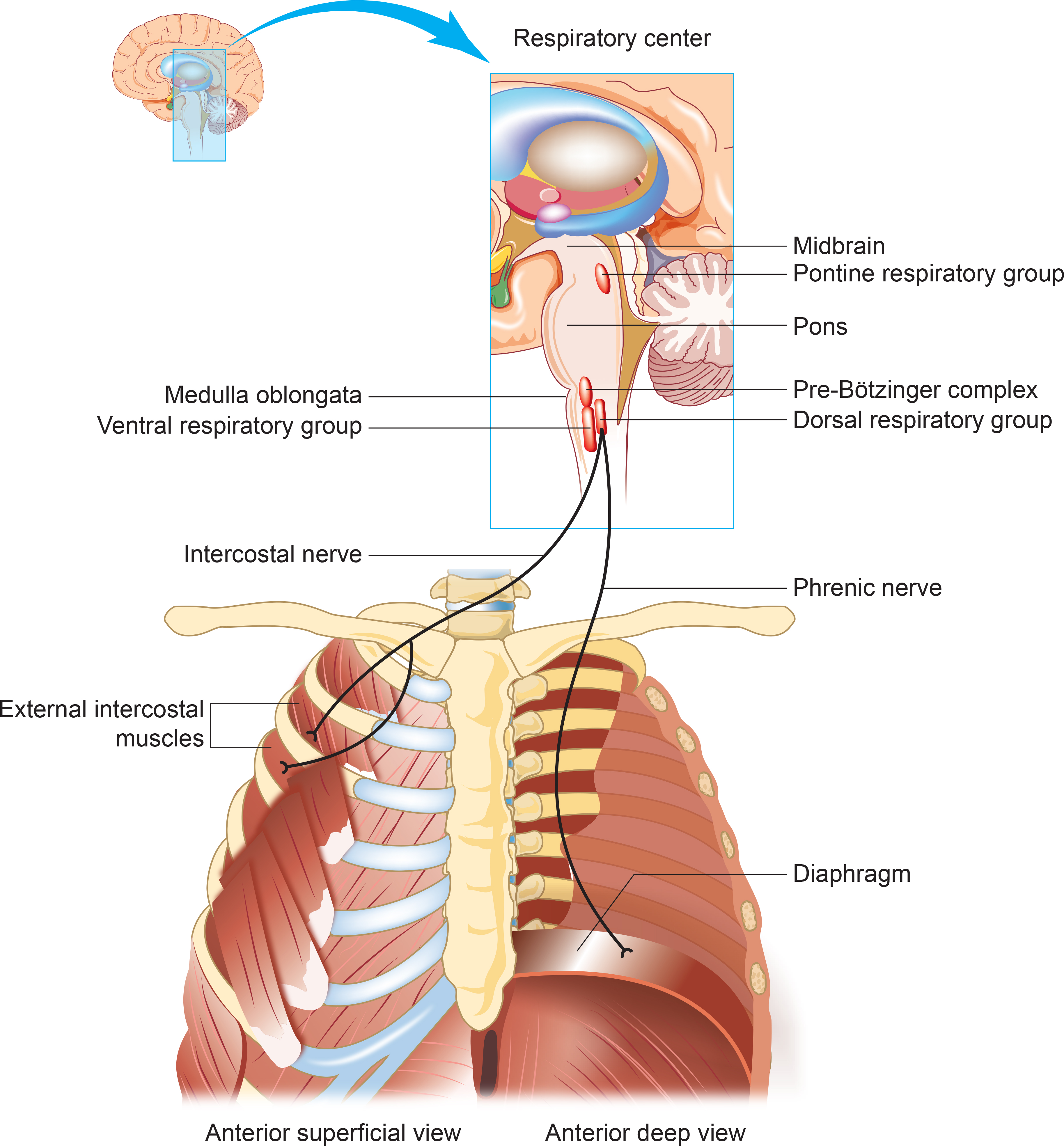
The Medulla: DRG and VRG
The DRG's Role in Breathing
The DRG collects information from peripheral stretch and chemoreceptors and distributes it to the VRG to modify its breathing rhythms. The DRG is responsible for normal quiet breathing.
The majority of VRG neurons are inactive at this time. DRG impulses to the diaphragm and external intercostals begin in weak bursts that strengthen for about 2 seconds and then stop. This stimulation causes muscle contraction and inhalation. After 2 seconds of DRG inactivity, the diaphragm and external intercostals relax for 3 seconds so that the lungs and chest wall can passively recoil to allow the next breathing cycle.
During forceful breathing, DRG neurons activate the VRG, which stimulates the diaphragm, sternocleidomastoid, pectoralis minor, scalene, and trapezius muscles to contract (Tortora & Derrickson, 2021).
The VRG's Role in Breathing
The phrenic and intercostal nerves transmit VRG inspiratory neuron action potentials to the diaphragm and external intercostal muscles. The contraction of these muscles expands the thoracic cavity and inflates the lungs.
VRG pacemaker cells influence the rate of DRG action potentials. VRG expiratory neurons inhibit DRG inspiratory neuron firing. Exhalation passively results from diaphragm and external intercostal muscle relaxation and recoil by the chest wall and lungs. The DRG and VRG neurons' continuous reciprocal activity results in a 12-15 bpm respiratory rate, with 2-second inspiratory and 3-second expiratory phases.
Why Drug Overdoses Can Be Lethal
An overdose of a CNS depressant like alcohol or morphine can completely inhibit VRG neurons in the medulla and stop breathing.
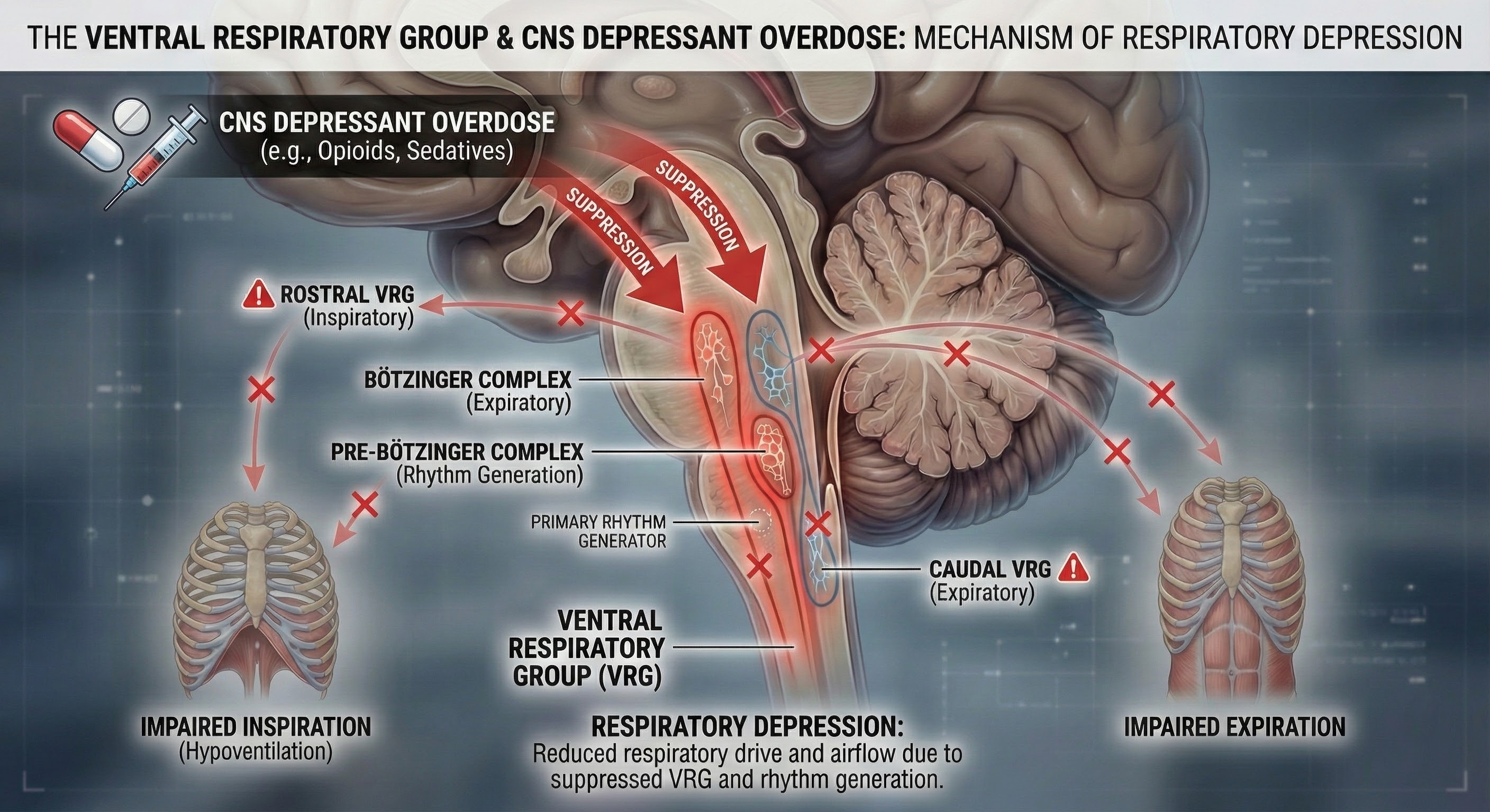
The Pons: The Respiratory Group's Role
The pontine respiratory group adjusts VRG breathing rhythms based on descending input from brain structures and peripheral sensory input.
Research has revealed that breathing biofeedback can modify these neural control mechanisms with lasting therapeutic effects. Tolin and colleagues (2017) conducted a multisite trial demonstrating that capnometry-guided respiratory intervention, which provides real-time feedback on end-tidal CO2 and respiration rate, achieved an 83% response rate and 54% remission rate for panic disorder, with benefits maintained at 12-month follow-up. This intervention works by helping patients recognize when their breathing exceeds metabolic needs and training them to restore healthy CO2 levels.
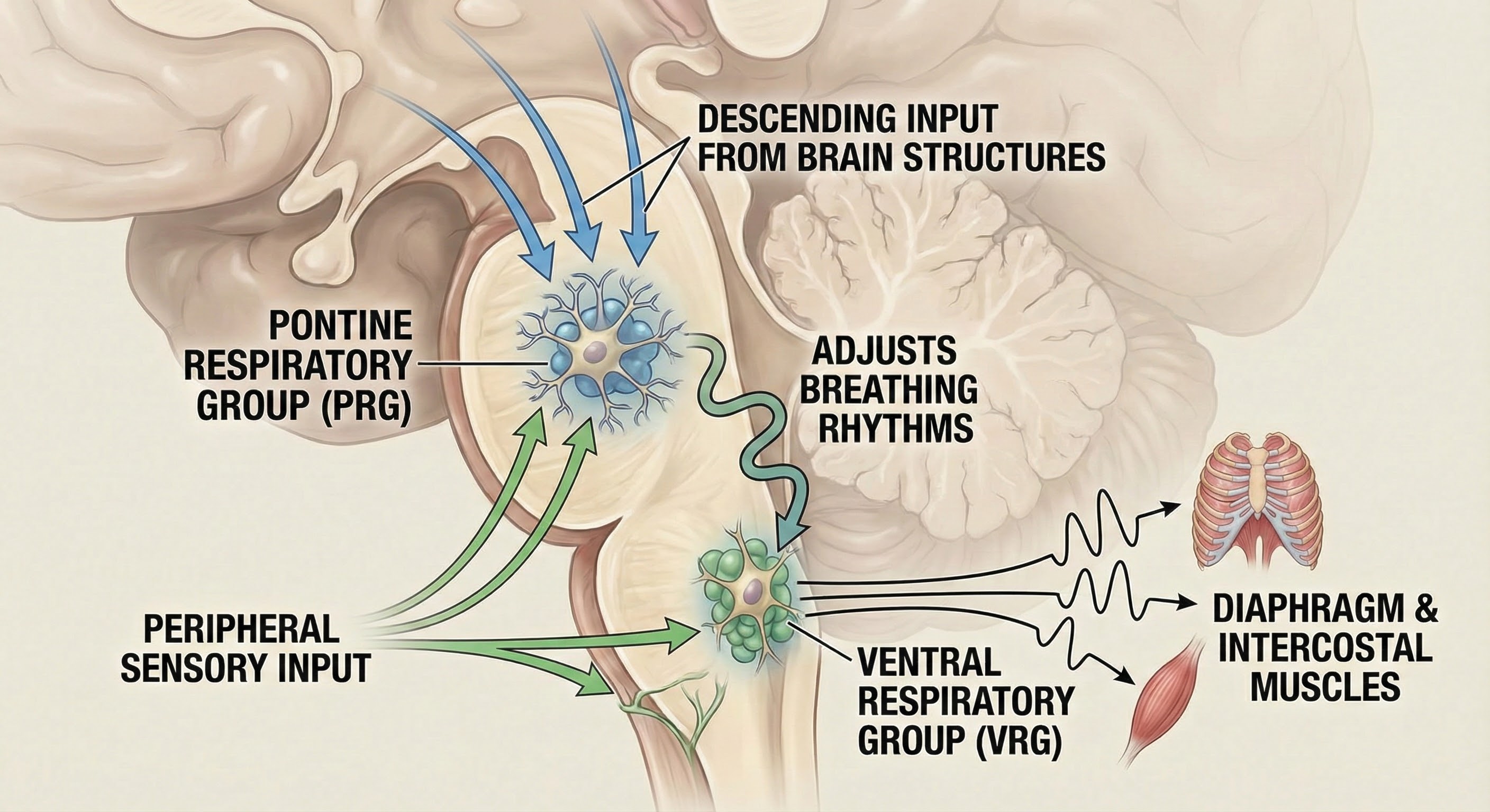
The pontine respiratory group modifies breathing during activities like exercise, sleep, and speech (Marieb & Hoehn, 2019). The body's cells require about 200 ml of oxygen at rest. This demand increases 15-20 times during strenuous exercise; 30 times for elite athletes.
The Cerebral Cortex's Role in Breathing
Cortical control of respiratory centers in the medulla and pons allows us to voluntarily stop or change our breathing pattern. This voluntary control protects against lung damage from water or toxic gases.
Why You Cannot Hold Your Breath Indefinitely
Have you ever tried to hold your breath until you passed out? You cannot do it, and here is why. The rise of CO2 and H+ in the blood limits our ability to suspend breathing by stimulating the inspiratory area when a critical level is reached. When chemoreceptors monitoring cerebrospinal fluid pH detect decreased pH (greater acidity), the dorsal respiratory group of the medulla initiates the next breath. This homeostatic mechanism prevents us from harming ourselves by holding our breath (Tortora & Derrickson, 2021).
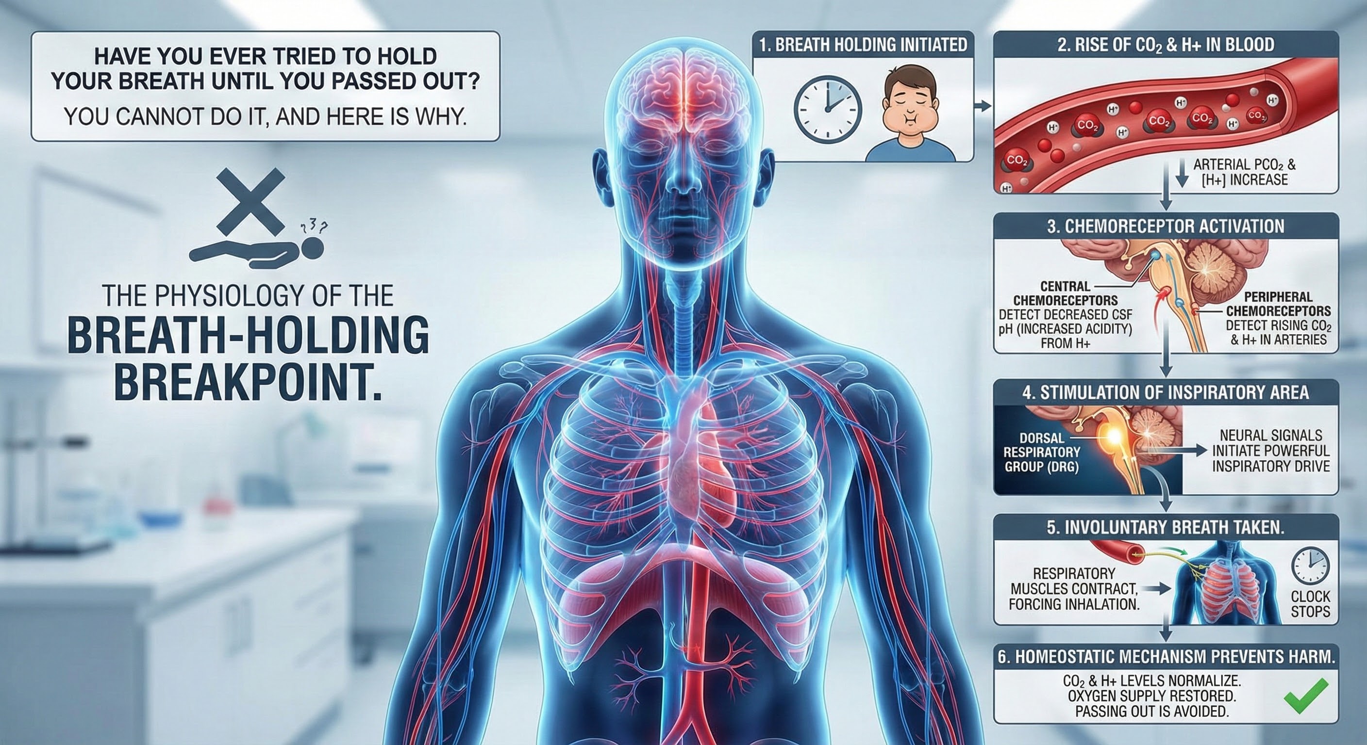
Mouth Versus Nose Breathing: What the Research Shows
Inhaling through the mouth results in greater dead space and airway resistance compared to nose breathing (Tanaka, Morikawa, & Honda, 1988).
Mouth breathing is fine when there is a need for large amounts of air, such as exercising, or when the nose is stuffed.
The nose acts as a natural filter, trapping dust, allergens, and other particulate matter before they can enter the lungs (Bjermer, 1999). Nasal passages also humidify and warm the air, protecting the respiratory tract from irritation and infection (Naclerio et al., 2007). Mouth breathing bypasses these natural defenses (Martel et al., 2020).
Inhaling through the nose releases nitric oxide, a gas that enhances the body's ability to transport oxygen by dilating blood vessels, to the lungs. This process improves oxygen uptake in the blood, contributing to better overall cardiovascular health. Mouth breathing does not offer this benefit (Kimberly et al., 1996; Watso et al., 2023).
Emerging research continues to highlight the cardiovascular benefits of nasal breathing. Watso and colleagues (2023) demonstrated that acute nasal breathing lowers blood pressure and increases parasympathetic contributions to heart rate variability in young adults. This effect appears mediated by nitric oxide released from the paranasal sinuses during nasal inhalation. Unlike mouth breathing, nasal breathing delivers this vasodilatory gas directly to the lungs, where it enhances oxygen uptake and improves overall cardiovascular function.
For individuals recovering from long COVID, slow breathing interventions show promise for restoring autonomic function. Marcella and colleagues (2024) found that healthcare workers with long COVID exhibited reduced heart rate variability compared to healthy controls, but slow-paced breathing significantly attenuated these abnormalities, suggesting that breathing exercises may help restore vascular function and reduce long-term cardiovascular risk in this population.
Chronic mouth breathing can lead to dry mouth, increasing the risk of dental decay and gum disease because saliva, which protects against bacteria and aids digestion, is reduced (Tamkin, 2020). Furthermore, the altered posture of the tongue and jaw can contribute to malformations in children, such as dental malocclusions and facial deformities (Lin et al., 2022).
Inhaling through the nose helps regulate the volume of inhaled air and maintains adequate levels of CO2 in the blood, which is necessary to efficiently release oxygen from hemoglobin to the body's tissues (Prisca et al., 2024). Mouth breathing can lead to overbreathing and a reduction in CO2 levels, impairing oxygen delivery (LaComb et al., 2017; Tanaka, Morikawa, & Honda, 1988).
Exhaling through the nose cannot be regulated in the way that pursed-lips breathing can. Exhaling through pursed lips reduces inappropriate breathing volume when the cause is emotion rather than exercise demand.
Many people favor practicing breathing by inhaling through the nose and exhaling through the mouth, so that is a compromise between the two that makes sense.
Inhalation through the nose, but not the mouth, activates the amygdala, hippocampus, and olfactory cortex neurons.
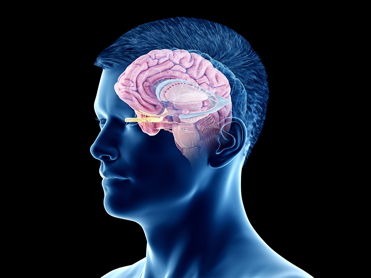
Nasal inhalation may accelerate our response to physical threats and impact fear and memory. During panic attacks, breathing is faster, and we spend more time inhaling (Zelano et al., 2016). Check out the YouTube video How you breathe affects memory and fear.

Disordered Breathing
Listen to a mini-lecture on Disordered BreathingClinicians encounter six abnormal breathing patterns which reduce oxygen delivery to the lungs: thoracic breathing, clavicular breathing, reverse breathing, overbreathing, hyperventilation, and apnea.
Thoracic Breathing: When the Chest Does All the Work
In thoracic breathing, the chest muscles are mainly responsible for breathing. The external intercostals lift the rib cage up and out. The diaphragm is pushed upward as the abdomen is drawn in. Upward and outward movement of the ribs enlarges the thoracic cavity producing a partial vacuum. Negative pressure expands the lungs but is too weak to ventilate their lower lobes. Thoracic breathing reduces ventilation since the lower lobes receive a disproportionate share of the blood supply due to gravity.
Thoracic breathing expends excessive energy, incompletely ventilates the lungs, and strains our accessory muscles.
In the BioGraph® Infiniti screen below, the abdominal (blue trace) strain gauge exhibits minimal excursion, and the respiration rate exceeds the desired 5-7 breaths-per-minute range.
Are you a thoracic breather? Place your left hand on your chest and your right hand on your navel. If both hands shallowly rise and fall at about the same time, you are breathing thoracically.
Clavicular Breathing: The Oxygen Thief
In clavicular breathing, the chest rises, and the collarbones are elevated to draw the abdomen in and raise the diaphragm (Khazan, 2021). Clavicular breathing may accompany thoracic breathing. Patients may breathe through their mouths to increase air intake. This pattern provides minimal pulmonary ventilation. Over time, the accessory muscles (sternocleidomastoid, pectoralis minor, scalene, and trapezius) use more oxygen than clavicular breathing provides.
Clavicular breathing may be accompanied by thoracic and mouth breathing, produce an oxygen deficit, reduce CO2, and cause overbreathing.
In the BioGraph® Infiniti screen below, the purple trace represents the chest strain gauge, and the red trace represents accessory SEMG activity. Note the rapid shallow chest movement and fluctuating accessory SEMG values increase with the shoulder elevation accompanying each inhalation.
Are you a clavicular breather? Have an observer lightly place one hand on your shoulder (the observer's shoulder must be relaxed). If this hand rises as you inhale, you are showing clavicular breathing.
Reverse Breathing: The Backwards Pattern
Reverse breathing, where the abdomen expands during exhalation and contracts during inhalation, often accompanies thoracic breathing and results in incomplete ventilation of the lungs.
In the BioGraph® Infiniti screen below, the client starts at the left with inhalation, followed by exhalation. Note how the stomach contracts during inhalation (falling blue trace) and expands during exhalation (rising blue trace). This pattern is the opposite of healthy breathing.
Are you a reverse breather? If the hand on your stomach falls and the hand on your chest rises when you inhale, you are reverse breathing. Reverse breathing expends excessive energy and incompletely ventilates the lungs.
Overbreathing: The Silent Saboteur
Overbreathing is a mismatch between breathing rate and depth (Khazan, 2021). This disparity may involve rapid breathing and/or increased tidal volume (the amount of air exhaled during a breath) as well as more subtle behaviors like gasps and sighs.
Here is a fact that surprises many people: clinical hyperventilation does not look like what most imagine. Forget the dramatic gasping and paper bag scenes from movies. Clinical hyperventilation is simply breathing that exceeds current metabolic needs, and it can be so subtle that even trained observers might miss it (Gilbert, 2005). The real problem is not that people breathe too fast, but that they exhale more carbon dioxide than their body is producing. This creates a state called hypocapnia, where CO2 levels drop below normal. The fascinating paradox here is that while people who hyperventilate often feel they cannot get enough air, they actually have plenty of oxygen. The sensation of air hunger comes not from oxygen deficiency but from the disruption of the CO2 balance that the body depends on for proper function.
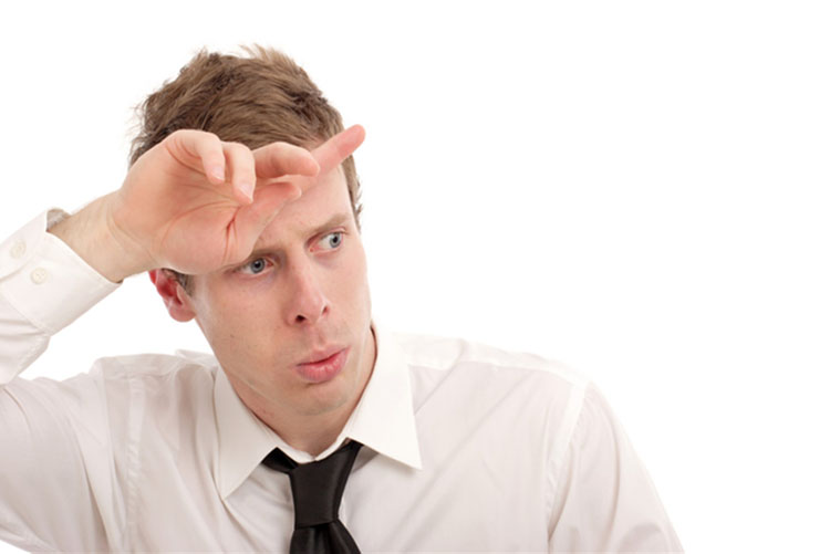
Gasps and sighs involve the quick intake of a large air volume, accompanied by breath-holding. They may comprise part of a defensive reaction. When clients expel excessive CO2, this lowers end-tidal CO2 (the percentage of CO2 at the end of exhalation) and causes hypocapnia, low CO2.
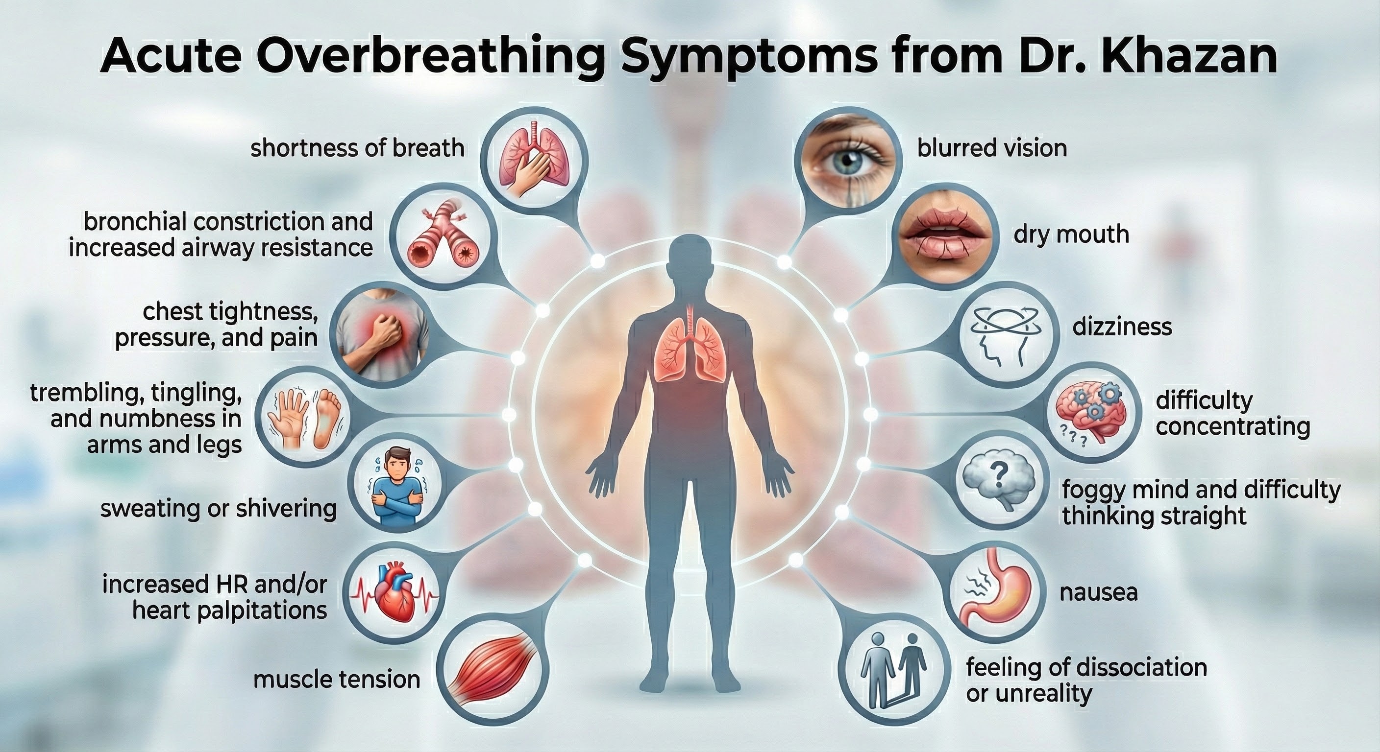
Acute overbreathing produces various symptoms.
How Hypocapnia Disrupts Homeostasis
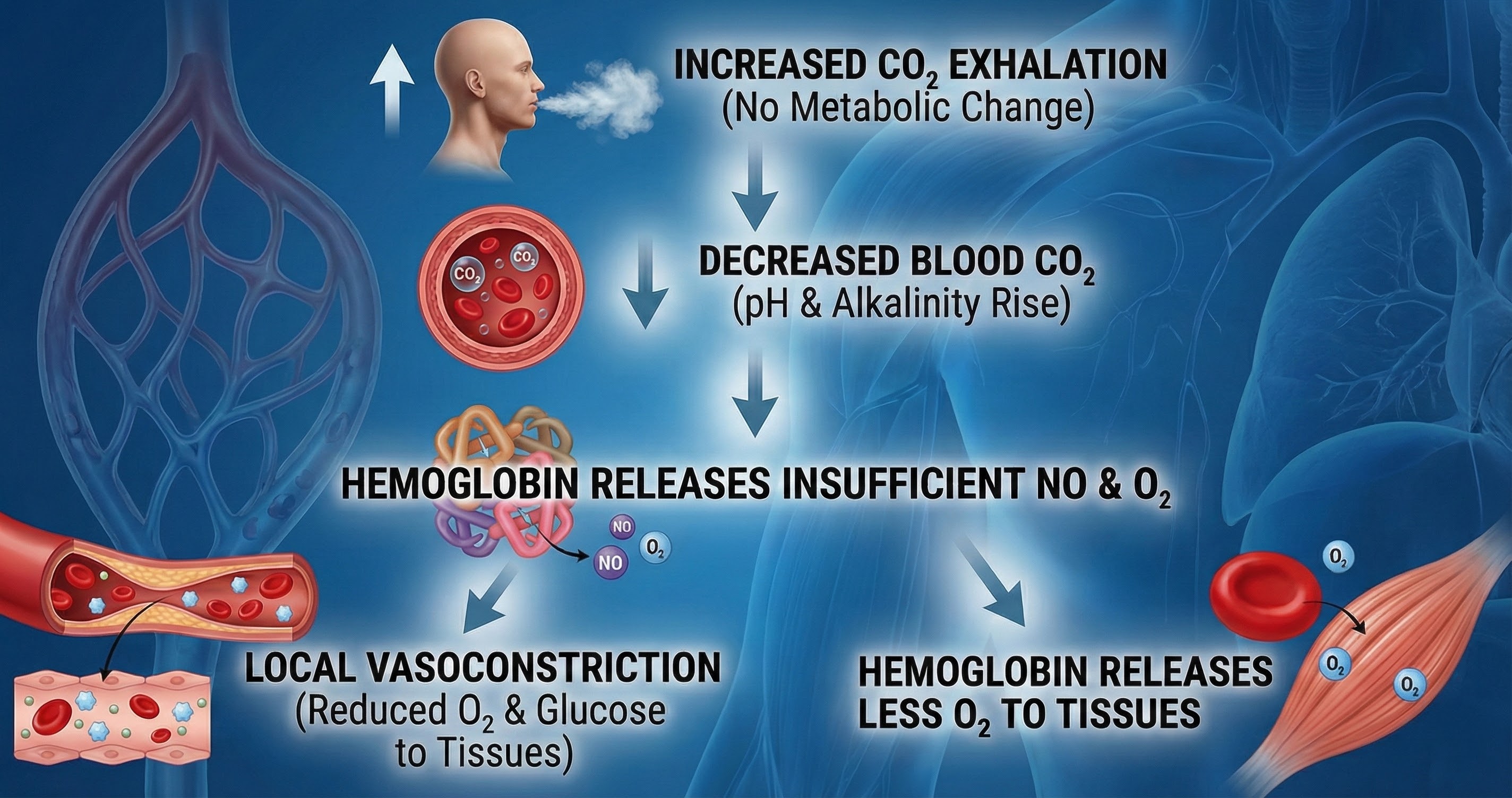
Hypocapnia disrupts homeostasis by disturbing the body's acid-base (pH) and electrolyte balance, blood flow, and oxygen delivery. Hypocapnia may force the kidneys to expel bicarbonates to restore pH balance (Khazan, 2021).
Electrolytes are substances like acids or salts that can dissociate into free ions when dissolved (for example, NaCl becomes Na+ + Cl-). Hypocapnia deprives cells (such as neurons, cardiac muscle, and skeletal muscle) of the ions (Ca+2 and Na+) required for typical membrane potentials and communication with other cells.
The Effects of Ca+2 Movement
Hypocapnia can move Ca+2 from the interstitial fluid into muscle cells with disastrous results. In skeletal muscle, calcium entry can cause spasms, fatigue, and weakness. In blood vessel smooth muscle, it can produce vasoconstriction. In the bronchioles of the lungs, it can trigger bronchoconstriction. Finally, in the GI tract smooth muscle, it can result in nausea and change motility.
The Effects of Na+ Movement
Na+ ion movement into neurons from extracellular fluid increases excitability, metabolism, and demand for oxygen while reducing oxygen availability for other organs. The brain can experience ischemia and excitotoxicity. Vasoconstriction due to Na+ ion entry and less nitric oxide release greatly diminishes glucose delivery to the tissues, especially to the outermost layers of the cortex (Gevirtz et al., 2016).
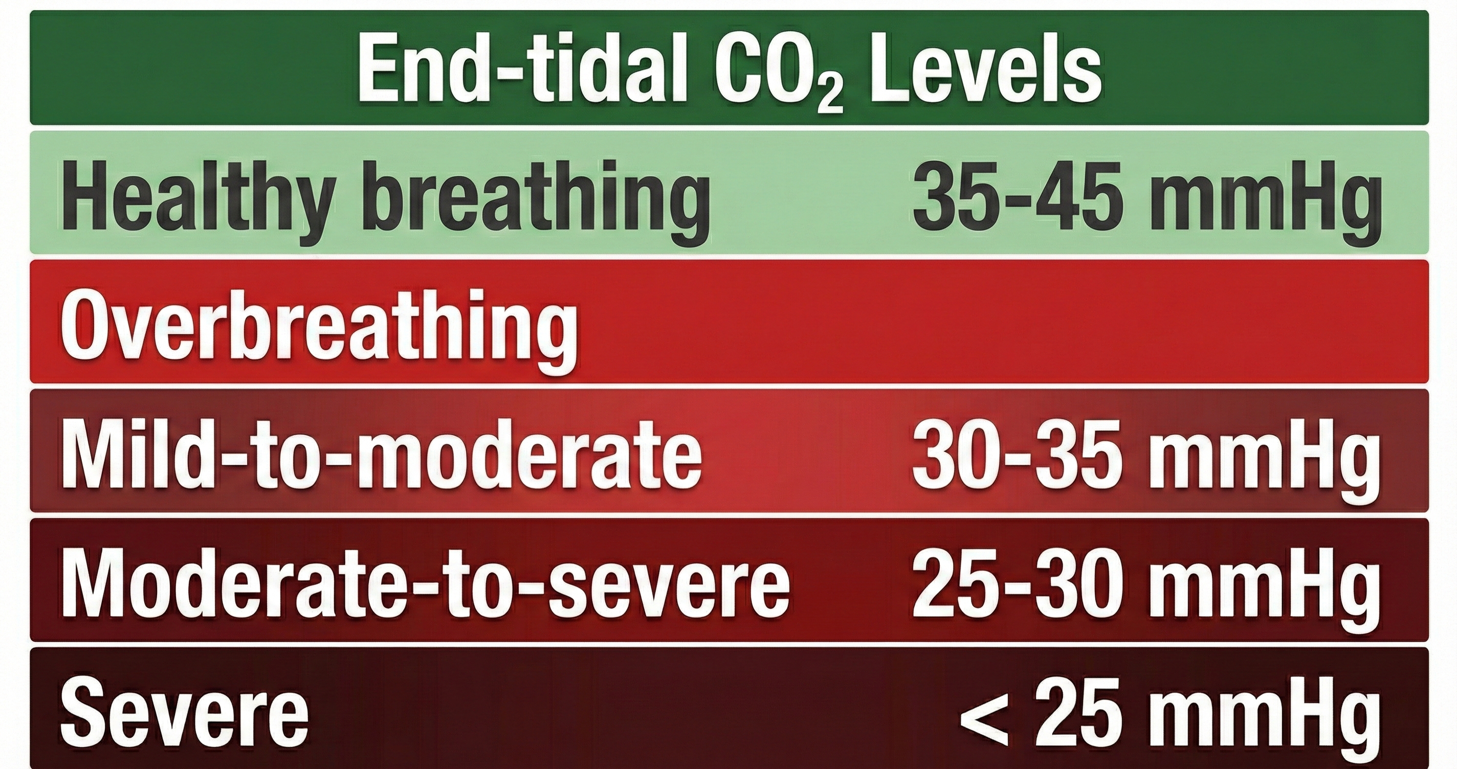
Healthy end-tidal CO2 values range from 35-45 mmHg. Moderate overbreathing can reduce oxygen delivery to the brain by 30%-40%, and severe overbreathing can reduce it by 60%.
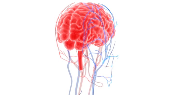
Overbreathing can produce acute and chronic vasoconstriction effects and reduced delivery of oxygen and glucose to body tissues, especially the brain (Khazan, 2013).
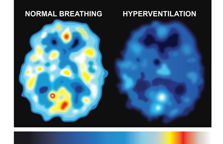
The SPECT scan created by Dr. Scott Woods below shows the effect of overbreathing on brain metabolism. Darker colors represent reduced metabolism and compromised cortical functioning.
The Overbreathing Cascade
Clinicians use the Nijmegen Questionnaire to screen for hyperventilation syndrome (HVS). For the general population, scores of 20 or higher have been suggested as optimal for predicting HVS, with a sensitivity of 0.91 and specificity of 0.92 (Looha et al., 2020). In asthma patients, a cutoff score of greater than 17 has been used to discriminate the presence of HVS, showing a sensitivity of 92.73% and specificity of 91.59% (Grammatopoulou et al., 2014). In a study involving dizzy patients, a cutoff score of 26.5 was found to be the most sensitive for diagnosing HVS (Talaat, Moaty, & Elsayed, 2019).
Detecting chronic hyperventilation presents unique challenges for healthcare providers. Unlike many medical conditions with clear visual markers, chronic overbreathing often goes unnoticed. The respiratory rate might appear normal, and the breathing pattern may seem unremarkable to casual observation. Clinicians must look for subtle clues: frequent sighing, upper chest breathing, breath-holding patterns, and a constellation of seemingly unrelated symptoms (Gilbert, 2005). The diversity of symptoms often leads people on long diagnostic journeys before anyone considers their breathing as the root cause.
For precise measurement, clinicians use capnometry to measure end-tidal CO2, the carbon dioxide concentration at the very end of exhalation. This provides real-time feedback about CO2 levels and can reveal patterns invisible to the naked eye. Studies have shown that individuals vary significantly in their response to the same CO2 drop, with some experiencing severe symptoms at levels that barely affect others (Gilbert, 2005). Davies and colleagues (2019) found that changes in perceived control and improvements in respiratory dysregulation for hypocapnic individuals specifically underlie symptom improvement from capnometry-guided respiratory interventions for panic disorder.
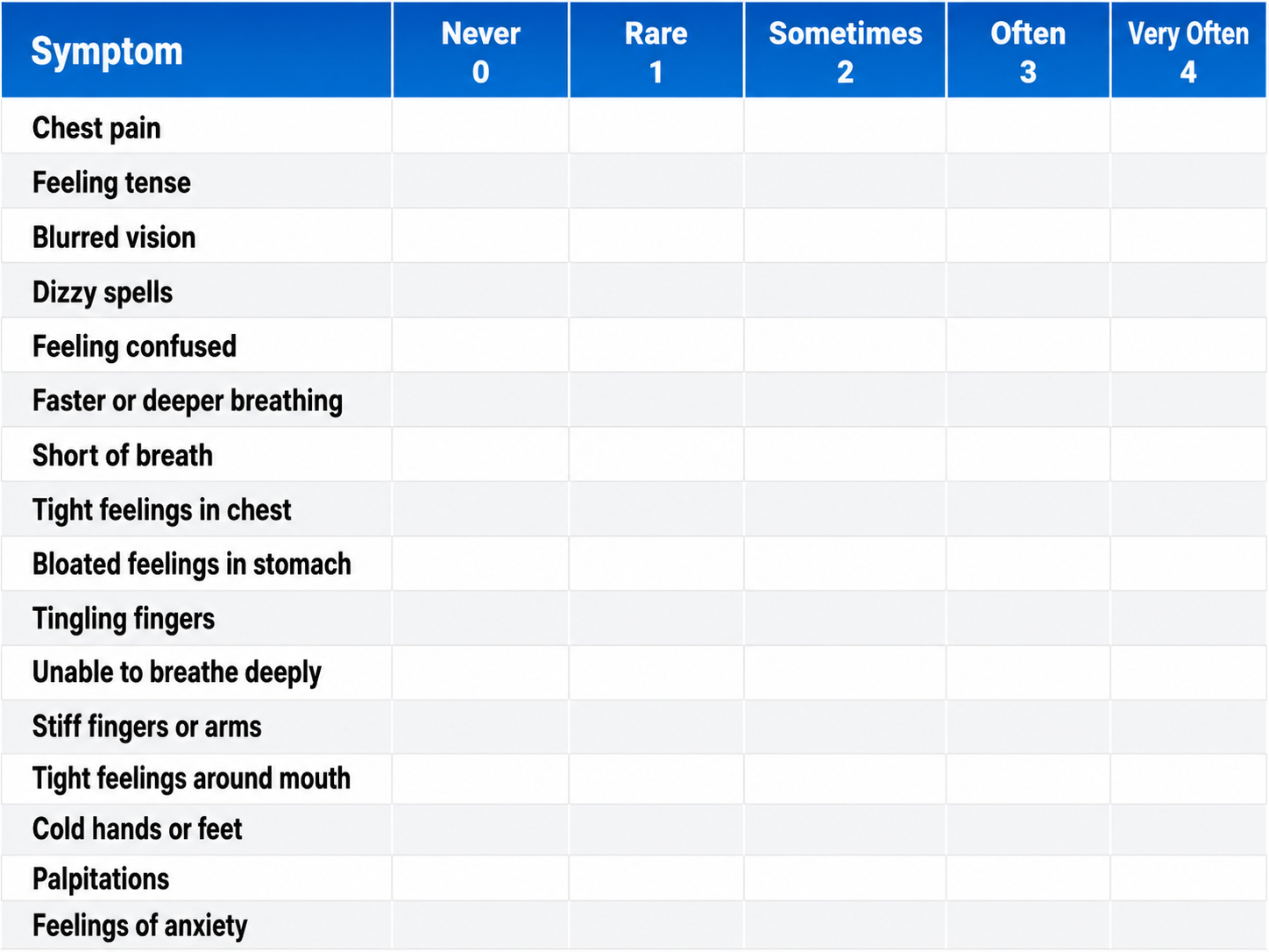
The Effects of Chronic Overbreathing
Clients who overbreathe may experience chronic hypocapnia. Since the body cannot function with sustained high pH, the kidneys excrete bicarbonates to return pH to near-normal levels. Bicarbonates are salts of carbonic acid that contain HCO3. Acid buffering can only restore homeostasis in the short run because increased metabolism raises acidity until needed bicarbonates are depleted. Clients experience fatigue, muscle pain, reduced physical endurance, and a sodium deficit when this happens. Acidosis may increase overbreathing in a failed attempt to reduce acidity (Khazan, 2021).
When someone maintains a pattern of slight overbreathing for weeks or months, their body attempts to compensate. The kidneys begin excreting bicarbonate to restore pH balance, creating what Gilbert (2005) calls a compensated state. This compensation comes at a significant cost: the body establishes a new, fragile equilibrium that depends on continued hyperventilation to maintain. These individuals live in a precarious state where any additional stress or change in breathing pattern can trigger severe symptoms. They often report persistent fatigue, muscle pain, difficulty concentrating, and ironically, a constant feeling that they cannot get enough air. Their breath-holding time typically drops to less than 20 seconds, compared to the normal 30 to 60 seconds most people can manage comfortably (Gilbert, 2005).
Why Do Clients Overbreathe?
Clients overbreathe as part of the fight-or-flight response in response to stressors, when they experience difficult emotions, and when they suffer chronic pain. They can learn this dysfunctional breathing pattern through classical and operant conditioning and social learning.
Gilbert (2005) reveals fascinating connections between emotional states and breathing patterns. He introduces the concept of action projection, where the body prepares for anticipated physical actions that never materialize. Imagine preparing to confront someone who upset you: your breathing accelerates as your body readies for action, but modern social constraints mean you likely will not engage in the physical confrontation your primitive systems anticipated. You are left in a state of respiratory alkalosis with nowhere for that preparation to go. This mismatch between physiological readiness and social behavior may explain why anxiety disorders are so common in modern society.
Research has demonstrated that hyperventilation and trauma are bidirectionally connected. Nixon and Bryant (2005) found that when individuals with Acute Stress Disorder deliberately hyperventilated for just three minutes, they experienced increased trauma-related memories and flashbacks. The physiological state created by overbreathing appears to directly trigger the re-experiencing of traumatic events, creating a vicious cycle where anxiety drives hyperventilation, which then intensifies psychological symptoms (Gilbert, 2005).
If clients practice overbreathing long enough, it can become a habit when this lowers the body's setpoint for CO2. Now, when breathing slows, the respiratory centers attempt to restore low CO2 levels by removing it through behaviors like breath-holding, sighing, and yawning (Gevirtz, Schwartz, & Lehrer, 2016). Reduced blood CO2 levels may contribute to asthma, panic, phobia, and pain disorders like chronic low back pain.
How Overbreathing and Hyperventilation Differ Clinically
Whereas overbreathing and hyperventilation produce the same physiological changes, hyperventilation involves distinctive behaviors and subjective sensations.
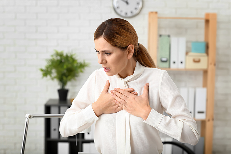
Hyperventilation syndrome (HVS) involves abnormal CO2 loss from the blood due to excessive breathing rate and depth. Here is a sobering statistic: HVS accounts for about 60% of major city ambulance calls due to frightening symptoms like chest pain, breathlessness, dizziness, and panic.
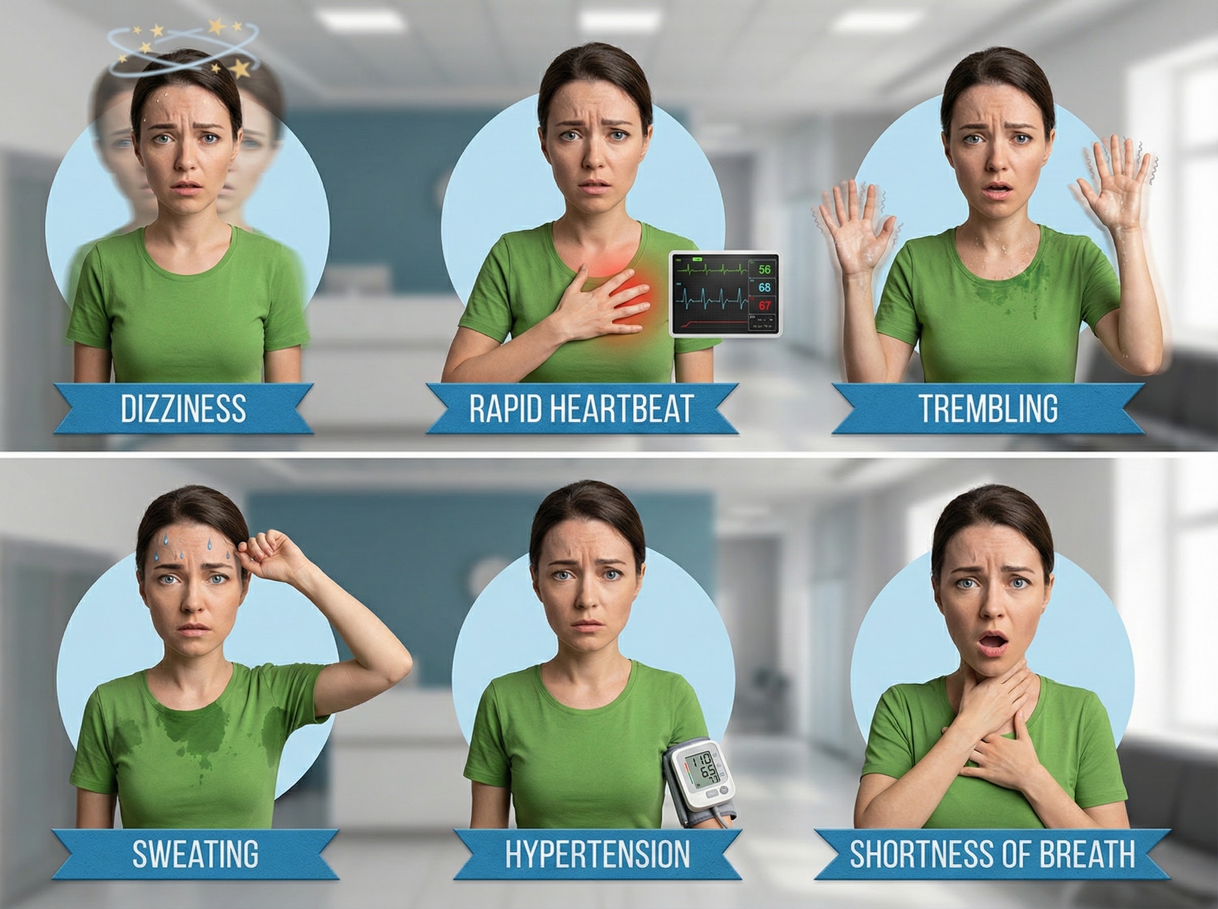
Clients who present with HVS breathe thoracically, deeply, and rapidly (over 20 bpm) using accessory muscles (the sternum moves forward and upward) and restricting diaphragm movement. Their rapid breathing can lower end-tidal CO2 from 5% to 2.5%, although many patients have normal values during attacks (Kern, 2021).
Like overbreathing, this pattern exceeds the body's need to eliminate CO2, reduces oxygen delivery to body tissues and NO release, and curtails their supply of glucose (Khazan, 2013). Check out the YouTube video Breathing Pattern Disorders Such as Hyperventilation.
The conventional advice to "take a deep breath" when stressed may actually worsen hyperventilation. Gilbert (2005) points out that people often interpret "deep" as meaning large volume, leading them to take massive inhalations that further deplete CO2. This well-intentioned but misguided instruction has probably triggered more hyperventilation episodes than it has prevented. Better breathing instructions should focus on pace and location rather than volume. Low and slow breathing, where the emphasis is on diaphragmatic movement and extended exhalation, allows CO2 to rebuild properly. The key is breathing at a rate that matches metabolic needs, typically around 6 to 10 breaths per minute at rest, with exhalation lasting longer than inhalation.
A comprehensive systematic review of 58 breathing intervention studies found that effective breath practices avoided fast-only breath paces and sessions shorter than 5 minutes, while including human-guided training, multiple sessions, and long-term practice (Bentley et al., 2023). These findings provide clinicians with evidence-based guidelines for designing breathing interventions: sessions should last at least 5 minutes, involve slow breathing rates, and include proper instruction rather than simply telling clients to "breathe deeply."
The BioGraph® Infiniti display below shows the shallow, rapid breathing that characterizes hyperventilation.
In contrast to HVS, overbreathing is usually so subtle that the patients are unaware that their sighs and yawns produce hypocapnia.
An important distinction concerns exercise and breathing. Heavy breathing during physical activity is not hyperventilation because increased muscle activity produces more CO2, which matches the increased respiratory rate (Gilbert, 2005). The body maintains its crucial balance even as production and elimination accelerate. This explains why the same breathing rate that would cause symptoms at rest feels perfectly normal during a workout. Understanding this distinction helps clinicians explain to anxious clients why their symptoms appear at rest but not during exercise.

Apnea: When Breathing Stops
Apnea involves the suspension of breathing. While commonly associated with sleep, breath-holding while awake may occur during stressful situations as part of a defensive response. A client may also hold breaths during ordinary activities like opening a jar, speaking, or writing a check. Episodes of apnea decrease ventilation and may increase blood pressure.
Do not confuse apnea with post-expiratory pauses.
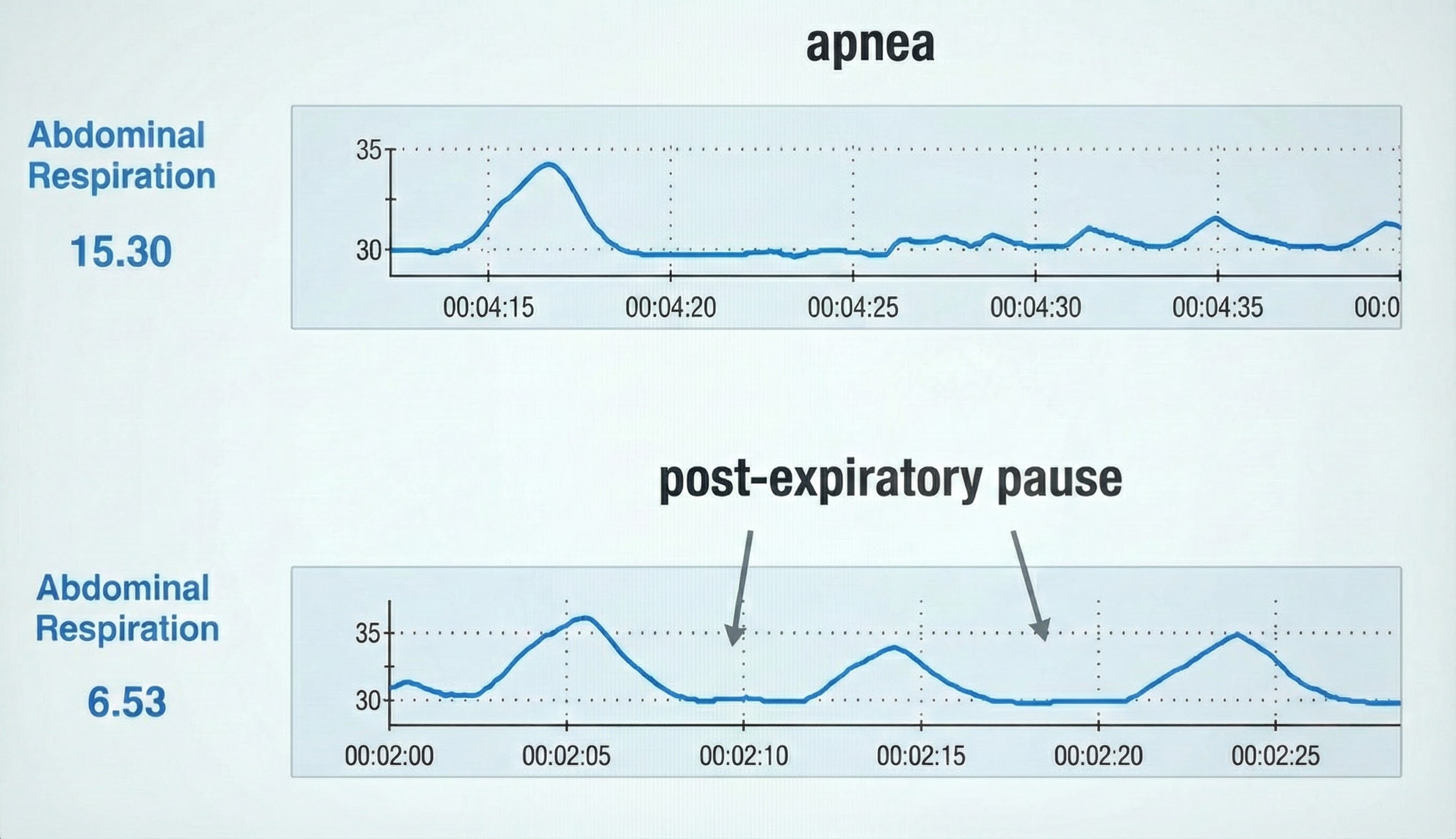
In the BioGraph® Infiniti display below, the blue abdominal strain gauge trace briefly flattens when the patient suspends breathing after the second breath.
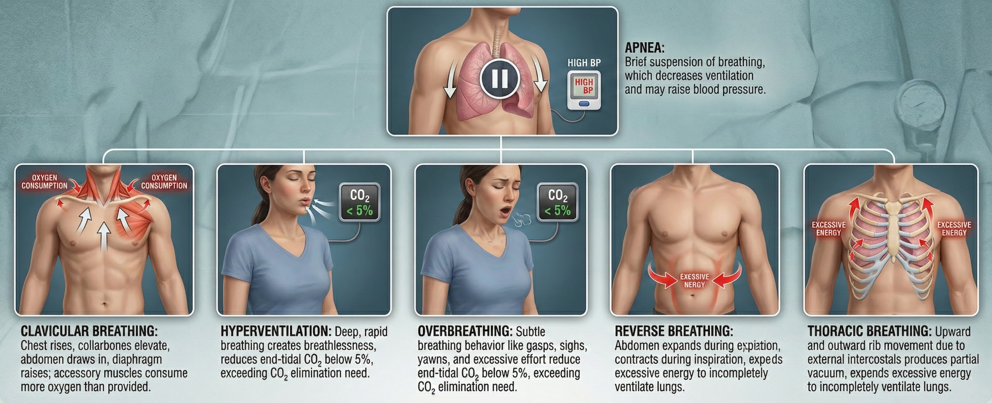
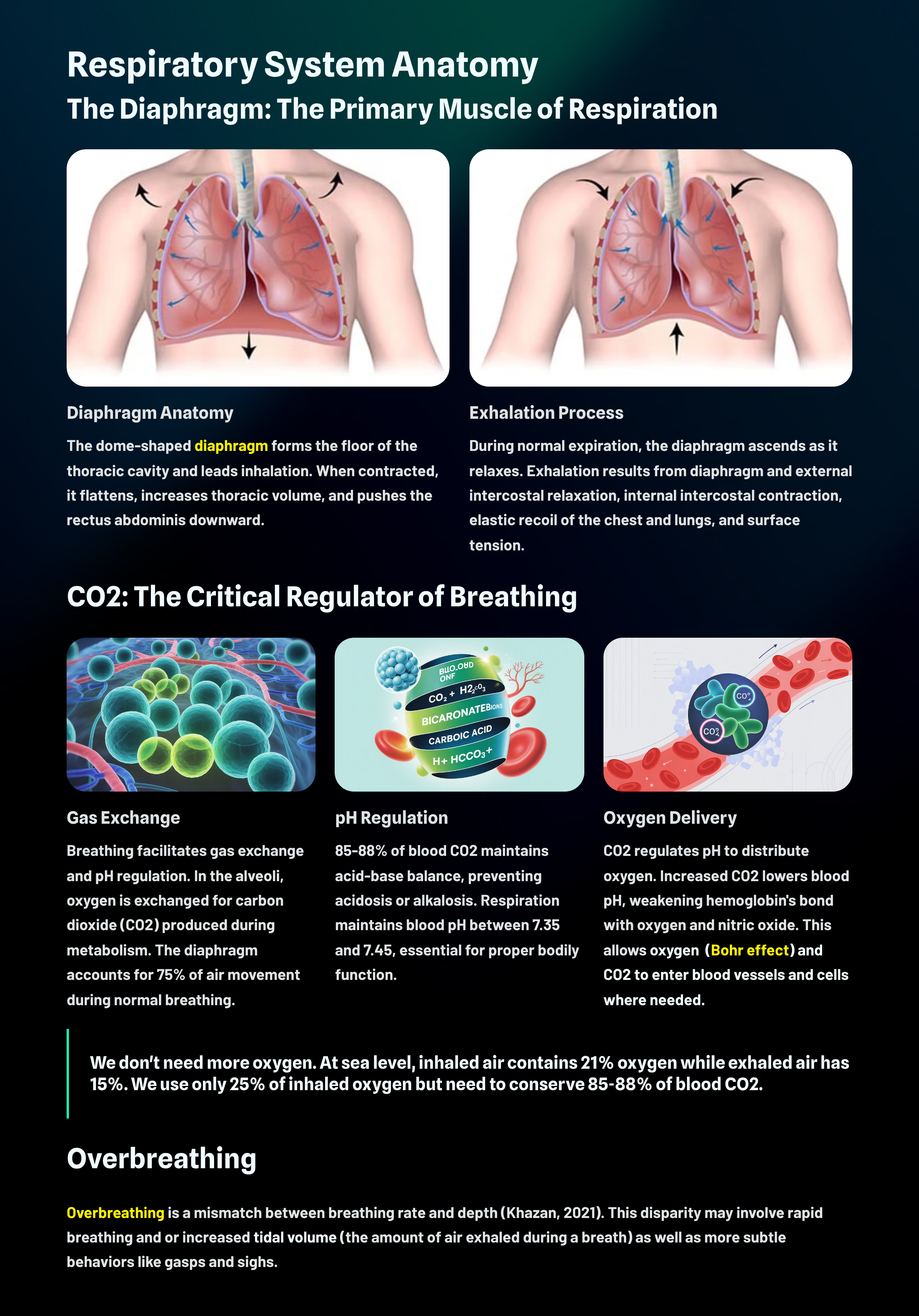
Healthy Breathing
Medical Cautions
Before teaching slow-paced breathing, you must screen for conditions that could make this intervention dangerous. Clients who chronically overbreathe may experience chronic hypocapnia, abnormally low CO2 levels in the blood. Since the body cannot function with sustained high pH, the kidneys compensate by excreting bicarbonates, the salts of carbonic acid that help buffer blood pH.
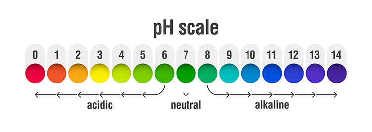
This acid buffering can only restore homeostasis in the short run. As metabolism increases acidity, needed bicarbonates become depleted. Clients then experience fatigue, muscle pain, reduced physical endurance, and sodium deficit. In a vicious cycle, acidosis may increase overbreathing in a failed attempt to reduce acidity (Khazan, 2021).
Slow-paced breathing could be hazardous for clients with metabolic acidosis, a pH imbalance in which the body has accumulated excessive acid and has insufficient bicarbonate to neutralize its effects. In conditions like diabetes and kidney disease, overbreathing represents the body's attempt to compensate for abnormal acid-base balance. Slowing the breathing rate in these clients could endanger their health by removing this compensatory mechanism.
Clients diagnosed with low blood pressure should also exercise caution since slow-paced breathing might further lower blood pressure. Finally, slow-paced breathing might produce a functional overdose if your client takes anti-hypertensive medication, insulin, or a thyroid supplement. If medication adjustment appears necessary, your client should consult the supervising physician before reducing dosage (Fried & Grimaldi, 1993).

Always screen before teaching slow-paced breathing. Clients who chronically overbreathe may develop hypocapnia, prompting the kidneys to excrete buffering bicarbonates. Slow-paced breathing can be hazardous for clients with metabolic acidosis, such as those with diabetes or kidney disease, because it removes a compensatory mechanism. Use caution with clients who have low blood pressure or who take anti-hypertensive medication, insulin, or thyroid supplements, and refer to the supervising physician when medication adjustment appears necessary.
Breathing Basics
Healthy breathing matches metabolic needs, balancing the production of CO2 with breath depth and rate. We should maintain optimal breathing chemistry for each activity level. Although rapid breathing does not always signal overbreathing and slow breathing does not always indicate health, there are strong correlations (Khazan, 2021).
Optimal breathing shares several characteristics: it is mindful with focus on the abdomen, effortless in quality, between 5-7 breaths per minute at rest, and supported by loose clothing, good posture, and ergonomics that promote healthy respiratory mechanics.
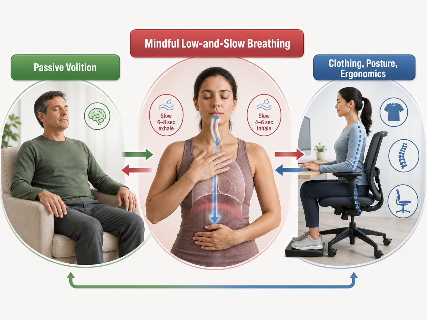
Breathe Effortlessly
Encourage your clients to breathe effortlessly (Peper & Tibbets, 1994). The subjective experience should be that the body is "breathing itself" without conscious control. Erik Peper developed the concept of effortless breathing, a relaxed breathing method in which the client uses about 70% of maximum effort, attention settles below the waist, and the volume of air moving through the lungs increases naturally.
The technical parameters of effortless breathing include a rate of 5-7 breaths per minute for adults, with pauses following expiration that are longer than those following inspiration. Tidal volume ranges between 1,000 and 3,000 ml. Airflow is smooth and continuous, with the stomach expanding and the lower ribs and back widening during inhalation.

Discourage Deep Breaths
Clients should breathe at a comfortable depth, like smelling a flower, exhaling longer than inhaling. Breathing will calm your client when its depth and rate satisfy the resting body's metabolic needs (Khazan, 2021). Do not encourage deeper or larger breaths.

Discourage typical deep breathing, where a client inhales a massive breath and inevitably exhales too quickly because they have taken in more air than needed. This pattern promotes overbreathing, subtle breathing behaviors like sighs and yawns that reduce end-tidal CO2 (the percentage of CO2 in exhaled air at the end of exhalation) below 5%, exceeding the body's need to eliminate CO2. My colleague Don Moss no longer uses the word "deep" when coaching breathing (Moss, 2022).
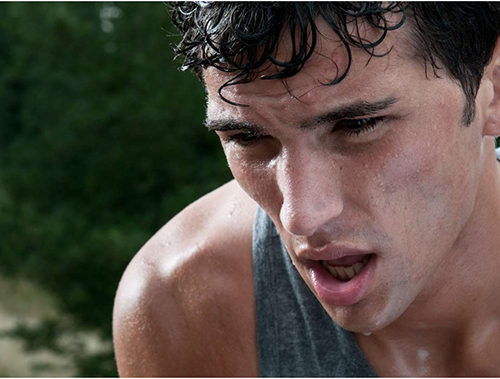

Encourage Your Clients to Dress for Success
Caution clients to avoid what Peper and Tibbets (1994) called the "designer jean syndrome," in which tight clothing prevents abdominal expansion and promotes thoracic breathing. This breathing pattern primarily relies on the external intercostals to inflate the lungs, resulting in a more rapid respiration rate, excessive energy consumption, and insufficient ventilation of the lungs.
Inhale Through the Nostrils
Encourage your clients to inhale through the nostrils to filter, moisten, and warm the air before it reaches the delicate lung tissue. Exhalation can occur through the mouth or nose depending on training goals and health considerations.

Great Posture Promotes Healthy Breathing
As Lagos (2020, p. 58) recommends: "Make sure you choose a comfortable chair that allows you to sit with a straight back, the vertebrae of your spine stacked neatly on top of one another. Your feet should be flat on the floor, legs uncrossed, knees at a 90-degree angle." This alignment allows the diaphragm, the dome-shaped muscle that accounts for about 75% of air movement during relaxed breathing, to descend freely and fully.

Enhance Your Clients' Respiratory Feedback
Teach your clients to exhale using pursed lips as if blowing out a candle (Khazan, 2021). This technique slows airflow and provides tactile feedback that helps clients monitor their breathing without instruments.
Invite clients to breathe with a weight on the abdomen while lying on the floor with knees bent. The weight, typically a book, rises during inhalation and falls during exhalation, providing clear visual and proprioceptive feedback about breathing location.

Alternatively, they can place their hands on their abdomens to feel the expansion and contraction of each breath.
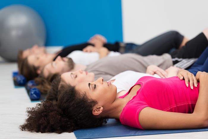
Healthy breathing basics include several key principles. Breathing should be effortless, feeling as if "the body breathes itself." Discourage deep breathing, which promotes overbreathing. Optimal respiration rate is 5-7 breaths per minute with smooth, continuous airflow. Loose clothing prevents the "designer jean syndrome" that restricts abdominal expansion. Nasal inhalation filters and warms air. Good posture supports proper diaphragm movement. Pursed-lip exhalation, abdominal weights, or hand placement provide feedback that helps clients shift from thoracic to diaphragmatic patterns.
Physiological Effects of Healthy Breathing
Hemoglobin, the iron-containing protein in red blood cells, transports oxygen and nitric oxide through the bloodstream. Understanding how hemoglobin works is essential for explaining the benefits of healthy breathing to your clients.
One hemoglobin molecule can carry up to four oxygen molecules. However, the critical question is not how much oxygen hemoglobin can carry, but when and where it releases that oxygen. This is where breathing patterns make a profound difference.
Relaxed Breathing
Relaxed diaphragmatic breathing increases metabolism and the carbon dioxide concentration of arterial blood compared to thoracic breathing. At rest, we only excrete 12-15% of blood CO2. This conservation of CO2 lowers blood pH, which triggers a crucial physiological response.
Lower pH weakens the bond between hemoglobin and oxygen, increasing oxygen delivery to body tissues. This phenomenon is called the Bohr effect, named after the Danish physiologist Christian Bohr. To explore this concept further, check out MEDCRAMvideos YouTube lecture Oxygen Hemoglobin Dissociation Curve Explained Clearly! The breathing chemistry graphics in this section were adapted from Inna Khazan.
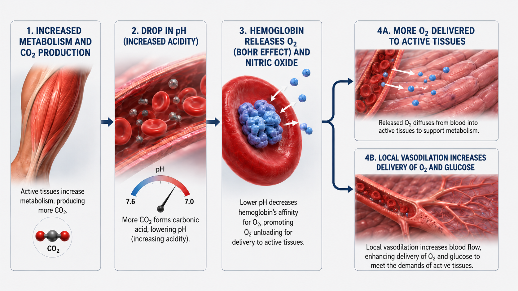
Overbreathing
Conversely, low CO2 levels due to overbreathing or hyperventilation raise blood pH and reduce oxygen delivery to body tissues. Paradoxically, despite breathing more, oxygen remains tightly bound to the hemoglobin molecules and less is available for the brain, muscles, and organs that need it (Fox & Rompolski, 2022).
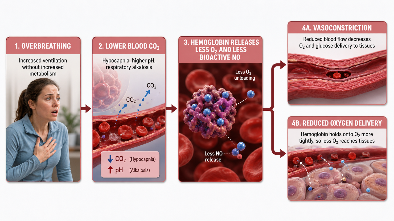
Healthy Breathing
The mantra "low and slow" captures the essence of healthy breathing. Breathing that originates low in the abdomen and proceeds at a slow pace optimizes blood chemistry for efficient oxygen delivery.
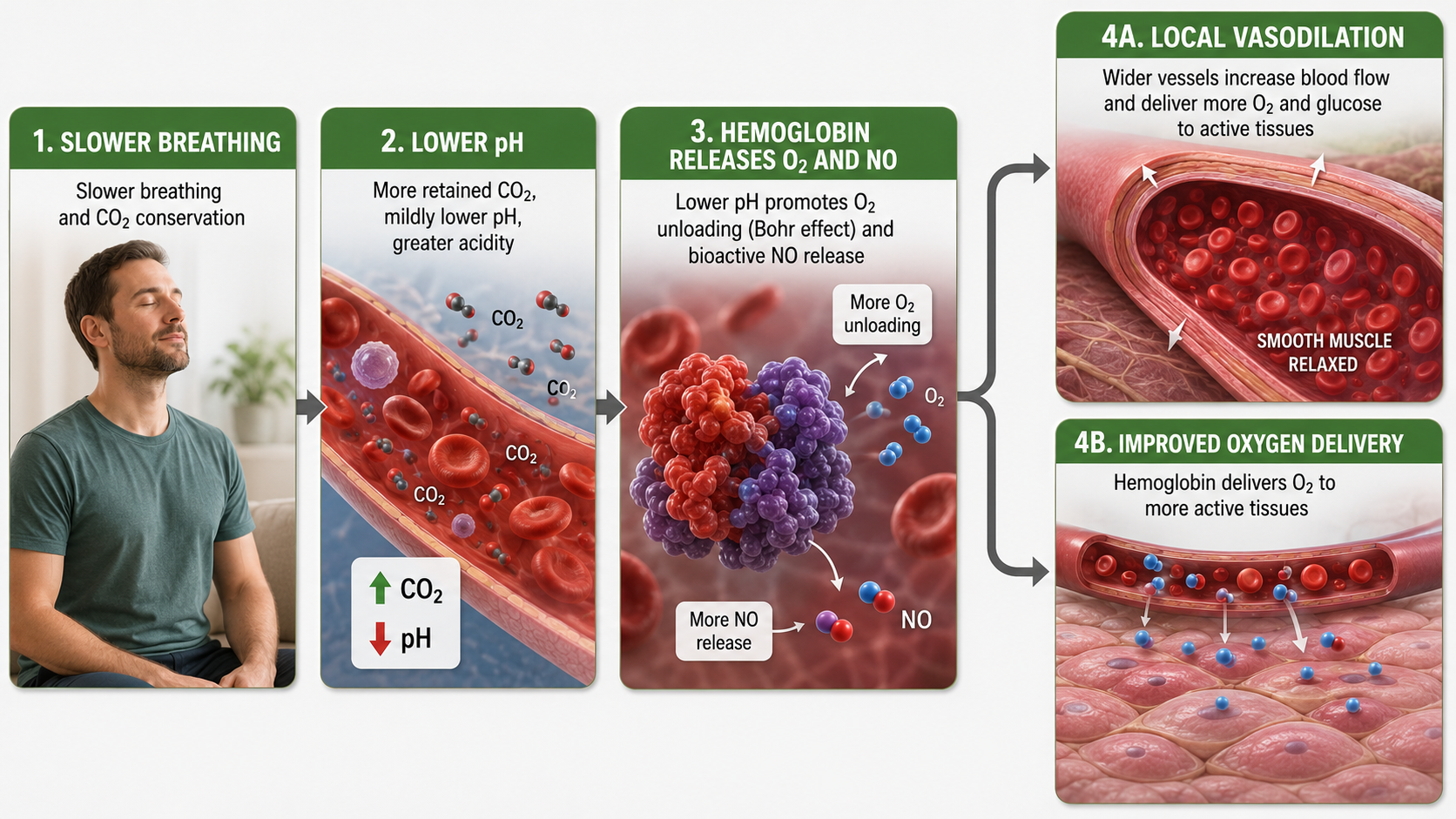
Healthy breathing can increase peripheral blood flow when the respiration rate slows to the resonance frequency range of 4.6 to 7.5 breaths per minute and end-tidal CO2 normalizes to 5% or 36 mmHg. Peripheral vasodilation increases perfusion and delivery of oxygen and glucose to the brain, reduces peripheral resistance, and promotes hand-warming.
These changes are crucial for executive functioning and treating hypertension, vascular headache, and Raynaud's disease. Breathing in the resonance frequency range can also slow heart rate and increase vagal tone and HRV. The interconnections between breathing, blood chemistry, and cardiovascular function explain why breathing training has such wide-ranging clinical applications.
Breathing chemistry directly affects oxygen delivery throughout the body. The Bohr effect explains how conserving CO2 through relaxed, slow breathing weakens the hemoglobin-oxygen bond and increases oxygen release to tissues. Overbreathing depletes CO2, raises blood pH, and keeps oxygen bound to hemoglobin, paradoxically starving tissues of oxygen. When respiration rate slows to the resonance frequency range of 4.6-7.5 breaths per minute and end-tidal CO2 normalizes around 36 mmHg, peripheral vasodilation increases blood flow to the brain and extremities while reducing blood pressure.
The Effects of Overbreathing on the EEG
Overbreathing causes cerebral vasoconstriction and reduced delivery of glucose, nitric oxide, and oxygen to the nervous system. Hyperventilation can increase frontal delta and theta activity (Thompson & Thompson, 2015)—the very EEG patterns that neurofeedback often aims to reduce. This means that an undetected overbreathing habit can directly undermine neurofeedback training, producing EEG abnormalities that mimic or worsen the dysregulation you are trying to correct. Hyperventilation graphic © EEGpedia.
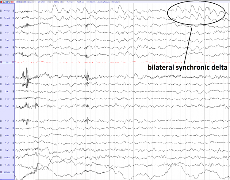
Healthy breathing matches metabolic needs with breath depth and rate. Key principles include effortless diaphragmatic breathing at 5–7 bpm, nasal inhalation, proper posture, and avoiding deep breaths that deplete CO2. Medical cautions apply for clients with metabolic acidoses, low blood pressure, or those on certain medications. Overbreathing can directly affect the EEG by increasing frontal delta and theta through cerebral vasoconstriction.
Hemoencephalography
This section introduces hemoencephalography as a complementary modality that can prepare clients for neurofeedback training. Hemoencephalography (HEG) uses passive infrared (pIR) and near-infrared (NIR) light to estimate cerebral blood flow (CBF) and metabolism. HEG indirectly measures frontal lobe activity based on the principle of neurovascular coupling—the mechanism by which a brief rise in neural activity triggers a proportional increase in CBF (Huneau et al., 2015). This large-scale CBF increase provides nutrients and removes wastes to support the operation of ion channels (Attwell & Laughlin, 2001) and metabolic regulation (Iadecola & Nedergaard, 2007) by neurons and their associated astrocytes.
HEG Instrumentation
Two forms of HEG instrumentation are available, each measuring a different proxy for cortical activity. pIR-HEG detects infrared light emitted from the frontal lobe to estimate the heat generated by cortical activity and the metabolic processes that support it. Greater frontal lobe activity correlates with increased metabolism and cortical perfusion, which produces more infrared radiation at the skin surface. A Thought Technology Ltd. pIR-HEG sensor is pictured below.
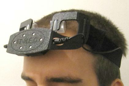
In NIR-HEG, diodes emit alternating infrared light at 660 nm (red light) and 850 nm (infrared light) into the skull, and photodetectors measure the refracted light at these wavelengths. Although the skull largely absorbs red and infrared wavelengths, oxygenated blood reflects red light. As hemoglobin oxygen saturation increases to support rising metabolism, blood backscatters more red light. The resulting change in the ratio of oxygenated (HbO2) to deoxygenated hemoglobin (Hb) provides the signal. Since the skull's absorption of infrared light does not notably change with perfusion, NIR-HEG uses the fraction of reflected infrared light as a relatively steady comparative baseline (Toomim, 2000). As cortical firing, metabolic demand, and blood oxygenation increase, the sensors detect proportionally more red than infrared light. The increased HEG ratio (refracted red vs. infrared light) indexes heightened frontal lobe blood oxygenation (Pecyna & Pokorski, 2013). Graphic © Biofeedback Institute of Los Angeles.
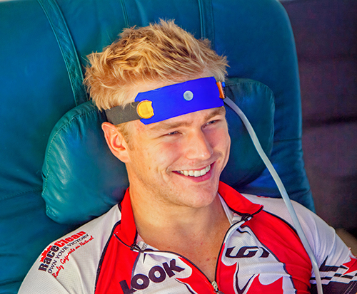
HEG Applications
John Demos (2019) trains forehead sites corresponding to the orbital gyrus, ventral lateral, and ventral medial prefrontal cortex. After removing oil from the skin with alcohol, sensors can be quickly positioned at sites like Fpz by adjusting their placement on an elastic band wrapped around the scalp. He recommends NIR-HEG for treating aggression, autism, depression, inattention, and impulsivity.
Michael and Lynda Thompson (2015) recommended combining pIR-HEG with neurofeedback and peripheral biofeedback modalities. In their ADD Centre experience, clients learn to increase forehead temperature fairly rapidly, which can teach them fundamental self-regulation skills like sustained attention, increase their perceived self-efficacy, and create positive expectancies as they progress to the more gradual learning process of neurofeedback. Because clients often see results quickly with HEG, it can build confidence and motivation early in treatment. As with all adjunctive procedures, the Thompsons could not quantify the specific percentage of client improvement attributable to pIR-HEG training itself.
The Biofeedback Institute of Toronto screen displays the left and right average temperature (pIR in degrees) as a green waveform at the top © Association for Applied Psychophysiology and Biofeedback. The client's goal is to increase forehead temperature through a "calm, relaxed yet intense focus" (p. 612).
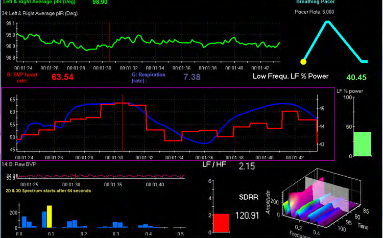
HEG Training for Migraine
Some of the most encouraging HEG evidence comes from migraine care. Jeffrey Carmen developed pIR-HEG specifically to interrupt migraines, and his clinical series of roughly 100 migraine sufferers reported that more than 90% of clients who completed at least six 30-minute forehead-sensor sessions experienced meaningful reductions in migraine activity, including people whose headaches had not responded to medication (Carmen, 2004). Early-session improvement tended to predict long-term success, which gives you an actionable rule of thumb: when a client is going to respond, you usually see the first signs within the opening handful of sessions.
A later study put Carmen's approach to a more standardized test. Walker and Lyle (2016) treated 31 adults who had migraine without aura (recurrent migraine that is not preceded by sensory warning signs such as visual disturbances) with a 10-week pIR-HEG protocol and measured outcomes with two validated instruments: the Headache Impact Test (HIT-6), a six-item questionnaire that gauges how much headaches interfere with daily life, and the Migraine Disability Assessment (MIDAS), which quantifies headache-related disability across work, home, and social activities. Both headache-impact and disability scores dropped significantly from baseline to the end of training (both p < .001). Because neither study used a control group, treat these results as promising rather than definitive, yet the convergence of a large case series and a standardized replication is reassuring.
HEG, Attention, and Executive Function
HEG also targets the prefrontal skills that clients with attention problems struggle to sustain. In the strongest controlled HEG study to date, Skalski, Pochwatko, and Balas (2021) randomly assigned 120 children aged 9 to 15, half with ADHD and half neurotypical, to HEG biofeedback or a physical-activity comparison condition. Ten sessions of HEG produced significant gains in vigilance, visual search, multitasking, inhibitory control, and working memory in both groups, while the activity condition produced no significant change. The children who improved most were those with higher intrinsic motivation, an internal locus of control, and positive mood, a useful reminder that engagement drives neurofeedback outcomes.
That engagement point cuts both ways. In a related study, Skalski (2022) gave 33 children with ADHD five HEG sessions and varied what they were told about the training. Children who received standard active-training instructions showed larger within-session prefrontal blood-oxygenation gains and better attention afterward than children given a placebo framing that downplayed the training. The lesson for your practice is concrete: how you set up expectations shapes what clients get out of HEG.
Physiological studies help explain these behavioral gains. Toomim and colleagues (2004) showed that people can intentionally raise the oxygenation of targeted prefrontal tissue using real-time HEG feedback. In a small imaging subset, single-photon emission computed tomography (SPECT), a nuclear imaging method that maps regional cerebral blood flow, documented increased local vascularity after about 30 sessions, with impulsivity scores improving after roughly 10 sessions.
HEG for Anxiety and Depression
Prefrontal HEG is now being explored for common mood and anxiety complaints. In the first formal documentation of pIR-HEG in a mental-health caseload, Tyrrell Baker (2023) tracked 66 private-practice clients and found statistically significant improvements in anxiety, depression, limbic overload, and coping self-efficacy emerging by the fifth session, with more robust change by 10 to 15 sessions. This was an uncontrolled practice-based study, so the numbers reflect real-world outcomes rather than a controlled experiment, yet they align with the dose that the migraine and attention research also points toward: plan for at least 10 sessions before judging whether HEG is helping.
Researchers are beginning to use HEG not only as a training tool but as a measurement method. Serra-Sala, Timoneda-Gallart, and Pérez-Álvarez (2016) used HEG to compare how adults and adolescents modulate prefrontal activity while viewing emotionally distressing images and found that adolescents showed less prefrontal regulation, a possible early marker of vulnerability to later depression and anxiety.
Hemoencephalography uses pIR and NIR sensors to estimate cerebral blood flow and metabolism through neurovascular coupling. Controlled and clinical studies suggest HEG can lower migraine impact, strengthen attention and executive function, and support mood, typically across about 10 or more 30-minute sessions, although most studies still lack control groups. Because early response and client engagement predict success, HEG works well as a confidence-building entry point that clinicians pair with neurofeedback.
Neuromodulation
This section distinguishes neuromodulation from neurofeedback and surveys the major stimulation-based approaches. Neuromodulation stimulates the nervous system to produce physiological change, but it is fundamentally different from neurofeedback because it acts on the nervous system rather than providing it with information about its own performance. This distinction matters clinically: neurofeedback promotes self-regulation through learning, while neuromodulation produces change through external stimulation. The two approaches can complement each other, but they operate through different mechanisms.
Audio and Visual Entrainment
Audio and visual entrainment (AVE) delivers visual and auditory stimuli pulsed at low frequencies (e.g., below 30 Hz), which can increase the amplitude of corresponding EEG frequencies and promote decreased arousal at lower frequencies or increased arousal at higher frequencies (Collura & Siever, 2009; Siever, 2007). These EEG changes are associated with shifts in mood, cognition, and behavior and may be clinically beneficial despite their typically short-term effects (Basu & Banerjee, 2020; Hanslmayr et al., 2019). Subliminal auditory stimulation has also been used to "drive" EEG toward states that can then be reinforced by simultaneous EEG neurofeedback (Swingle, 2015). The MindAlive Inc. David Delight is shown below.
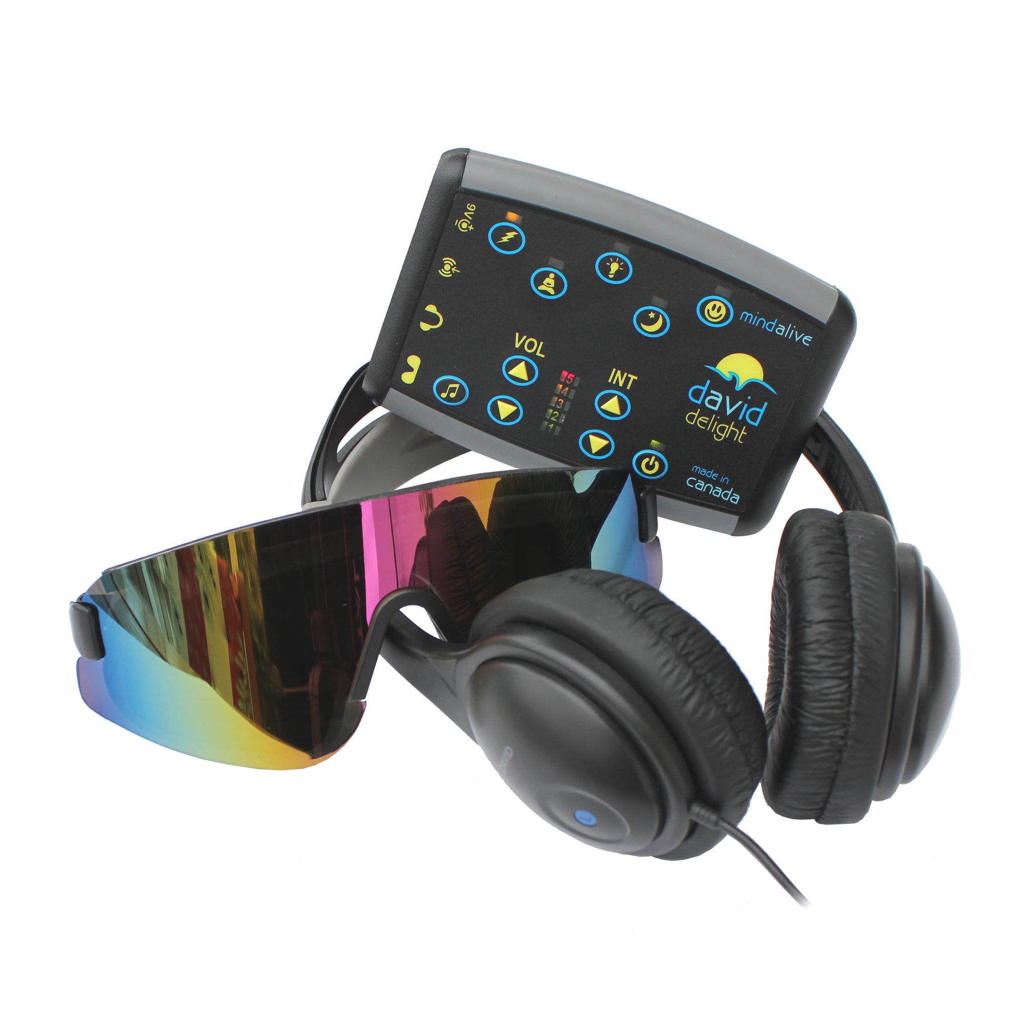
How strong is the evidence? A systematic review of 20 brainwave-entrainment studies found preliminary support for improved cognition and stress relief, with reported benefits for pain and migraine, but noted that most studies were small (Huang & Charyton, 2008). A recent review was more cautious, concluding that AVE findings for depression, seasonal affective disorder, and insomnia are heterogeneous and include null effects, so premature claims of efficacy are not warranted (Rahmani, Romero Lauro, & Pisoni, 2025). Treat AVE as a low-risk adjunct that some clients find calming, not a stand-alone treatment.
Transcranial Alternating Current Stimulation
Transcranial alternating current stimulation (tACS) uses an alternating electrical current to entrain brain oscillations—essentially synchronizing neural activity to an external rhythm (Tavakoli & Yun, 2017). It is beginning to be applied to various psychiatric conditions including ADHD, depression, OCD, schizophrenia, and dementia (Elyamany et al., 2020). Graphic © The Scientist Magazine.
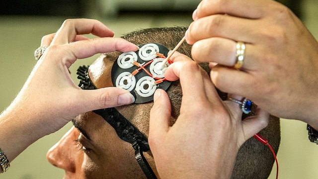
Early clinical trials show both the promise and the limits of this approach. In a double-blind, sham-controlled pilot, Alexander and colleagues (2019) delivered 10 Hz tACS to target alpha oscillations in adults with major depression. The primary outcome did not differ across groups, but an exploratory analysis found more responders in the alpha-targeted group two weeks later, along with reduced left-frontal alpha power. These are proof-of-concept signals, not proof of efficacy, and they show why matching the stimulation frequency to each client's own rhythms remains an open question.
Cranial Electrotherapy Stimulation
Cranial electrotherapy stimulation (CES) applies low-intensity microcurrent to the head, usually through ear-clip electrodes, to treat anxiety, PTSD, depression, insomnia, and pain. Low-intensity current (50 μA to 4 mA) is typically applied to the earlobes. Although shortcomings in research designs have been documented (Brunye et al., 2021), a consistent body of evidence suggests safe and beneficial effects for many clients (Shekelle et al., 2018).
Independent reviews temper the enthusiasm while confirming safety. The most rigorous synthesis, conducted for the Department of Veterans Affairs, judged the evidence insufficient for pain, depression, PTSD, or insomnia and found only low-strength evidence of modest benefit for anxiety, with no serious adverse effects (Shekelle et al., 2018). A randomized trial is more positive: Barclay and Barclay (2014) treated 115 adults with five weeks of daily one-hour CES and found significantly greater reductions in anxiety and comorbid depression than sham. CES is best framed as a safe option with modest, still-uncertain benefits, strongest for anxiety.
Transcranial Magnetic Stimulation
Repetitive transcranial magnetic stimulation (rTMS) uses electromagnetic coils held above the scalp to deliver pulses that activate or inhibit local brain tissue depending on the stimulation frequency (Marques et al., 2019). The standard target for depression is the dorsolateral prefrontal cortex (DLPFC), a frontal region central to mood regulation, working memory, and executive control. The Food and Drug Administration cleared the first TMS device for major depression in 2008; the conventional protocol applies 10 Hz stimulation to the left DLPFC across roughly 20 to 30 daily sessions over four to six weeks, with each session lasting about 37 minutes.
Two developments have made TMS far more practical. First, intermittent theta burst stimulation (iTBS), a patterned protocol that delivers short bursts of pulses at theta frequency and compresses a session to about three minutes, proved just as effective as standard 37-minute stimulation in a non-inferiority trial of 414 patients with treatment-resistant depression (Blumberger et al., 2018). Second, accelerated protocols pack many sessions into a few days. A double-blind randomized trial of Stanford Neuromodulation Therapy (SNT), which delivered 50 iTBS sessions over five days, found that 79% of the actively treated group remitted compared with 13% of the sham group (Cole et al., 2020, 2022). These samples are small, so the striking numbers await larger replication, but they point toward rapid, individualized treatment.
TMS is no longer limited to depression. The FDA cleared deep TMS as an adjunct for obsessive-compulsive disorder in 2018 (U.S. Food and Drug Administration, 2018) and as an aid for short-term smoking cessation in 2020, its first clearance for an addiction (BrainsWay, 2020).
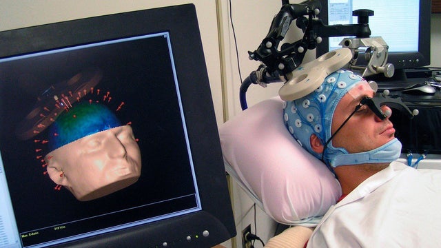
Direct Current and Electromagnetic Field Stimulation
In transcranial direct current stimulation (tDCS), a weak constant current, usually 1 to 2 milliamps for 20 to 30 minutes, flows between a positive electrode over the left DLPFC and a negative electrode elsewhere on the head, gently biasing neurons toward or away from firing (Lefaucheur et al., 2017). The antidepressant evidence is real but modest: bifrontal tDCS outperformed placebo but not the antidepressant escitalopram in a three-arm trial (Brunoni et al., 2017), and a meta-analysis of nine trials found it roughly doubled the odds of response over sham (Moffa et al., 2020). Do not expect tDCS to sharpen a healthy client's memory, however, since single-session gains are slight and appear mainly when it is paired with cognitive training (Mancuso et al., 2016).
Pulsed electromagnetic field therapy (PEMF) operates at lower power than TMS and delivers stimulation through electromagnetic coils often arranged in a helmet or a mat. Added to stable medication, transcranial PEMF produced a moderate advantage over sham for treatment-resistant depression, with a between-group effect size of 0.62 (Martiny, Lunde, & Bech, 2010), and a larger single-arm cohort reported a remission rate near 24% after eight weeks (Larsen et al., 2020). Because the strongest PEMF trials are small or lack sham controls, treat it as promising but unproven.
Near-Infrared Light Stimulation
Photobiomodulation (PBM) uses near-infrared light from lasers or light-emitting diodes in the range of 600–1100 nm, delivered either transcranially or intranasally, to increase mitochondrial activity in the brain. PBM also modulates EEG activity (Jahan et al., 2019), stimulates blood flow and oxygenation, reduces inflammation, promotes antioxidative activity and neurogenesis, and supports the healing of injured tissue (Cardoso et al., 2021; Hamblin, 2018).
Research suggests that PBM activates intrinsic brain networks. Among healthy individuals, these effects translate into improved mood and cognitive function, while those who have sustained a traumatic brain injury show improvements in executive function and sleep. Benefits have also been reported for neurodegenerative conditions (e.g., Alzheimer's disease), stroke, and psychiatric disorders including depression, anxiety, and PTSD (Berman & Nichols, 2019; Gutierrez-Menendez et al., 2020; Hamblin, 2016, 2018). For clinicians in military or VA settings, PBM may offer a non-invasive adjunct for TBI rehabilitation.
Controlled and case data are beginning to define where transcranial photobiomodulation (tPBM), the delivery of red or near-infrared light through the scalp to reach the brain, actually helps. In the sham-controlled ELATED-2 pilot, near-infrared light to the prefrontal cortex reduced depression symptoms significantly more than sham over eight weeks (Cassano et al., 2018). For brain injury, Naeser and colleagues (2014) gave 11 people with chronic mild traumatic brain injury 18 sessions of red and near-infrared light and documented significant gains in executive function and verbal memory. Typical transcranial dosing uses red light near 630 to 670 nm or near-infrared near 810 to 870 nm for about 20 minutes per site, roughly three times a week over six weeks.
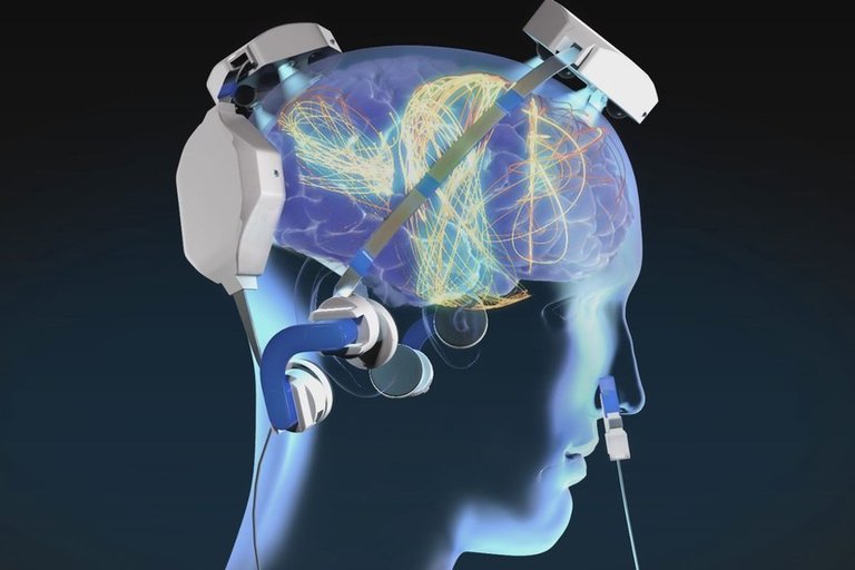
Can You Combine Neuromodulation with Neurofeedback?
Because neuromodulation and neurofeedback influence the brain in complementary ways, pairing them is appealing, and clinicians sometimes use audio-visual entrainment or subliminal stimulation to nudge the EEG toward a state that is then reinforced with neurofeedback (Swingle, 2015). The controlled evidence, however, is not yet encouraging. A randomized trial comparing neurofeedback and tDCS for attention found no advantage of either active condition over sham (Rêgo, Gonçalves, & Boggio, 2022). For now, combining the two is a reasonable clinical experiment rather than an evidence-based protocol.
Neuromodulation approaches, including AVE, tACS, TMS, tDCS, PEMF, and photobiomodulation, stimulate the nervous system to produce change but are not neurofeedback because they do not provide performance feedback. The best-supported option is TMS for depression, where accelerated protocols such as SNT have achieved high remission rates, while tDCS, PEMF, CES, tPBM, tACS, and AVE show modest or preliminary benefits. These modalities may serve as adjuncts to neurofeedback, though controlled evidence for combining them is still limited.
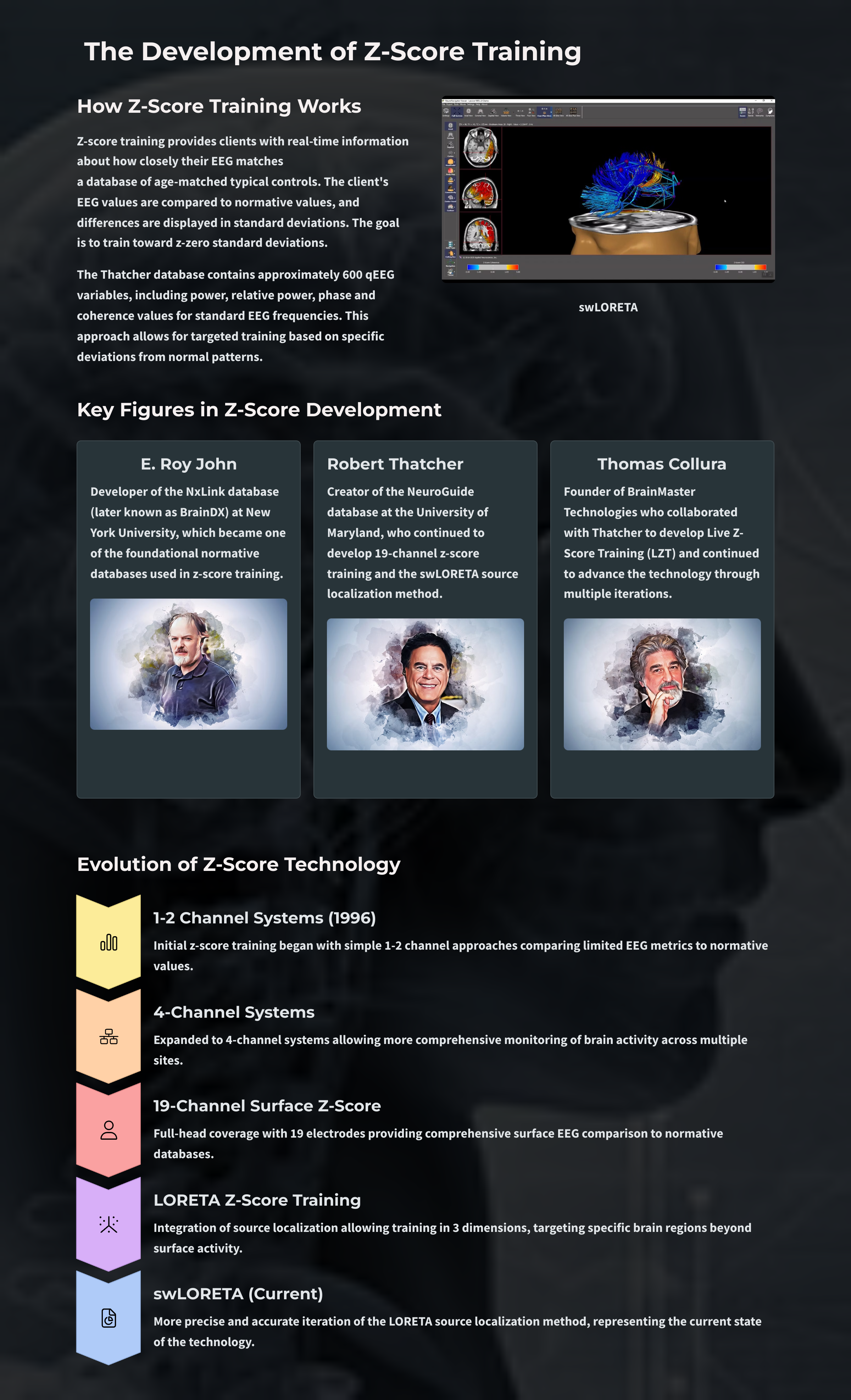
fMRI-EEG Integration
Recent research has begun combining real-time fMRI neurofeedback with simultaneous EEG neurofeedback (Bezmaternykh et al., 2021). This integration may offer the spatial precision of fMRI with the temporal resolution of EEG, potentially advancing post-stroke motor rehabilitation and other clinical applications.
Connectivity Neurofeedback
Yamashita et al. (2017) demonstrated that connectivity neurofeedback training can differentially change functional connectivity and cognitive performance. This approach trains communication between brain regions rather than activity at single sites, representing a shift from traditional neurofeedback paradigms.
Implicit Neurofeedback
Dobrushina et al. (2020) showed that implicit EEG neurofeedback, where participants are unaware they are receiving neurofeedback, can modulate intrinsic brain connectivity. This finding challenges assumptions about the role of conscious awareness in neurofeedback training and opens new possibilities for clients who struggle with traditional explicit training approaches.
Check Your Understanding
- How did the development of normative EEG databases and the FFT algorithm contribute to the emergence of qEEG-based neurofeedback training?
- What is the goal of z-score training, and how does it differ from traditional amplitude-based neurofeedback protocols?
- Why is the validity of infra-low and infra-slow frequency training signals questioned, and what artifacts occupy this frequency range?
- How can HRV biofeedback and healthy breathing training complement and enhance neurofeedback outcomes?
- What distinguishes neuromodulation approaches (such as AVE, rTMS, and photobiomodulation) from neurofeedback, and why is this distinction clinically important?
Assignment
Now that you have completed this module, explain how heart rate variability biofeedback and healthy breathing training can complement neurofeedback. How might hemoencephalography prepare clients for neurofeedback training?
Glossary
accessory muscles: the sternocleidomastoid, pectoralis minor, scalene, and trapezius muscles, which are used during forceful breathing, as well as during clavicular and thoracic breathing.
acidosis: a decrease in the normal alkalinity of the blood or tissues, leading to a lower pH level. It can be caused by an increase in acid production, a decrease in acid excretion, or a loss of bicarbonate, which is a base that helps neutralize acids in the body.
allostatic load: the cumulative physiological wear and tear that results from repeated or chronic activation of the body's stress-response systems.
alveoli: tiny, thin-walled gas exchange sacs in the lungs where oxygen and carbon dioxide are exchanged with the blood.
apnea: a dysfunctional respiratory pattern in which an individual suspends breathing.
audio and visual entrainment (AVE): the delivery of visual and auditory stimuli pulsed at low frequencies (e.g., below 30 Hz) to increase the amplitude of corresponding EEG frequencies.
Autonomic Space Theory: Berntson and colleagues challenged the reciprocal model of autonomic control, proposing that sympathetic and parasympathetic systems can function independently. It identifies three modes: reciprocal activation, coactivation, and coinhibition, highlighting the complex interplay of autonomic regulation in stress, cognition, and physiological adaptability.
bicarbonates: salts of carbonic acid that contain HCO3−.
Bohr effect: the influence of carbon dioxide on hemoglobin release of nitric oxide and oxygen.
carbon dioxide: a gas produced by cellular metabolism, crucial for regulating breathing, maintaining blood pH balance, and facilitating oxygen delivery to tissues.
central autonomic network (CAN): an interconnected set of brainstem and forebrain structures, including the nucleus of the solitary tract, anterior cingulate, insula, ventromedial prefrontal cortex, amygdala, and hypothalamus, that regulates autonomic, endocrine, and behavioral responses and modulates heart rate variability.
cerebral cortex: the 2-4 millimeter-thick outer layers that cover the cerebral hemispheres that contain circuitry essential to complex brain functions like cognition and consciousness.
cholinergic anti-inflammatory pathway: a vagally mediated reflex in which efferent vagal activity releases acetylcholine that suppresses the production of pro-inflammatory cytokines, linking parasympathetic activity to reduced inflammation.
clavicular breathing: a dysfunctional breathing pattern that primarily relies on the external intercostals and the accessory muscles to inflate the lungs, resulting in a more rapid respiration rate, excessive energy consumption, and incomplete ventilation of the lungs.
cranial electrotherapy stimulation (CES): the application of low-intensity microcurrent to the head, usually through ear-clip electrodes, to treat anxiety, insomnia, depression, and pain.
cycle length dependence: the phenomenon where faster HRs reduce the time between successive beats and the opportunity for the interbeat intervals (IBIs) to vary, resulting in lower HRV.
diaphragm: a dome-shaped muscle whose contraction enlarges the vertical diameter of the chest cavity and accounts for about 75% of air movement into the lungs during relaxed breathing.
dorsal respiratory group (DRG): neuron clusters in the medulla of the brainstem that collect information from peripheral stretch and chemoreceptors and distribute this information to the VRG to modify its breathing rhythms.
dorsolateral prefrontal cortex (DLPFC): a frontal brain region involved in mood regulation, working memory, and executive control that serves as the standard target for stimulation treatments of depression.
effortless breathing: Erik Peper's relaxed breathing method in which the client uses about 70% of maximum effort, attention settles below the waist, and the volume of air moving through the lungs increases. The subjective experience is that "my body breathes itself."
electrolytes: substances such as acids or salts that dissociate into free ions when dissolved and are required for normal membrane potentials and intercellular communication.
electromagnetic stimulation: cranial microcurrent electrical stimulation of the brain.
end-tidal CO2: the percentage of CO2 in exhaled air at the end of exhalation.
external intercostals: the muscles of inhalation that pull the ribs upward and enlarge the thoracic cavity. The external intercostals account for about 25% of air movement into the lungs during relaxed breathing.
Headache Impact Test (HIT-6): a validated six-item questionnaire that measures how much headaches interfere with daily functioning.
heart rate: the number of heartbeats per minute.
heart rate variability (HRV): beat-to-beat changes in heart rate, including changes in the R-R intervals between consecutive heartbeats.
heartbeat event-related potentials (HERPs): negative EEG potentials appearing approximately 200-300 ms after each R-spike that index cardiac afferent communication with the brain.
HEG ratio: the ratio of refracted red vs. infrared light.
hemoencephalography (HEG): using passive infrared (pIR) and near infrared (NIR) to estimate cerebral blood flow (CBF) and metabolism.
hemoglobin: red blood cell protein that carries oxygen throughout the circulatory system.
hypercapnia: a condition characterized by an abnormally high level of carbon dioxide (CO2) in the blood.
hyperventilation (HV): a dysfunctional breathing pattern in which deep and rapid breathing results in breathlessness and reduces end-tidal CO2 below 5%, exceeding the body's need to eliminate CO2.
hyperventilation syndrome (HVS): a condition involving abnormal loss of CO2 from the blood due to excessive breathing rate and depth, producing symptoms such as chest pain, breathlessness, dizziness, and panic.
hypocapnia: decreased CO2 in arterial blood.
hypothalamic-pituitary-adrenal (HPA) axis: the neuroendocrine system that governs the body's response to stress through the release of cortisol from the adrenal cortex.
inspiratory muscles: the diaphragm and external intercostals.
interbeat interval (IBI): the time interval between the peaks of successive R-spikes (initial upward deflections in the QRS complex). This is also called the NN (normal-to-normal) interval after removing artifacts.
intermittent theta burst stimulation (iTBS): a patterned form of transcranial magnetic stimulation that delivers short bursts of pulses at theta frequency, shortening a treatment session to about three minutes.
intrinsic nervous system: the network of interconnected cardiac ganglia within the heart, often called the heart's "little brain," that can process information and influence the cardiac pacemakers independently of the brain.
metabolic acidosis: a disturbance characterized by a decrease in the body's bicarbonate levels or an increase in the production of acids, leading to a reduction in the arterial blood pH below 7.35. This condition can result from increased acid production (such as ketoacidosis or lactic acidosis), reduced kidney acid secretion, or significant bicarbonate losses.
Migraine Disability Assessment (MIDAS): a validated questionnaire that quantifies headache-related disability across work, household, and social activities.
migraine without aura: a recurrent migraine headache that is not preceded by sensory warning signs such as visual disturbances.
near-infrared hemoencephalography (NIR-HEG): projecting alternating infrared light at 660 nm (red light) and 850 nm (infrared light) into the skull and measuring the refracted light at these wavelengths to estimate the metabolic activity that supports cortical activity.
neuromodulation: stimulating the nervous system to produce physiological change.
neurovascular coupling: the mechanism in which vascular blood flow increases to provide nutrients and remove wastes to satisfy the greater metabolic demands of accelerated neuronal activity.
neurovisceral integration model: Thayer and Lane's theoretical framework describing the role of the central autonomic network (CAN) in regulating autonomic, cognitive, and emotional processes. It emphasizes the prefrontal cortex's top-down control over heart rate variability (HRV) via the vagus nerve, linking HRV to self-regulation and mental health.
nitric oxide (NO): a gaseous neurotransmitter that promotes vasodilation and long-term potentiation.
overbreathing: mismatch between breathing rate and depth due to excessive breathing effort and subtle breathing behaviors, like sighs and yawns. Overbreathing can reduce arterial CO2.
oxygen saturation: a measure of the percentage of hemoglobin binding sites in the bloodstream occupied by oxygen.
passive infrared hemoencephalography (pIR-HEG): a pIR sensor measures skin surface temperature that indexes cortical blood flow and related cerebral processing.
pH: the power of hydrogen; the acidity or basicity of an aqueous solution determined by the concentration of hydrogen ions.
photobiomodulation (PBM): the delivery of near-infrared light from lasers or light-emitting diodes in the range of 600-1100 nm either transcranially or intranasally to increase brain mitochondrial activity.
pons: the brainstem structure above the medulla that contains breathing centers that adjust VRG breathing rhythms based on descending input from brain structures and peripheral sensory input.
pontine respiratory group (PRG): neurons located in the pons that communicate with the dorsal respiratory group (DRG) in the medulla to modify the basic breathing rhythm.
pulsed electromagnetic field therapy (PEMF therapy): stimulation at lower power than rTMS by electromagnetic coils arrayed in a helmet or a mat.
rectus abdominis: the muscle of forceful expiration that depresses the inferior ribs and compresses the abdominal viscera to push the diaphragm upward.
repetitive transcranial magnetic stimulation (rTMS): a technique that uses electromagnetic coils held above the scalp to deliver repetitive magnetic pulses that either activate or inhibit local brain tissue depending on the stimulation frequency.
respiratory acidosis: a state in which decreased ventilation (hypoventilation) leads to an increase in carbon dioxide concentration and a decrease in blood pH. This condition is often due to impaired lung function, chest injuries, or diseases that affect the respiratory muscles or control of breathing.
respiratory alkalosis: a condition characterized by elevated blood pH due to excessive carbon dioxide exhalation, typically caused by hyperventilation.
respiratory amplitude: the excursion of an abdominal strain gauge.
respiratory cycle: consists of an inspiratory phase, inspiratory pause, expiratory phase, and expiratory pause.
respiratory membrane: the site of respiratory gas exchange that is comprised by alveolar and capillary walls.
reverse breathing: a dysfunctional breathing pattern in which the abdomen expands during exhalation and contracts during inhalation, often resulting in incomplete ventilation of the lungs.
single-photon emission computed tomography (SPECT): a nuclear imaging technique that measures regional cerebral blood flow.
Stanford Neuromodulation Therapy (SNT): an accelerated, individually targeted intermittent theta burst stimulation protocol that delivers many sessions over a few days.
sympathetically-mediated HRV (smHRV): HRV components influenced by the sympathetic nervous system, typically assessed through measures such as low-frequency (LF) power in HRV analysis. However, the interpretation of LF power as a direct marker of sympathetic activity remains debated.
thoracic breathing: a breathing pattern that primarily relies on the external intercostals to inflate the lungs, resulting in a more rapid respiration rate, excessive energy consumption, and insufficient lung ventilation.
tidal volume: the amount of air inhaled or exhaled during a normal breath.
transcranial alternating current stimulation (tACS): using an alternating electrical current to entrain brain oscillations.
transcranial photobiomodulation (tPBM): the delivery of red or near-infrared light through the scalp to stimulate mitochondrial activity and cerebral blood flow in the brain.
transdiagnostic biomarker: a physiological indicator, such as reduced vagally-mediated HRV, that is associated with multiple disorders rather than being specific to a single diagnosis.
Vagal Tank Theory: Laborde and colleagues view cardiac vagal control as a dynamic resource for self-regulation, comprising resting vagal tone, reactivity to stress, and recovery. A higher vagal tank supports stress resilience, cognitive flexibility, and emotional regulation, while depletion leads to dysregulation and health risks.
vagally-mediated HRV (vmHRV): the high-frequency component of HRV, controlled by the parasympathetic nervous system via the vagus nerve. Indexed by the RMSSD and LF power during normal breathing, vmHRV serves as a biomarker of autonomic flexibility, with higher vmHRV indicating better cognitive control, emotional regulation, and stress resilience, while lower vmHRV is associated with poor health outcomes.
ventral respiratory group (VRG): neurons located in the medulla that initiate inhalation and exhalation.
References
Abdullahi, A., Wong, T. W. L., & Ng, S. S. M. (2024). Efficacy of diaphragmatic breathing exercise on respiratory, cognitive, and motor function outcomes in patients with stroke: A systematic review and meta-analysis. Frontiers in Neurology, 14, 1233408. https://doi.org/10.3389/fneur.2023.1233408
Akselrod, S., Gordon, D., Ubel, F. A., Shannon, D. C., Berger, A. C., & Cohen, R. J. (1981). Power spectrum analysis of heart rate fluctuation: A quantitative probe of beat-to-beat cardiovascular control. Science, 213, 220-222. https://doi.org/10.1126/science.6166045
Alexander, M. L., Alagapan, S., Lugo, C. E., Mellin, J. M., Lustenberger, C., Rubinow, D. R., & Fröhlich, F. (2019). Double-blind, randomized pilot clinical trial targeting alpha oscillations with transcranial alternating current stimulation (tACS) for the treatment of major depressive disorder (MDD). Translational Psychiatry, 9, Article 106. https://doi.org/10.1038/s41398-019-0439-0
Attwell, D., & Laughlin, S. B. (2001). An energy budget for signaling in the grey matter of the brain. Journal of Cerebral Blood Flow & Metabolism, 21, 1133-1145. https://doi.org/10.1097/00004647-200110000-00001
Barclay, T. H., & Barclay, R. D. (2014). A clinical trial of cranial electrotherapy stimulation for anxiety and comorbid depression. Journal of Affective Disorders, 164, 171-177. https://doi.org/10.1016/j.jad.2014.04.029
Basu, S., & Banerjee, B. (2020). Prospect of brainwave entrainment to promote well-being in individuals: A brief review. Psychological Studies, 65, 296-306. https://doi.org/10.1007/s12646-020-00555
Beauchaine, T. P., & Thayer, J. F. (2015). Heart rate variability as a transdiagnostic biomarker of psychopathology. International Journal of Psychophysiology, 98(2 Pt 2), 338-350. https://doi.org/10.1016/j.ijpsycho.2015.08.004
Benichou, T., Pereira, B., Mermillod, M., Tauveron, I., Pfabigan, D., Maqdasy, S., & Dutheil, F. (2018). Heart rate variability in type 2 diabetes mellitus: A systematic review and meta-analysis. PLoS ONE, 13(4), e0195166. https://doi.org/10.1371/journal.pone.0195166
Bentley, T. G. K., D'Andrea-Penna, G., Rakic, M., Arce, N., LaFaille, M., Berman, R., Cooley, K., & Sprimont, P. (2023). Breathing practices for stress and anxiety reduction: Conceptual framework of implementation guidelines based on a systematic review of the published literature. Brain Sciences, 13(12), 1612. https://doi.org/10.3390/brainsci13121612
Berman, M. H., & Nichols, T. W. (2019). Treatment of neurodegeneration: Integrating photobiomodulation and neurofeedback in Alzheimer's dementia and Parkinson's: A review. Photobiomodulation, Photomedicine, and Laser Surgery, 37(10), 623-634. https://doi.org/10.1089/photob.2019.4685
Berntson, G. G., Bigger, J. T., Eckberg, D. L., Grossman, P., Kaufmann, P. G., Malik, M., Nagaraja, H. N., Porges, S. W., Saul, J. P., Stone, P. H., & van der Molen, M. W. (1997). Heart rate variability: Origins, methods, and interpretive caveats. Psychophysiology, 34(6), 623-648. https://doi.org/10.1111/j.1469-8986.1997.tb02140.x
Berntson, G. G., Cacioppo, J. T., Quigley, K. S., & Fabro, V. T. (1994). Autonomic space and psychophysiological response. Psychophysiology, 31(1), 44-61. https://doi.org/10.1111/j.1469-8986.1994.tb01024.x
Bezmaternykh, D. D., Kalgin, K. V., Maximova, P. E., Mel'nikov, M. Y., Petroskii, E. D., Predtechenskaya, E. V., Savelov, A. A., Semenikhina, A. A., Tsaplina, T. N., Shtark, M. B., & Shurunova, A. V. (2021). Application of fMRI and simultaneous fMRI-EEG neurofeedback in post-stroke motor rehabilitation. Bulletin of Experimental Biological Medicine, 171, 379-383. https://doi.org/10.1007/s10517-021-05232-1
Billman, G. E. (2013). The LF/HF ratio does not accurately measure cardiac sympatho-vagal balance. Frontiers in Physiology, 4, 26. https://doi.org/10.3389/fphys.2013.00026
Bjermer, L. (1999). The nose as an air conditioner for the lower airways. Allergy, 54. https://doi.org/10.1111/j.1398-9995.1999.tb04403.x
Blumberger, D. M., Vila-Rodriguez, F., Thorpe, K. E., Feffer, K., Noda, Y., Giacobbe, P., Knyahnytska, Y., Kennedy, S. H., Lam, R. W., Daskalakis, Z. J., & Downar, J. (2018). Effectiveness of theta burst versus high-frequency repetitive transcranial magnetic stimulation in patients with depression (THREE-D): A randomised non-inferiority trial. The Lancet, 391(10131), 1683-1692. https://doi.org/10.1016/S0140-6736(18)30295-2
BrainsWay. (2020, August 24). BrainsWay receives FDA clearance for smoking addiction in adults [Press release]. https://investors.brainsway.com/news-releases/news-release-details/brainsway-receives-fda-clearance-smoking-addiction-adults
Brunoni, A. R., Moffa, A. H., Sampaio-Junior, B., Borrione, L., Moreno, M. L., Fernandes, R. A., Veronezi, B. P., Nogueira, B. S., Aparicio, L. V. M., Razza, L. B., Chamorro, R., Tort, L. C., Fraguas, R., Lotufo, P. A., Gattaz, W. F., Fregni, F., & Benseñor, I. M. (2017). Trial of electrical direct-current therapy versus escitalopram for depression. New England Journal of Medicine, 376(26), 2523-2533. https://doi.org/10.1056/NEJMoa1612999
Brunye, T. T., Patterson, J. E., Wooten, T., & Hussey, E. K. (2021). Critical review of cranial electrotherapy stimulation for neuromodulation in clinical and non-clinical samples. Frontiers in Human Neuroscience: Brain Imaging and Stimulation, 15, 625321. https://doi.org/10.3389/fnhum.2021.625321
Byrd, D. L., Reuther, E. T., McNamara, J. P. H., DeLucca, T. L., & Berg, W. K. (2015). Age differences in high frequency phasic heart rate variability and performance response to increased executive function load in three executive function tasks. Frontiers in Psychology, 5, 1470. https://doi.org/10.3389/fpsyg.2014.01470
Cardoso, F. D. S., Gonzalez-Lima, F., & da Silva, S. G. (2021). Photobiomodulation for the aging brain. Ageing Research Reviews, 70, 101415. https://doi.org/10.1016/j.arr.2021.101415
Carmen, J. A. (2004). Passive infrared hemoencephalography: Four years and 100 migraines. Journal of Neurotherapy, 8(3), 23-51. https://doi.org/10.1300/J184v08n03_03
Cassano, P., Petrie, S. R., Mischoulon, D., Cusin, C., Katnani, H., Yeung, A., De Taboada, L., Archibald, A., Bui, E., Baer, L., Chang, T., Chen, J., Pedrelli, P., Fisher, L., Farabaugh, A., Hamblin, M. R., Alpert, J. E., Fava, M., & Iosifescu, D. V. (2018). Transcranial photobiomodulation for the treatment of major depressive disorder: The ELATED-2 pilot trial. Photomedicine and Laser Surgery, 36(12), 634-646. https://doi.org/10.1089/pho.2018.4490
Chouchou, F., & Desseilles, M. (2014). Heart rate variability: A tool to explore the sleeping brain? Frontiers in Neuroscience, 8, 402. https://doi.org/10.3389/fnins.2014.00402
Coben, R., Middlebrooks, M., Lightstone, H., & Corbell, M. (2018). Four channel multivariate coherence training: Development and evidence in support of a new form of neurofeedback. Frontiers in Neuroscience, 12, 729. https://doi.org/10.3389/fnins.2018.00729
Cole, E. J., Phillips, A. L., Bentzley, B. S., Stimpson, K. H., Nejad, R., Barmak, F., Veerapal, C., Khan, N., Cherian, K., Felber, E., Brown, R., Choi, E., King, S., Pankow, H., Bishop, J. H., Gulser, M., Gansner, M., Bentzley, C., Schatzberg, A. F., … Williams, N. R. (2022). Stanford Neuromodulation Therapy (SNT): A double-blind randomized controlled trial. American Journal of Psychiatry, 179(2), 132-141. https://doi.org/10.1176/appi.ajp.2021.20101429
Cole, E. J., Stimpson, K. H., Bentzley, B. S., Gulser, M., Cherian, K., Tischler, C., Nejad, R., Pankow, H., Choi, E., Aaron, H., Espil, F. M., Pannu, J., Xiao, X., Duvio, D., Solvason, H. B., Hawkins, J., Guerra, A., Jo, B., Raj, K. S., … Williams, N. R. (2020). Stanford Accelerated Intelligent Neuromodulation Therapy for treatment-resistant depression. American Journal of Psychiatry, 177(8), 716-726. https://doi.org/10.1176/appi.ajp.2019.19070720
Collura, T. F., & Siever, D. (2009). Audio-visual entrainment in relation to mental health and EEG. In T. H. Budzynski, H. K. Budzynski, J. R. Evans, & A. Abarbanel (Eds.), Introduction to quantitative EEG and neurofeedback: Advanced theory and applications (pp. 195-224). Academic Press.
Davies, C. D., McGrath, P. B., Hale, L. R., Weiner, D. N., & Gueorguieva, R. (2019). Mediators of change in capnometry guided respiratory intervention for panic disorder. Applied Psychophysiology and Biofeedback, 44, 97-102. https://doi.org/10.1007/s10484-018-9424-2
Demos, J. N. (2019). Getting started with EEG neurofeedback (2nd ed.). W. W. Norton & Company.
Dobrushina, O. R., Vlasova, R. M., Rumshiskaya, A. D., Litvinova, L. D., Mershina, E. A., Sinitsyn, V. E., & Pechenkova, E. (2020). Modulation of intrinsic brain connectivity by implicit electroencephalographic neurofeedback. Frontiers in Human Neuroscience, 14. https://doi.org/10.3389/fnhum.2020.00192
Eckberg, D. L. (1997). Sympathovagal balance: A critical appraisal. Circulation, 96(9), 3224-3232. https://doi.org/10.1161/01.CIR.96.9.3224
Elyamany, O., Leicht, G., Herrmann, C. S., & Mulert, C. (2020). Transcranial alternating current stimulation (tACS): From basic mechanisms towards first applications in psychiatry. European Archives of Psychiatry and Clinical Neuroscience, 271, 135-156. https://doi.org/10.1007/s00406-020-01209-9
Fox, S. I., & Rompolski, K. (2022). Human physiology (16th ed.). McGraw-Hill.
Fried, R. (1987). The hyperventilation syndrome: Research and clinical treatment. Johns Hopkins University Press.
Fried, R., & Grimaldi, J. (1993). The psychology and physiology of breathing. Springer.
Gevirtz, R. N., Lehrer, P. M., & Schwartz, M. S. (2016). Cardiorespiratory biofeedback. In M. S. Schwartz & F. Andrasik (Eds.), Biofeedback: A practitioner's guide (4th ed.). The Guilford Press.
Gilbert, C. (2005). Better chemistry through breathing: The story of carbon dioxide and how it can go wrong. Biofeedback, 33(3), 100-104.
Goldstein, D. S., Bentho, O., Park, M. Y., & Sharabi, Y. (2011). Low frequency power of heart rate variability is not a measure of cardiac sympathetic tone but may be a measure of modulation of cardiac autonomic outflows by baroreflexes. Experimental Physiology, 96(12), 1255-1261. PMID: 21642439
Grammatopoulou, E., Skordilis, E., Georgoudis, G., Haniotou, A., Evangelodimou, A., Fildissis, G., Katsoulas, T., & Kalagiakos, P. (2014). Hyperventilation in asthma: A validation study of the Nijmegen Questionnaire - NQ. Journal of Asthma, 51, 839-846. https://doi.org/10.3109/02770903.2014.922190
Gutierrez-Menendez, A., Marcos-Nistal, M., Mendez, M., & Arias, J. L. (2020). Photobiomodulation as a promising new tool in the management of psychological disorders: A systematic review. Neuroscience and Biobehavioral Reviews, 119, 242-254. https://doi.org/10.1016/j.neubiorev.2020.10.002
Guyenet, P., & Bayliss, D. (2015). Neural control of breathing and CO2 homeostasis. Neuron, 87, 946-961. https://doi.org/10.1016/j.neuron.2015.08.001
Hamblin, M. R. (2016). Shining light on the head: Photobiomodulation for brain disorders. BBA Clinical, 6, 113-124. https://doi.org/10.1016/j.bbacli.2016.09.002
Hamblin, M. R. (2018). Photobiomodulation for traumatic brain injury and stroke. Journal of Neuroscience Research, 96(4), 731-743. https://doi.org/10.1002/jnr.24190
Hanslmayr, S., Axmacher, N., & Inman, C. S. (2019). Modulating human memory via entrainment in brain oscillations. Trends in Neurosciences, 42, 485-499. https://doi.org/10.1016/j.tins.2019.04.004
Hillebrand, S., Gast, K. B., de Mutsert, R., Swenne, C. A., Jukema, J. W., Middeldorp, S., & Dekkers, O. M. (2013). Heart rate variability and first cardiovascular event in populations without known cardiovascular disease: Meta-analysis and dose-response meta-regression. EP Europace, 15(5), 742-749. https://doi.org/10.1093/europace/eus341
Holloway, E. A. (1994). The role of the physiotherapist in the treatment of hyperventilation. In B. H. Timmons & R. Ley (Eds.), Behavioral and psychological approaches to breathing disorders. Plenum Press.
Hopkins, E., Sanvictores, T., & Sharma, S. (2022). Physiology, acid base balance. StatPearls. StatPearls Publishing. PMID: 29939584
Hsia, C. (2023). Tissue perfusion and diffusion and cellular respiration: Transport and utilization of oxygen. Seminars in Respiratory and Critical Care Medicine, 44, 594-611. https://doi.org/10.1055/s-0043-1770061
Huang, T. L., & Charyton, C. (2008). A comprehensive review of the psychological effects of brainwave entrainment. Alternative Therapies in Health and Medicine, 14(5), 38-50.
Huneau, C., Benali, H., & Chabriat, H. (2015). Investigating human neurovascular coupling using functional neuroimaging: A critical review of dynamic models. Frontiers in Neuroscience, 9. https://doi.org/10.3389/fnins.2015.00467
Iadecola, C., & Nedergaard, M. (2007). Glial regulation of the cerebral microvasculature. Nature Neuroscience, 10, 1369-1376. https://doi.org/10.1038/nn2003
Imahori, Y., Vetrano, D. L., Xia, X., Grande, G., Ljungman, P., Fratiglioni, L., & Qui, C. (2021). Association of resting heart rate with cognitive decline and dementia in older adults: A population-based cohort study. Alzheimer's & Dementia. https://doi.org/10.1002/alz.12495
Jahan, A., Nazari, M. A., Mahmoudi, J., Salehpour, F., & Salimi, M. M. (2019). Transcranial near-infrared photobiomodulation could modulate brain electrophysiological features and attentional performance in healthy young adults. Lasers in Medical Science, 34(6), 1193-1200. https://doi.org/10.1007/s10103-018-02710-3
Jog, M. V., Wang, D. J. J., & Narr, K. L. (2019). Review of transcranial direct current stimulation (tDCS) for the individualized treatment of depressive symptoms. Personalized Medicine in Psychiatry, 17-18, 17-22. https://doi.org/10.1016/j.pmip.2019.03.001
Jung, W., Jang, K. I., & Lee, S. H. (2024). Heart rate variability biofeedback and its effect on emotional and cognitive systems: A meta-analysis. Neuroscience and Biobehavioral Reviews, 156, 105477. https://doi.org/10.1016/j.neubiorev.2023.105477
Kern, B. (2021). Hyperventilation syndrome treatment & management. eMedicine. Retrieved from Medscape. https://emedicine.medscape.com/article/807277-treatment
Khazan, I. (2021). Respiratory anatomy and physiology. BCIA HRV Biofeedback Certificate of Completion Didactic workshop.
Khazan, I. Z. (2013). The clinical handbook of biofeedback: A step-by-step guide for training and practice with mindfulness. John Wiley & Sons, Ltd.
Khazan, I. Z. (2019). A guide to normal values for biofeedback. In D. Moss & F. Shaffer (Eds.), Physiological recording technology and applications in biofeedback and neurofeedback (pp. 2-6). Association for Applied Psychophysiology and Biofeedback.
Kimberly, B., Nejadnik, B., Giraud, G., Holden, W., & Holden, W. (1996). Nasal contribution to exhaled nitric oxide at rest and during breathholding in humans. American Journal of Respiratory and Critical Care Medicine, 153(2), 829-836. https://doi.org/10.1164/AJRCCM.153.2.8564139
Kluetsch, R. C., Ros, T., Theberge, J., Frewen, P. A., Calhoun, V. D., Schmahl, C., Jetly, R., & Lanius, R. A. (2014). Plastic modulation of PTSD resting-state networks and subjective wellbeing by EEG neurofeedback. Acta Psychiatrica Scandinavica, 130, 123-136. https://doi.org/10.1111/acps.12229
Laborde, S., Ackermann, S., Borges, U., D'Agostini, M., Giraudier, M., Iskra, M., Mosley, E., Ottaviani, C., Salvotti, C., Schmaußer, M., Szeska, C., Van Diest, I., Ventura-Bort, C., Voigt, L., Wendt, J., & Weymar, M. (2023). Leveraging vagally mediated heart rate variability as an actionable, noninvasive biomarker for self-regulation: Assessment, intervention, and evaluation. Policy Insights from the Behavioral and Brain Sciences, 10(2), 212-220. https://doi.org/10.1177/23727322231196789
Laborde, S., Allen, M. S., Borges, U., Dosseville, F., Hosang, T. J., Iskra, M., Mosley, E., Salvotti, C., Spolverato, L., Zammit, N., & Javelle, F. (2022). Effects of voluntary slow breathing on heart rate and heart rate variability: A systematic review and a meta-analysis. Neuroscience and Biobehavioral Reviews, 138, 104711. https://doi.org/10.1016/j.neubiorev.2022.104711
Laborde, S., Mosley, E., & Mertgen, A. (2018). Vagal tank theory: The three Rs of cardiac vagal control functioning - resting, reactivity, and recovery. Frontiers in Neuroscience, 12, 458. https://doi.org/10.3389/fnins.2018.00458
Laborde, S., Mosley, E., & Thayer, J. F. (2017). Heart rate variability and cardiac vagal tone in psychophysiological research - Recommendations for experiment planning, data analysis, and data reporting. Frontiers in Psychology. https://doi.org/10.3389/fpsyg.2017.00213
LaComb, C., Tandy, R., Lee, S., Young, J., & Navalta, J. (2017). Oral versus nasal breathing during moderate to high intensity submaximal aerobic exercise. International Journal of Kinesiology and Sports Sciences, 5, 8-16. https://doi.org/10.7575//AIAC.IJKSS.V.5N.1P.8
Lagos, L. (2020). Heart breath mind: Train your heart to conquer stress and achieve success. HarperCollins Publishers.
Larsen, E. R., Licht, R. W., Nielsen, R. E., Lolk, A., Borck, B., Sorensen, C., Christensen, E. M., Bizik, G., Ravn, J., Martiny, K., Vinberg, M., Jankuviene, O., Jorgensen, P. B., Videbech, P., & Bech, P. (2020). Transcranial pulsed electromagnetic fields for treatment-resistant depression: A multicenter 8-week single-arm cohort study. European Psychiatry: The Journal of the Association of European Psychiatrists, 63(1), e18. https://doi.org/10.1192/j.eurpsy.2020.3
Lefaucheur, J.-P., Antal, A., Ayache, S. S., Benninger, D. H., Brunelin, J., Cogiamanian, F., Cotelli, M., De Ridder, D., Ferrucci, R., Langguth, B., Marangolo, P., Mylius, V., Nitsche, M. A., Padberg, F., Palm, U., Poulet, E., Priori, A., Rossi, S., Schecklmann, M., … Paulus, W. (2017). Evidence-based guidelines on the therapeutic use of transcranial direct current stimulation (tDCS). Clinical Neurophysiology, 128(1), 56-92. https://doi.org/10.1016/j.clinph.2016.10.087
Legarda, S. B., McMahon, D., Othmer, S., & Othmer S. (2011). Clinical neurofeedback: Case studies, proposed mechanism, and implications for pediatric neurology practice. Journal of Child Neurology, 26(8), 1045-1051. https://doi.org/10.1177/0883073811405052
Leong, S. L., Vanneste, S., Lim, J., Smith, M., Manning, P., & De Ridder, D. (2018). A randomised, double-blind, placebo-controlled parallel trial of closed-loop infraslow brain training in food addiction. Sci Rep, 8(11659). https://doi.org/10.1038/s41598-018-30181-7
Lin, L., Zhao, T., Qin, D., Hua, F., & He, H. (2022). The impact of mouth breathing on dentofacial development: A concise review. Frontiers in Public Health, 10, 929165. https://doi.org/10.3389/fpubh.2022.929165
Linhartova, P., Latalova, A., Kosa, B., Kasparek, T., Schmahl, C., & Paret, C. (2019). fMRI neurofeedback in emotional regulation: A literature review. NeuroImage, 193, 75-92. https://doi.org/10.1016/j.neuroimage.2019.03.011
Long, K. (2016, February 9). The science behind the sigh. The Wall Street Journal, D3.
Looha, M., Masaebi, F., Abedi, M., Mohseni, N., & Fakharian, A. (2020). The optimal cut-off score of the Nijmegen Questionnaire for diagnosing hyperventilation syndrome using a Bayesian model in the absence of a gold standard. Galen Medical Journal, 9, e1738. https://doi.org/10.31661/gmj.v9i0.1738
Lorig, T. S. (2007). The respiratory system. In J. T. Cacioppo, L. G. Tassinary, & G. G. Berntson (Eds.), Handbook of psychophysiology (3rd ed.). Cambridge University Press.
Lubar, J. F., & Bahler, W. W. (1976). Behavioral management of epileptic seizures following EEG biofeedback training of the sensorimotor rhythm. Biofeedback Self Regul, 1(1), 77-104. https://doi.org/10.1007/BF00998692
Lubar, J. F., Shabsin, H. S., Natelson, S. E., Holder, G. S., Whitsett, S. F., Pamplin, W. E., & Krulikowski, D. I. (1981). EEG operant conditioning in intractable epileptics. Archives of Neurology, 38, 700-704. https://doi.org/10.1001/archneur.1981.00510110060009
MacKinnon, S., Gevirtz, R., McCraty, R., & Brown, M. (2013). Utilizing heartbeat evoked potentials to identify cardiac regulation of vagal afferents during emotion and resonant breathing. Applied Psychophysiology and Biofeedback, 38, 241-255. https://doi.org/10.1007/s10484-013-9226-5
Mancuso, L. E., Ilieva, I. P., Hamilton, R. H., & Farah, M. J. (2016). Does transcranial direct current stimulation improve healthy working memory? A meta-analytic review. Journal of Cognitive Neuroscience, 28(8), 1063-1089. https://doi.org/10.1162/jocn_a_00956
Marcella, M., Negro, C., Ronchese, F., Rui, F., Bovenzi, M., & Filon, F. L. (2024). Heart rate variability modulation through slow-paced breathing in health care workers with long COVID: A case-control study. The American Journal of Medicine, 137(9), 876-882. https://doi.org/10.1016/j.amjmed.2024.05.019
Marieb, E. N., & Hoehn, K. (2019). Human anatomy and physiology (11th ed.). Pearson Benjamin Cummings.
Marques, R. C., Vieira, L., Marques, D., & Cantilino, A. (2019). Transcranial magnetic stimulation of the medial prefrontal cortex for psychiatric disorders: A systematic review. Brazilian Journal of Psychiatry, 41, 447-457. https://doi.org/10.1590/1516-4446-2019-0344
Martel, J., Ko, Y., Young, J., & Ojcius, D. (2020). Could nasal nitric oxide help to mitigate the severity of COVID-19? Microbes and Infection, 22, 168-171. https://doi.org/10.1016/j.micinf.2020.05.002
Martiny, K., Lunde, M., & Bech, P. (2010). Transcranial low voltage pulsed electromagnetic fields in patients with treatment-resistant depression. Biological Psychiatry, 68, 163-169. https://doi.org/10.1016/j.biopsych.2010.02.017
Marzbani, H., Marateb, H. R., & Mansourian, M. (2016). Neurofeedback: A comprehensive review on system design, methodology and clinical applications. Basic and Clinical Neuroscience, 7(2), 143-158.
Mather, M., & Thayer, J. (2018). How heart rate variability affects emotion regulation brain networks. Current Opinion in Behavioral Sciences, 19, 98-104. https://doi.org/10.1016/j.cobeha.2017.12.017
McCraty, R., & Shaffer, F. (2015). Heart rate variability: New perspectives on physiological mechanisms, assessment of self-regulatory capacity, and health risk. Glob Adv Health Med, 4(1), 46-61. https://doi.org/10.7453/gahmj.2014.073
Milic-Emili, J., & Tyler, J. (1963). Relation between work output of respiratory muscles and end-tidal CO2 tension. Journal of Applied Physiology, 18(3), 497-504. https://doi.org/10.1152/JAPPL.1963.18.3.497
Moffa, A. H., Martin, D., Alonzo, A., Bennabi, D., Blumberger, D. M., Benseñor, I. M., Daskalakis, Z., Fregni, F., Haffen, E., Lisanby, S. H., Padberg, F., Palm, U., Razza, L. B., Sampaio-Junior, B., Loo, C., & Brunoni, A. R. (2020). Efficacy and acceptability of transcranial direct current stimulation (tDCS) for major depressive disorder: An individual patient data meta-analysis. Progress in Neuro-Psychopharmacology & Biological Psychiatry, 99, 109836. https://doi.org/10.1016/j.pnpbp.2019.109836
Monastra, V. J., Lubar, J. F., Linden, M., VanDeusen, P., Green, G., Wing, W., Phillips, A., & Fenger, T. N. (1999). Assessing attention deficit hyperactivity disorder via quantitative electroencephalography: An initial validation study. Neuropsychology, 13(3), 424-433. https://doi.org/10.1037/0894-4105.13.3.424
Moss, D. (2022). HRV biofeedback bootcamp. Association for Applied Psychophysiology and Biofeedback.
Naclerio, R., Pinto, J., Assanasen, P., & Baroody, F. (2007). Observations on the ability of the nose to warm and humidify inspired air. Rhinology, 45(2), 102-111. PMID: 17708456
Naeser, M. A., Zafonte, R., Krengel, M. H., Martin, P. I., Frazier, J., Hamblin, M. R., Knight, J. A., Meehan, W. P., III, & Baker, E. H. (2014). Significant improvements in cognitive performance post-transcranial, red/near-infrared light-emitting diode treatments in chronic, mild traumatic brain injury: Open-protocol study. Journal of Neurotrauma, 31(11), 1008-1017. https://doi.org/10.1089/neu.2013.3244
Nixon, R. D. V., & Bryant, R. A. (2005). Induced arousal and reexperiencing in acute stress disorder. Journal of Anxiety Disorders, 19(6), 587-594. https://doi.org/10.1016/j.janxdis.2004.05.001
Nuwer, M. R., & Coutin-Churchman, P. (2014). Brain mapping and quantitative electroencephalogram. Encyclopedia of the Neurological Sciences (2nd ed.). Elsevier.
Oken, B. S., & Chiappa, K. H. (1988). Short-term variability in EEG frequency analysis. Electroencephalography and Clinical Neurophysiology, 69, 191-198. https://doi.org/10.1016/0013-4694(88)90128-9
Pagani, M., Lombardi, F., Guzzetti, S., Rimoldi, O., Furlan, R., Pizzinelli, P., Sandrone, G., Malfatto, G., Dell'Orto, S., Piccaluga, E., Turiel, M., Baselli, G., Cerutti, S., & Malliani, A. (1986). Power spectral analysis of heart rate and arterial pressure variabilities as a marker of sympatho-vagal interaction in man and conscious dog. Circulation Research, 59(2), 178-193. https://doi.org/10.1161/01.RES.59.2.178
Pagani, M., Montano, N., Porta, A., Malliani, A., Abboud, F. M., Birkett, C., & Somers, V. K. (1997). Relationship between spectral components of cardiovascular variabilities and direct measures of muscle sympathetic nerve activity in humans. Circulation, 95(6), 1441-1448. https://doi.org/10.1161/01.CIR.95.6.1441
Pecyna, M. B., & Pokorski, M. (2013). Near-infrared hemoencephalography for monitoring blood oxygenation in prefrontal cortical areas in diagnosis and therapy of developmental dyslexia. Advances in Experimental Medicine and Biology, 788, 175-180. https://doi.org/10.1007/978-94-007-6627-3_26
Peniston, E. G., & Kulkosky, P. J. (1989). Alpha-theta brain wave training and beta-endorphin levels in alcoholics. Alcoholism, Clinical and Experimental Research, 13, 271-279. https://doi.org/10.1111/j.1530-0277.1989.tb00325.x
Peniston, E. G., & Kulkosky, P. J. (1990). Alcoholic personality and alpha-theta brain wave training. Medical Psychotherapy, 3, 37-55.
Peper, E., & Tibbets, V. (1994). Effortless diaphragmatic breathing. Physical Therapy Products, 6(2), 67-71.
Peper, E., & Tibbetts, V. (1992). Fifteen-month follow-up with asthmatics utilizing EMG/incentive inspirometer feedback. Biofeedback and Self-Regulation, 17(2), 143-151. https://doi.org/10.1007/BF01000104
Pomeranz, B., Macaulay, R. J., Caudill, M. A., Kutz, I., Adam, D., Gordon, D., Kilborn, K. M., Barger, A. C., Shannon, D. C., Cohen, R. J., & Benson, H. (1985). Assessment of autonomic function in humans by heart rate spectral analysis. American Journal of Physiology, 248(1 Pt 2), H151-H153. https://doi.org/10.1152/ajpheart.1985.248.1.H151
Prisca, E., Pietro, C., Anja, K., Laura, S., Sarina, H., Dominic, K., Sabina, G., & Matthias, W. (2024). Improved exercise ventilatory efficiency with nasal compared to oral breathing in cardiac patients. Frontiers in Physiology, 15. https://doi.org/10.3389/fphys.2024.1380562
Putman, J. A., Othmer, S. F., Othmer, S., & Pollock, V. E. (2005). TOVA results following inter-hemispheric bipolar EEG training. Journal of Neurotherapy: Investigations in Neuromodulation, Neurofeedback and Applied Neuroscience, 9(1), 37-52. https://doi.org/10.1300/J184v09n01_04
Rahmani, M., Romero Lauro, L. J., & Pisoni, A. (2025). Audio-visual entrainment neuromodulation: A review of technical and functional aspects. Brain Sciences, 15(10), Article 1070. https://doi.org/10.3390/brainsci15101070
Reyes del Paso, G. A., Langewitz, W., Mulder, L. J. M., van Roon, A., & Duschek, S. (2013). The utility of low frequency heart rate variability as an index of sympathetic cardiac tone: A review with emphasis on a reanalysis of previous studies. Psychophysiology, 50(5), 477-487. https://doi.org/10.1111/psyp.12027
Ribas, V. R., Ribas, R., & Martins, H. (2016). The learning curve in neurofeedback of Peter Van Deusen: A review article. Dementia & Neuropsychologia, 10(2), 98-103. https://doi.org/10.1590/S1980-5764-2016DN1002005
Riggs, A. (1988). The Bohr effect. Annual Review of Physiology, 50, 181-204. https://doi.org/10.1146/ANNUREV.PH.50.030188.001145
Rogala, J., Jurewicz, K., Paluch, K., Kublik, E., Cetnarski, R., & Wrobel, A. (2016). The do's and don'ts of neurofeedback training: A review of the controlled studies using healthy adults. Frontiers in Human Neuroscience, 10, 301. https://doi.org/10.3389/fnhum.2016.00301
Rêgo, G. G., Gonçalves, Ó. F., & Boggio, P. S. (2022). Attention neuroenhancement through tDCS or neurofeedback: A randomized, single-blind, controlled trial. Scientific Reports, 12(1), Article 17613. https://doi.org/10.1038/s41598-022-22245-6
Saatchi, R., Holloway, A., Travis, J., Elphick, H., Daw, W., Kingshott, R. N., Hughes, B., Burke, D., Jones, A., & Evans, R. L. (2025). Design, development, and evaluation of a contactless respiration rate measurement device utilizing a self-heating thermistor. Technologies, 13(6), 237. https://doi.org/10.3390/technologies13060237
Serra-Sala, M., Timoneda-Gallart, C., & Pérez-Álvarez, F. (2016). Clinical usefulness of hemoencephalography beyond the neurofeedback. Neuropsychiatric Disease and Treatment, 12, 1173-1180. https://doi.org/10.2147/NDT.S105476
Shaffer, F., McCraty, R., & Zerr, C. L. (2014). A healthy heart is not a metronome: An integrative review of the heart's anatomy and heart rate variability. Frontiers in Psychology, 5, Article 1040. https://doi.org/10.3389/fpsyg.2014.01040
Shaffer, F., Meehan, Z. M., & Zerr, C. L. (2020). A critical review of ultra-short-term heart rate variability norms research. Frontiers in Neuroscience, 14, 594880. https://doi.org/10.3389/fnins.2020.594880
Shekelle, P., Cook, I., Miake-Lye, I., Mak, S., Booth, M. S., Shanman, R., & Beroes, J. M. (2018). Effectiveness and risks of cranial electrical stimulation for the treatment of pain, depression, PTSD, and insomnia: A systematic review. Department of Veterans Affairs (US). https://www.ncbi.nlm.nih.gov/books/NBK493132/
Shouse, M. N., & Lubar, J. F. (1979). Operant conditioning of EEG rhythms and ritalin in the treatment of hyperkinesis. Biofeedback and Self-Regulation, 4, 299-312. https://doi.org/10.1007/BF00998960
Siever, D. (2007). Audio-visual entrainment: History, physiology, and clinical studies. In J. R. Evans (Ed.), Handbook of neurofeedback: Dynamics and clinical applications (pp. 155-183). Haworth Medical Press.
Sinha, M., Sinha, R., Ghate, J., & Sarnik, G. (2020). Impact of altered breathing patterns on interaction of EEG and heart rate variability. Annals of Neurosciences, 27(2). https://doi.org/10.1177/0972753120950075
Sittenfeld, P., Budzynski, T., & Stoyva, J. (1976). Differential shaping of EEG theta rhythms. Biofeedback and Self-Regulation, 1, 31-45. https://doi.org/10.1007/BF00998689
Skalski, S. (2022). Impact of placebo-related instruction on HEG biofeedback outcomes in children with ADHD. Applied Neuropsychology: Child, 11(3), 383-390. https://doi.org/10.1080/21622965.2020.1861546
Skalski, S., Pochwatko, G., & Balas, R. (2021). Effect of HEG biofeedback on selected cognitive functions—Randomized study in children with ADHD and neurotypical children. Infant and Child Development, 30(4), e2242. https://doi.org/10.1002/icd.2242
Smith, M. L., Collura, T. F., Ferrara, J., & de Vries, J. (2014). Infra-slow fluctuation training in clinical practice: A technical history. NeuroRegulation, 1(2), 187-207.
Smith, M. L., Leiderman, L., & de Vries, J. (2017). Infra-slow fluctuation (ISF) for autism spectrum disorders. In T. F. Collura & J. A. Frederick (Eds.), Handbook of clinical QEEG and neurotherapy. Routledge Taylor and Francis Group.
Sterman, M. B. (1976). Effects of brain surgery and EEG operant conditioning on seizure latency following monomethylhydrazine in the cat. Experimental Neurology, 50, 757-765.
Sterman, M. B., & Friar, L. (1972). Suppression of seizures in an epileptic following sensorimotor EEG feedback training. Electroencephalogr Clin Neurophysiol, 33(1), 89-95. https://doi.org/10.1016/0013-4694(72)90028-4
Sterman, M. B., Lopresti, R. W., & Fairchild, M. D. (1969). Electroencephalographic and behavioral studies of monomethylhydrazine toxicity in the cat. AMRL-TR-69-3, Aerospace Medical Research Laboratory, Air Force Systems Command, Wright-Patterson Air Force Base, Ohio.
Sterman, M. B., LoPresti, R. W., & Fairchild, M. D. (2010). Electroencephalographic and behavioral studies of monomethyl hydrazine toxicity in the cat. Journal of Neurotherapy, 14(4), 293-300. https://doi.org/10.1080/10874208.2010.523367
Stern, R. M., Ray, W. J., & Quigley, K. S. (2001). Psychophysiological recording (2nd ed.). Oxford University Press.
Strehl, U. (2014). What learning theories can teach us in designing neurofeedback treatments. Frontiers in Human Neuroscience, 8(894). https://doi.org/10.3389/fnhum.2014.00894
Swenson, E. (2017). Carbon dioxide elimination by cardiomyocytes: A tale of high carbonic anhydrase activity and membrane permeability. Acta Physiologica, 221. https://doi.org/10.1111/apha.12922
Swingle, P. (2014). Clinical versus normative databases: Case studies of clinical Q assessments. NeuroConnections.
Swingle, P. G. (2015). Adding neurotherapy to your practice: Clinician's guide to the ClinicalQ, neurofeedback, and braindriving. Springer.
Talaat, H., Moaty, A., & Elsayed, M. (2019). Arabization of Nijmegen questionnaire and study of the prevalence of hyperventilation in dizzy patients. Hearing, Balance and Communication, 17, 182-188. https://doi.org/10.1080/21695717.2019.1590989
Tamkin, J. (2020). Impact of airway dysfunction on dental health. Bioinformation, 16(1), 26-29. https://doi.org/10.6026/97320630016026
Tanaka, T., Sato, H., & Kasai, K. (2020). Lethal physiological effects of carbon dioxide exposure at high concentration in rats. Legal Medicine, 47, 101746. https://doi.org/10.1016/j.legalmed.2020.101746
Tanaka, Y., Morikawa, T., & Honda, Y. (1988). An assessment of nasal functions in control of breathing. Journal of Applied Physiology, 65(4), 1520-1524. https://doi.org/10.1152/JAPPL.1988.65.4.1520
Task Force of the European Society of Cardiology and the North American Society of Pacing and Electrophysiology. (1996). Heart rate variability: Standards of measurement, physiological interpretation, and clinical use. Circulation, 93, 1043-1065. PMID: 8737210
Tavakoli, A. V., & Yun, K. (2017). Transcranial alternating current stimulation (tACS) mechanisms and protocols. Frontiers in Cellular Neuroscience, 11, 00214.
Terrasa, J. L., Alba, G., Cifre, I., Rey, B., Montoya, P., & Munoz, M. A. (2019). Power spectral density and functional connectivity changes due to a sensorimotor neurofeedback training: A preliminary study. Neural Plast., 2019, 7647204. https://doi.org/10.1155/2019/7647204
Thatcher, R. W. (1998). Normative EEG databases and EEG biofeedback. Journal of Neurotherapy, 2(4), 8-39. https://doi.org/10.1300/J184v02n04_02
Thatcher, R. W. (2005). Normative EEG databases and neurofeedback: Commentary on Johnstone et al. (2005). Journal of Neurotherapy, 9(1), 61-64. https://doi.org/10.1300/J184v09n01_08
Thatcher, R. W., Lubar, J. F., & Koberda, J. L. (2019). Z-Score EEG biofeedback: Past, present, and future. Biofeedback, 47(4), 89-103. https://doi.org/10.5298/1081-5937-47.4.04
Thayer, J. F., & Lane, R. D. (2000). A model of neurovisceral integration in emotion regulation and dysregulation. Journal of Affective Disorders, 61, 201-216. https://doi.org/10.1016/s0165-0327(00)00338-4
Thayer, J. F., Ahs, F., Fredrikson, M., Sollers, J. J., & Wager, T. D. (2012). A meta-analysis of heart rate variability and neuroimaging studies: Implications for heart rate variability as a marker of stress and health. Neuroscience and Biobehavioral Reviews, 36, 747-756. https://doi.org/10.1016/j.neubiorev.2011.11.009
Thayer, J. F., Hansen, A. L., Saus-Rose, E., & Johnsen, B. H. (2009). Heart rate variability, prefrontal neural function, and cognitive performance: The neurovisceral integration perspective on self-regulation, adaptation, and health. Annals of Behavioral Medicine, 37(2), 141-153. https://doi.org/10.1007/s12160-009-9101-z
Thayer, J. F., Yamamoto, S. S., & Brosschot, J. F. (2010). The relationship of autonomic imbalance, heart rate variability and cardiovascular disease risk factors. International Journal of Cardiology, 141(2), 122-131. https://doi.org/10.1016/j.ijcard.2009.09.543
Thompson, M., & Thompson, L. (2015). The neurofeedback book (2nd ed.). Association for Applied Psychophysiology and Biofeedback.
Tolin, D. F., McGrath, P. B., Hale, L. R., Weiner, D. N., & Gueorguieva, R. (2017). A multisite benchmarking trial of capnometry guided respiratory intervention for panic disorder in naturalistic treatment settings. Applied Psychophysiology and Biofeedback, 42(1), 51-58. https://doi.org/10.1007/s10484-017-9354-4
Toomim, H. (2000). A report of preliminary data: QEEG, SPECT, and HEG; Targeted treatment positions for neurofeedback. Applied Psychophysiology and Biofeedback, 25(4), 253-254.
Toomim, H., Mize, W., Kwong, P. C., Toomim, M., Marsh, R., Kozlowski, G. P., Kimball, M., & Rémond, A. (2004). Intentional increase of cerebral blood oxygenation using hemoencephalography (HEG): An efficient brain exercise therapy. Journal of Neurotherapy, 8(3), 5-21. https://doi.org/10.1300/J184v08n03_02
Tortora, G. J., & Derrickson, B. H. (2021). Principles of anatomy and physiology (16th ed.). John Wiley & Sons, Inc.
Tyrrell Baker, C. (2023). Documenting the effects of noninvasive prefrontal pIR HEG neurofeedback in the treatment of common mental health problems. NeuroRegulation, 10(3), 207. https://doi.org/10.15540/nr.10.3.207
U.S. Food and Drug Administration. (2018, August 17). FDA permits marketing of transcranial magnetic stimulation for treatment of obsessive compulsive disorder [Press release]. https://www.fda.gov/news-events/press-announcements/fda-permits-marketing-transcranial-magnetic-stimulation-treatment-obsessive-compulsive-disorder
Vranich, B., Lee, C., Zapanta, P., Shoen, J., Houghton, A., Ruelas, S., Wahlfeldt, R., Kerr, J., Pripotnev, S., Goorahoo, V., Baptiste, L., & Elliot, J. (2025). The Breathing IQ: An anthropometric index of diaphragmatic breathing efficiency. Frontiers in Sports and Active Living, 6, 1507001. https://doi.org/10.3389/fspor.2024.1507001
Walker, A. K., & Lyle, R. (2016). Passive infrared hemoencephalography (pIR HEG) for the treatment of migraine without aura. NeuroRegulation, 3(2), 78. https://doi.org/10.15540/nr.3.2.78
Walker, J. E., & Horvat, J. (2010). Is it better to train power first or coherence first? Journal of Neurotherapy: Investigations in Neuromodulation, Neurofeedback and Applied Neuroscience, 14(2), 102-106. https://doi.org/10.1080/10874201003767239
Wang, Z., Luo, Y., Zhang, Y., Chen, L., Zou, Y., Xiao, J., & Zou, Z. (2023). Heart rate variability in generalized anxiety disorder, major depressive disorder and panic disorder: A network meta-analysis and systematic review. Journal of Affective Disorders, 330, 259-266. https://doi.org/10.1016/j.jad.2023.03.018
Watanabe, T., Sasaki, Y., Shibata, K., & Kawato, M. (2017). Advances in fMRI real-time neurofeedback. Trends in Cognitive Sciences, 21, 997-1010. https://doi.org/10.1016/j.tics.2017.09.010
Watso, J., Cuba, J., Boutwell, S., Moss, J., Bowerfind, A., Fernandez, I., Cassette, J., May, A., & Kirk, K. (2023). Acute nasal breathing lowers blood pressure and increases parasympathetic contributions to heart rate variability in young adults. Hypertension, 80(Suppl 1), P342. https://doi.org/10.1161/hyp.80.suppl_1.p342
Williams, D. P., Koenig, J., Carnevali, L., Sgoifo, A., Jarczok, M. N., Sternberg, E. M., & Thayer, J. F. (2019). Heart rate variability and inflammation: A meta-analysis of human studies. Brain, Behavior, and Immunity, 80, 219-226. https://doi.org/10.1016/j.bbi.2019.03.009
Yamashita, A., Hayasaka, S., Kawato, M., & Imamizu, H. (2017). Connectivity neurofeedback training can differentially change functional connectivity and cognitive performance. Cerebral Cortex, 27(10), 4960-4970. https://doi.org/10.1093/cercor/bhx177
Zelano, C., Jiang, H., Zhou, G., Arora, N., Schuele, S., Rosenow, J., & Gottfried, J. A. (2016). Nasal respiration entrains human limbic oscillations and modulates cognitive function. Journal of Neuroscience, 36(49), 12448. https://doi.org/10.1523/JNEUROSCI.2586-16.2016
Zhang, D., Shen, X., & Qi, X. (2016). Resting heart rate and all-cause and cardiovascular mortality in the general population: A meta-analysis. CMAJ, 188(3), E53-63. https://doi.org/10.1503/cmaj.150535
Return to Top