History
What You Will Learn in This Chapter
Where did neurofeedback come from, and how did a fringe idea about controlling brain waves become an evidence-based clinical practice? This unit traces that story from its earliest roots, beginning with the physiologists who first recognized that the body actively defends its own internal stability. You will see how Bernard, Cannon, and Selye built the concepts of homeostasis and stress that still anchor biofeedback today.
You will then follow the two theoretical streams that made self-regulation thinkable: Wiener's cybernetic model of feedback and the operant conditioning research of Thorndike and Skinner. From there you will track the long technical arc of EEG science, from Galvani's twitching frog leg and Berger's first human recording through the pioneers who turned brain electrical activity into a measurable, mappable signal.
Finally, you will meet the researchers who transformed laboratory demonstrations into clinical neurofeedback, including Kamiya, Brown, Sterman, the Lubars, and the developers of modern quantitative EEG. Along the way you will learn why the field nearly collapsed under overpromise in its "Dark Ages" and how digital technology powered its renaissance, giving you the historical context to understand both the promise and the cautions that shape practice today.
BCIA Blueprint Coverage: This unit addresses I. Orientation to Neurofeedback - B. History and development of neurofeedback.
History depends on who writes it and who survives it. It is shaped by those who promote it and those who contribute to it. The official history of American biofeedback started in 1969 at the Surf Rider Inn in Santa Monica, California. Barbara Brown, a Veterans Administration (VA) electroencephalography (EEG) researcher, organized this meeting and placed her feisty stamp on the field. Here, the separate threads of scientific research into the possibility of autoregulation and the autoregulation practices of millennia-old meditative techniques coalesced.
(Peper & Shaffer, 2010, p. 142)
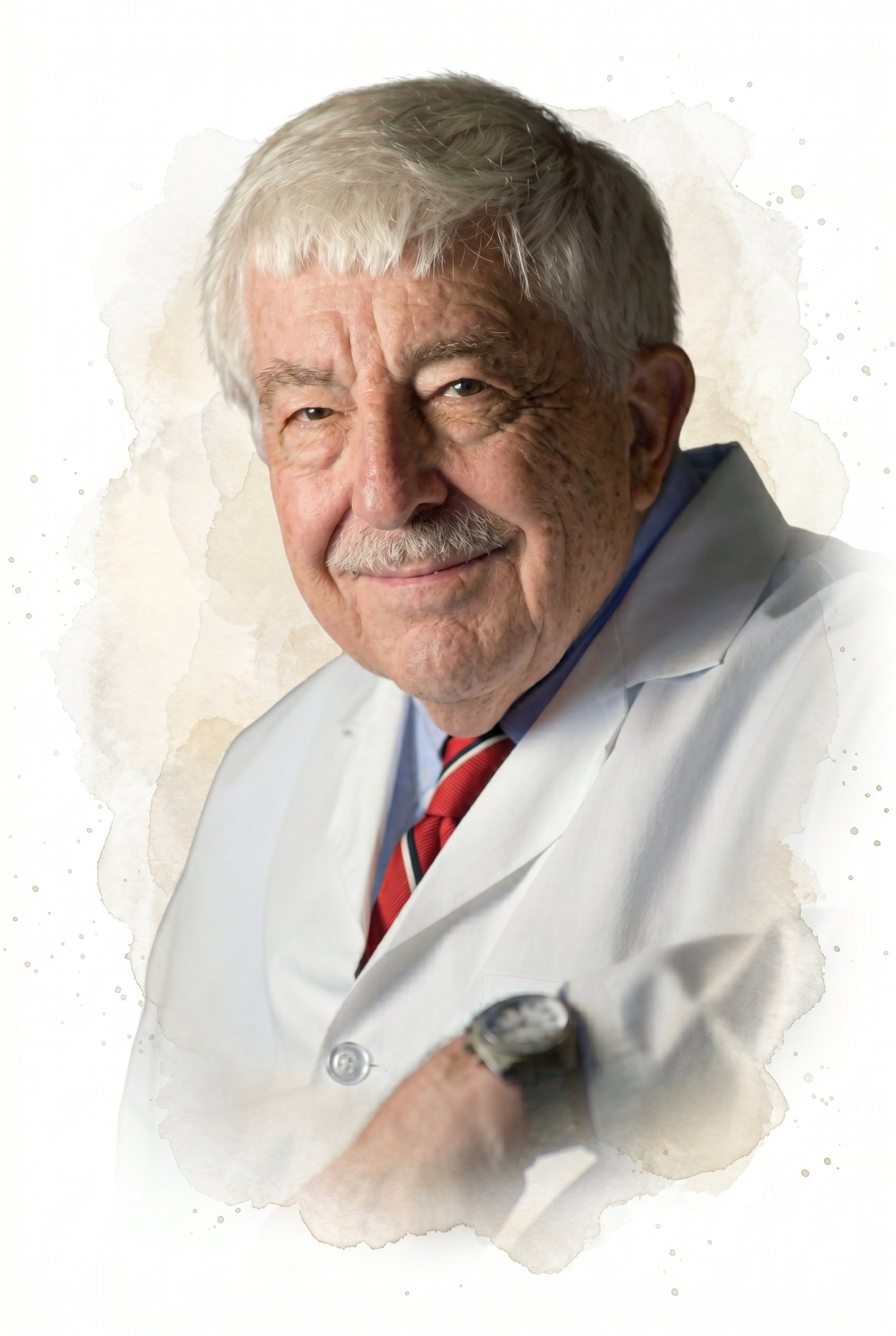
Jay Gunkelman has been an indispensable educator, leader, and mentor. He has generously taught generations of teachers and researchers about neurofeedback and qEEG. The field is more inclusive and vibrant because of Jay's tireless advocacy, especially on behalf of students and our international colleagues.
BCIA Blueprint Coverage
This unit addresses the I. Orientation to Neurofeedback - B. History and development of neurofeedback.

This unit covers Early Antecedents, Cybernetic Theory, Operant Conditioning, The Evolution of EEG Research and Practice, Contributors To EEG Research, and Neurofeedback Research.
Early Antecedents
This section traces the ancient roots of self-regulation and the foundational concepts that made modern biofeedback possible. We will explore how pioneering physiologists recognized the body's drive toward internal stability and how their insights laid the groundwork for understanding voluntary control of biological processes.
Springing from the cultural ferment of the 1960s, these efforts frequently engaged questions of self-awareness and consciousness. As researchers turned toward the latter, occasional speculation about mystical outcomes drew skepticism from the scientific community, a reaction that likely slowed the adoption of neurofeedback methods.
The concept of self-regulation, the voluntary monitoring and adjustment of internal states, has deep historical roots despite its relatively recent formalization in Western science. Practitioners of yoga in India developed sophisticated systems for controlling autonomic functions more than 3,000 years ago. Long before the term "biofeedback" gained widespread recognition in 1969, researchers and practitioners repeatedly demonstrated this learning capacity without fully grasping its implications or potential for broader application across clinical and performance domains.
Claude Bernard (1865) was a French physiologist widely regarded as a founder of modern experimental medicine. Working in Paris, he clarified the liver's role in storing and releasing glucose, traced how the vasomotor nerves control blood vessels, and championed the controlled experiment as the engine of physiological discovery. He set out these ideas in his influential An Introduction to the Study of Experimental Medicine (Bernard, 1957/1865).
Bernard made his most enduring contribution to biofeedback when he proposed that the body actively strives to maintain a steady internal state, which he termed the milieu interieur (internal environment). This idea challenged the prevailing view of the organism as a passive system buffeted by its surroundings. His insight that living systems actively defend their physiological stability gave later researchers the conceptual foundation for understanding how feedback mechanisms operate within the body.
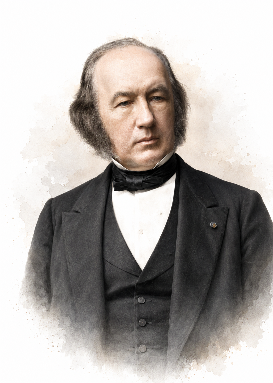
Walter Cannon (1914) was an American physiologist who chaired the Department of Physiology at Harvard Medical School for more than three decades. Studying how the body mobilizes for emergencies, he described the fight-or-flight response, the pattern of sympathetic and adrenal arousal that prepares an organism to confront or flee a threat (Cannon, 1914). His work showed that strong emotions trigger the release of adrenaline and rapid shifts in circulation, heart rate, and blood sugar.
Cannon expanded Bernard's framework, using stress in its engineering sense as a force acting on the organism, with strain as the resulting deformation. Selye later gave stress its modern biological meaning as the body's nonspecific response, and credited Bernard and Cannon as his principal influences. He coined the term homeostasis in 1926 to describe the body's dynamic equilibrium, emphasizing that a steady internal state requires continuous physiological adjustment rather than static, unchanging conditions, and developed the idea for a general readership in The Wisdom of the Body (Cannon, 1932). This insight remains clinically relevant because neurofeedback can help clients restore homeostatic balance.
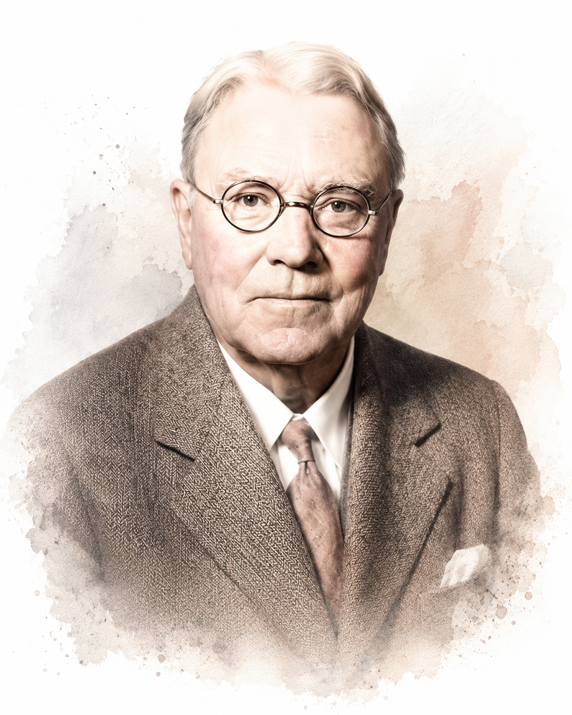
Hans Selye (1936) was an Austrian-born endocrinologist who spent most of his career in Canada at McGill University and the Université de Montréal. He refined our understanding of stress through research on how organisms react to persistent demands, first reporting the pattern in a brief letter to Nature (Selye, 1936). He defined stress as a nonspecific response to various stimuli, called stressors, recognizing that different challenges can produce similar physiological reactions.
Selye's most influential contribution was the general adaptation syndrome (GAS), a three-stage model of how organisms respond to ongoing stress: an initial alarm reaction, a resistance phase during which the body attempts to adapt, and an exhaustion stage if stress persists beyond adaptive capacity. He later distinguished helpful eustress from harmful distress, noting that the same arousal can energize or damage depending on its intensity and duration. This model remains clinically significant because it explains how chronic stress can lead to pathology, a principle that informs biofeedback interventions designed to prevent stress-related illness.
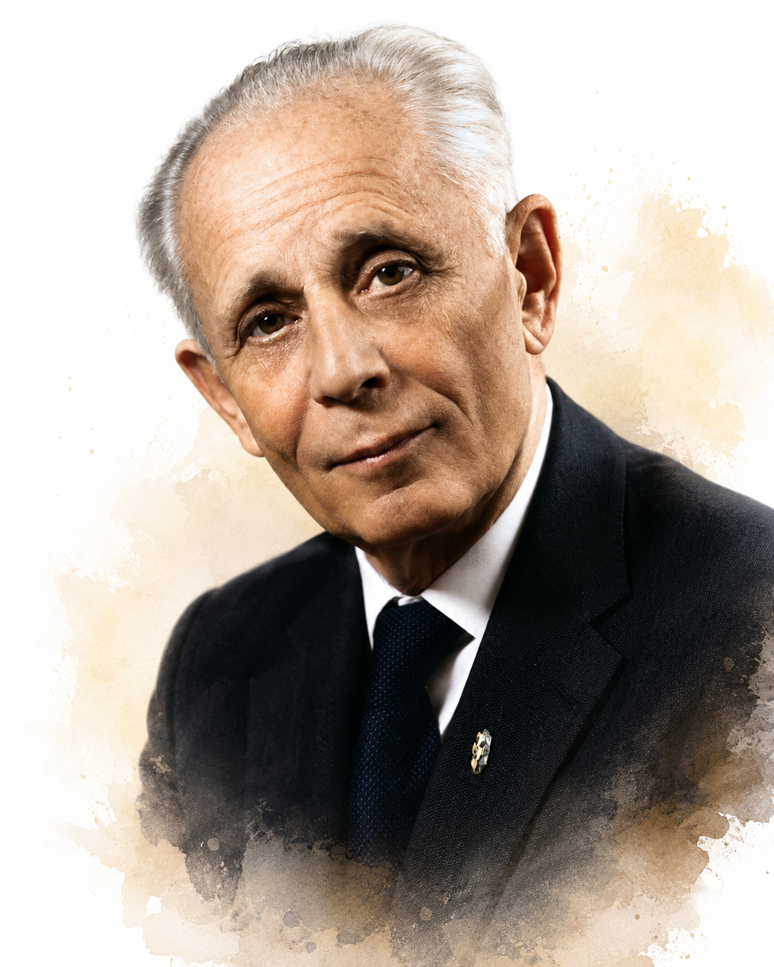
Early physiologists established that the body actively maintains internal stability through homeostatic mechanisms. Bernard identified the internal environment requiring protection, Cannon named this process homeostasis and defined stress as its disruption, and Selye mapped how organisms respond to persistent stressors through predictable stages. These foundational concepts created the theoretical framework necessary for understanding how biofeedback helps clients learn to regulate their physiological responses.
Check Your Understanding
- What did Claude Bernard mean by the milieu interieur, and why was this concept foundational for biofeedback?
- How did Walter Cannon extend Bernard's work through the concepts of homeostasis and the fight-or-flight response?
- What are the three stages of Selye's general adaptation syndrome, and why does this model remain clinically relevant?
Cybernetic Theory
This section examines how cybernetics, the study of control and communication in systems, provided the theoretical framework for modern biofeedback. We will explore Wiener's feedback concept and how it was adapted to create biofeedback as a clinical modality.
Norbert Wiener (1948) was a child prodigy who earned his Harvard doctorate at 18 and spent his career as a mathematician at the Massachusetts Institute of Technology. He founded the field of cybernetics, the study of control and communication in both living organisms and machines, taking the name from the Greek kybernetes, meaning steersman. His most crucial contribution to biofeedback was introducing the concept of feedback, which describes how systems use information about their current state to self-correct and maintain desired conditions. Without Wiener's cybernetic framework, the theoretical foundation for biofeedback would not exist.
Wiener's cybernetic model works through several components acting together. A comparator continuously measures the difference between a regulated variable's actual value and its desired target, or setpoint. That difference is an error signal. The comparator's job ends there: it detects the discrepancy and passes the error signal onward.
Separate effectors, or control mechanisms, use that signal to adjust the system variable and drive the error toward zero. Each adjustment is then measured again, so the loop repeats until the actual and target values converge.
A household thermostat perfectly illustrates this feedback process: the thermostat's comparator constantly compares actual room temperature with your desired temperature setting and automatically adjusts heating or cooling to reduce any difference. This same principle underlies biofeedback, where clients learn to adjust their physiological responses by observing the difference between their current state and their target state.
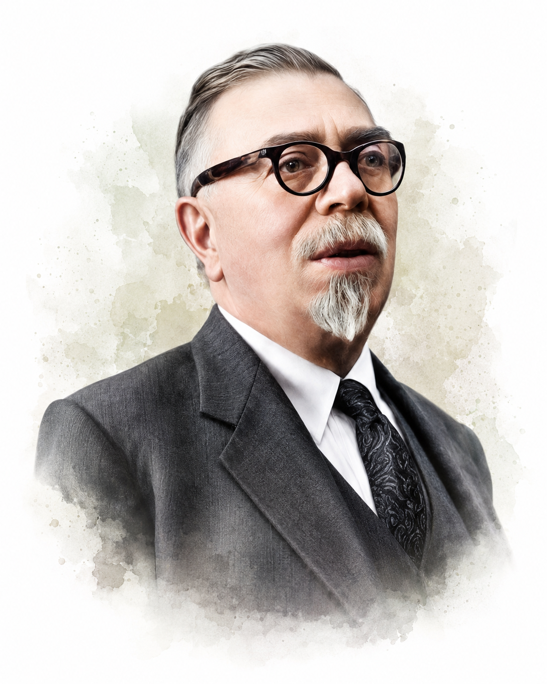
At the landmark 1969 conference at the Surfrider Inn in Santa Monica, participants coined the term biofeedback directly from Wiener's feedback concept (Moss, 1998). This naming decision had lasting implications for how the field would be understood and practiced.
This group needed a name, and the two candidates were biofeedback and autoregulation. Just before the final vote, someone in the audience yelled out that autoregulation sounded like government control of cars. This spontaneous comment created a tipping point, the consensus shifted to biofeedback, and the Biofeedback Research Society (BRS) was born.
(Peper & Shaffer, 2010, p. 142)
Cybernetic theory provided the conceptual scaffolding that made biofeedback possible as a systematic approach to self-regulation. Wiener's feedback model demonstrated how systems use information about their current state to make corrective adjustments to achieve a target value, a principle that translates directly into biofeedback's core mechanism, where clients use real-time physiological information to learn voluntary control to achieve their training goals.
Check Your Understanding
- How does Wiener's feedback model use a comparator and an error signal to maintain a desired state?
- Why does a household thermostat serve as a useful analogy for how biofeedback works?
- How did Wiener's cybernetic framework shape the naming of the field at the 1969 Santa Monica conference?
Operant Conditioning
This section explores how operant conditioning theory shaped our understanding of voluntary learning and its application to biofeedback. We will examine how Thorndike's and Skinner's research on reinforcement established the learning principles underlying biofeedback practice and how these theories had to overcome entrenched beliefs about autonomic control.
Edward Thorndike (1911) was an American psychologist at Columbia University whose puzzle-box experiments with cats laid the groundwork for the study of learning. His work founded the study of what later became known as instrumental learning, the shaping of voluntary responses by the outcomes they produce, although the term itself came into general use later, through Hilgard and Marquis (1940). Thorndike's own vocabulary was trial-and-error learning and the law of effect. His law of effect proposed a mechanistic explanation: responses followed by satisfying consequences are "stamped in" and become more likely to recur. This principle, that any response producing favorable results increases in frequency, proved essential for understanding how biofeedback training works.
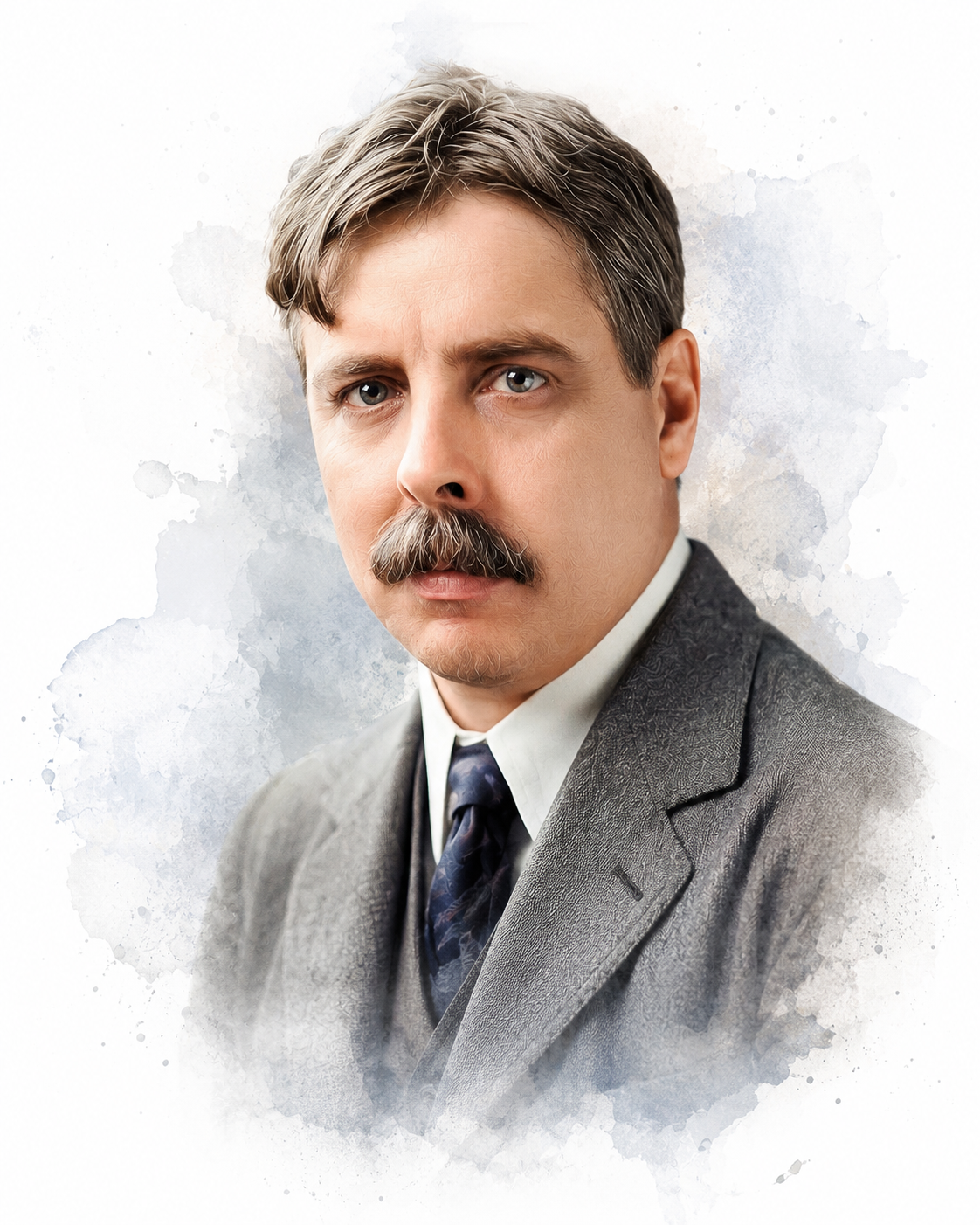
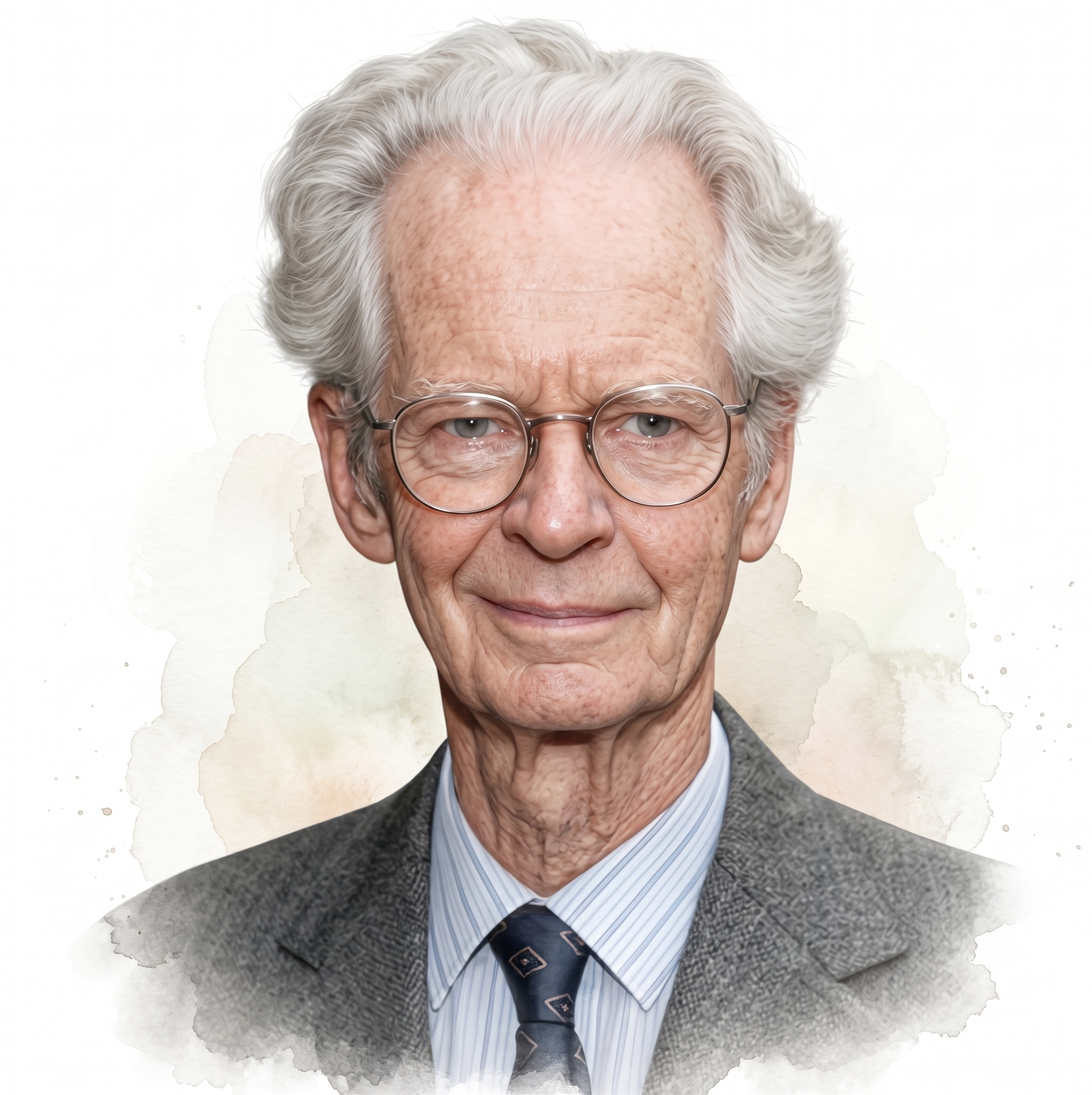
B. F. Skinner's (1938) operant conditioning research significantly expanded Thorndike's law of effect through extensive laboratory studies at Harvard University. Using the operant chamber he invented, now widely called the Skinner box, he showed that animals reliably repeat responses that produce favorable consequences. He mapped how different schedules of reinforcement shape the strength and persistence of behavior. His work established operant conditioning as the dominant framework for understanding voluntary behavior modification.
Skinner introduced several foundational concepts that directly influenced how we conceptualize biofeedback today. Reinforcement describes the process by which consequences of voluntary responses increase the likelihood those responses will be repeated. Consequences that strengthen responses are called reinforcers; in biofeedback, these often take the form of auditory tones or visual displays that change when clients successfully modify their physiological state.
Conversely, punishment describes a process that actively suppresses responses. When a negative consequence follows a voluntary response, it decreases the probability that response will recur. Consequences that weaken responses are termed punishers. Understanding the distinction between reinforcement and punishment remains critical for designing effective biofeedback protocols.
Building on Skinner's framework, early researchers applied operant theory to voluntary musculoskeletal responses. They assumed that the autonomic viscera (internal organs like the heart, blood vessels, lungs, stomach, and intestines) lay beyond the reach of operant conditioning and that classical or respondent conditioning governed these involuntary processes. This assumption created a major obstacle that biofeedback would later need to overcome.
From its inception, biofeedback had to overcome the entrenched paradigm that individuals could not voluntarily control autonomic functions. Researchers who applied B. F. Skinner's work to biofeedback used operant theory to determine which responses could be voluntarily controlled and which could not. For example, Kimble (1961) argued that although subjects could learn to consciously control skeletal muscle responses, autonomic processes (such as heart rate) were involuntary, could be only classically conditioned, and were forever outside of conscious control. This perspective ignored the almost 3,000-year-old yogic practice of autonomic control and research by Lisina (1958); Lapides, Sweet, and Lewis (1957); and Kimmel (1967) that demonstrated voluntary control of autonomic responses.
(Peper & Shaffer, 2010, p. 143)
Neal Miller (1969) was an American experimental psychologist at Yale University, and later Rockefeller University, who directly challenged the belief that autonomic responses lie beyond operant control. Working with Leo DiCara, he reasoned that if an animal could not move its skeletal muscles, then any learned change in an internal organ could not be dismissed as a byproduct of voluntary muscle activity. To test this, the team paralyzed rats with curare, maintained them on artificial respiration, and reinforced small changes in autonomic activity with pleasurable electrical brain stimulation (Miller & DiCara, 1967). Their curarized rats appeared to learn specific increases or decreases in heart rate, blood pressure, and intestinal contraction, results that implied operant conditioning could reach the viscera (Miller, 1969).
This work electrified the young field, but its story carries an important caution. Miller himself was later unable to reliably reproduce the dramatic visceral learning effects, and he reported these replication difficulties with notable candor (Miller & Dworkin, 1974). The original experiments therefore remain unconfirmed, yet their influence proved profound. By making autonomic self-regulation seem possible, Miller's research inspired the human biofeedback studies that followed and helped dissolve the assumption that the viscera were forever outside conscious control (Miller, 1978).
Operant conditioning provided the learning theory that explains how biofeedback works. Thorndike established that successful responses are strengthened by their consequences, while Skinner refined this into a systematic framework of reinforcement and punishment. Despite initial resistance based on assumptions that autonomic functions could not be voluntarily controlled, operant principles proved applicable to physiological self-regulation, validating what yogic practitioners had demonstrated for millennia. Neal Miller's curarized-rat experiments dramatized this possibility, even though his own difficulty replicating them is a reminder that bold early findings require careful confirmation.
Check Your Understanding
- How does Thorndike's law of effect explain why successful responses become more frequent over time?
- What is the difference between reinforcement and punishment in Skinner's framework, and how does each appear in biofeedback?
- What entrenched assumption about autonomic control did early biofeedback have to overcome, and what evidence challenged it?
The Evolution of EEG Research and Practice
This section traces three major phases in EEG development, showing how technological advances transformed neurofeedback from a laboratory curiosity into a practical clinical tool. Understanding this evolution helps explain both current capabilities and ongoing challenges in the field.
1800s to 1940s: The Pioneering Era
Early EEG equipment suffered from severe technical limitations that restricted its usefulness for studying brain structure and function. The available amplifiers were imprecise, artifacts easily contaminated recordings, and the technology could provide only minimal insights into neuroanatomy and neurophysiology. Despite these obstacles, pioneering researchers persisted in developing the foundational knowledge that would later enable clinical applications.
1960 to 1985: The Dark Ages
The dates below mark the periods of sharpest change rather than a continuous partition; the intervening years saw steady but less dramatic development.
This period presented formidable challenges for EEG biofeedback that nearly derailed the field entirely. Clinical EEG biofeedback devices remained highly susceptible to environmental artifacts, making reliable recordings difficult in typical clinical settings. Excessive media hype and unrealistic public expectations surrounding alpha training created a backlash when promised benefits failed to materialize consistently. As disillusionment spread, the biofeedback field largely shifted its focus toward autonomic and skeletal muscle applications, which offered more reliable and repeatable results. The small minority of professionals who continued investigating and applying EEG biofeedback found themselves working in an often unsupportive and sometimes hostile scientific environment that questioned the validity of their entire endeavor.
2005 to Present: The Renaissance
Several technological breakthroughs converged to transform neurofeedback into an accessible clinical modality. The development of affordable, powerful digital computer technology enabled complex signal processing previously possible only in research laboratories. Advances in micro-electronics produced more accurate sensors with better signal quality and less susceptibility to artifacts. The creation of normative databases provided clinicians with reference standards for comparison, while improved software made systems more user-friendly.
These innovations collectively enabled average clinicians, not just EEG specialists, to provide effective neurofeedback training. The increased portability and turn-key operation of modern EEG systems have extended neurofeedback beyond traditional clinic settings into homes, schools, and performance training facilities.
EEG technology evolved through three distinct phases: an early period of primitive equipment, a challenging middle period when technical limitations and overpromise nearly destroyed the field's credibility, and a modern renaissance enabled by digital technology that has made neurofeedback practical for widespread clinical use. This evolution demonstrates how technological advancement can transform scientific possibility into clinical reality.
Check Your Understanding
- What technical limitations constrained EEG biofeedback during its early pioneering era?
- What factors nearly derailed the field during the "Dark Ages" of EEG biofeedback?
- Which technological advances enabled the modern renaissance of neurofeedback, and how did they broaden access to training?
Contributors To EEG Research
This section chronicles the remarkable individuals who built our understanding of brain electrical activity from its earliest detection to modern clinical applications. We will trace the evolution from basic discoveries of bioelectricity through the identification of EEG rhythms to the development of sophisticated recording and analysis techniques that made neurofeedback possible. The section closes with a reappraisal of that history in light of recent archival scholarship.
Luigi Galvani (1791) was an Italian physician and anatomist at the University of Bologna who reported electrical currents in animals (Swartz & Goldensohn, 1998). Observing that a frog's leg twitched when touched by metal instruments, he proposed that living tissue stored a vital animal electricity. His rival Alessandro Volta argued the current came from the contact of dissimilar metals rather than the tissue itself, a dispute that helped launch the science of electrophysiology. Galvani's name survives in the term galvanism, the stimulation of muscle by electric current.
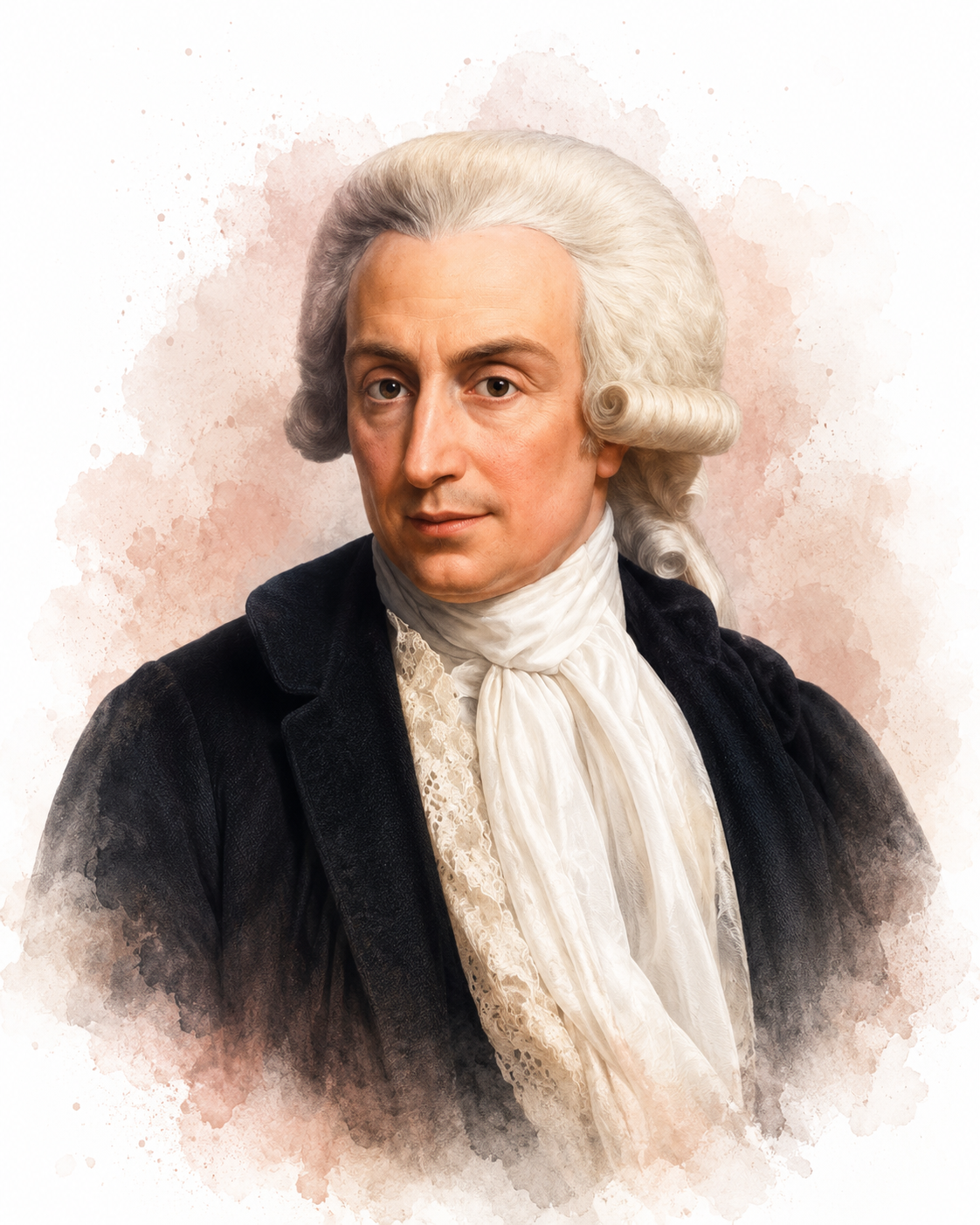
Galvani's findings were confirmed by Giovanni Aldini (1794) and Freiherr (baron) von Humboldt (1797). Aldini, Galvani's nephew, staged dramatic public demonstrations in which he applied current to the bodies of executed criminals, producing grimaces and twitching limbs that captivated audiences and helped inspire Mary Shelley's Frankenstein. Von Humboldt, the Prussian naturalist, took a more cautious experimental approach, performing hundreds of careful trials, including on his own body, to separate genuine bioelectricity from the effects Volta described.
Ernst Fleischl von Marxow (1883) was an Austrian physiologist in Vienna who recorded visual cortical potentials, the cortical potentials evoked by visual stimuli, but did not describe rhythmic oscillation (Niedermeyer, 1993). To establish priority while continuing his work, he deposited a sealed letter describing his observations with the Imperial Academy of Sciences in 1883.
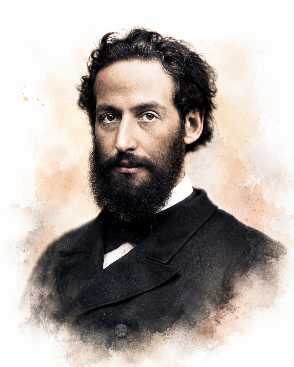
Emil du Bois-Reymond (1849) was a German physiologist in Berlin and a founder of experimental electrophysiology who reported electrical conduction in muscles and peripheral nerves (Schomer & Lopes da Silva, 2011). Building sensitive galvanometers, he was the first to detect the electrical basis of the nerve impulse, establishing that nerves signal through measurable changes in electrical potential.
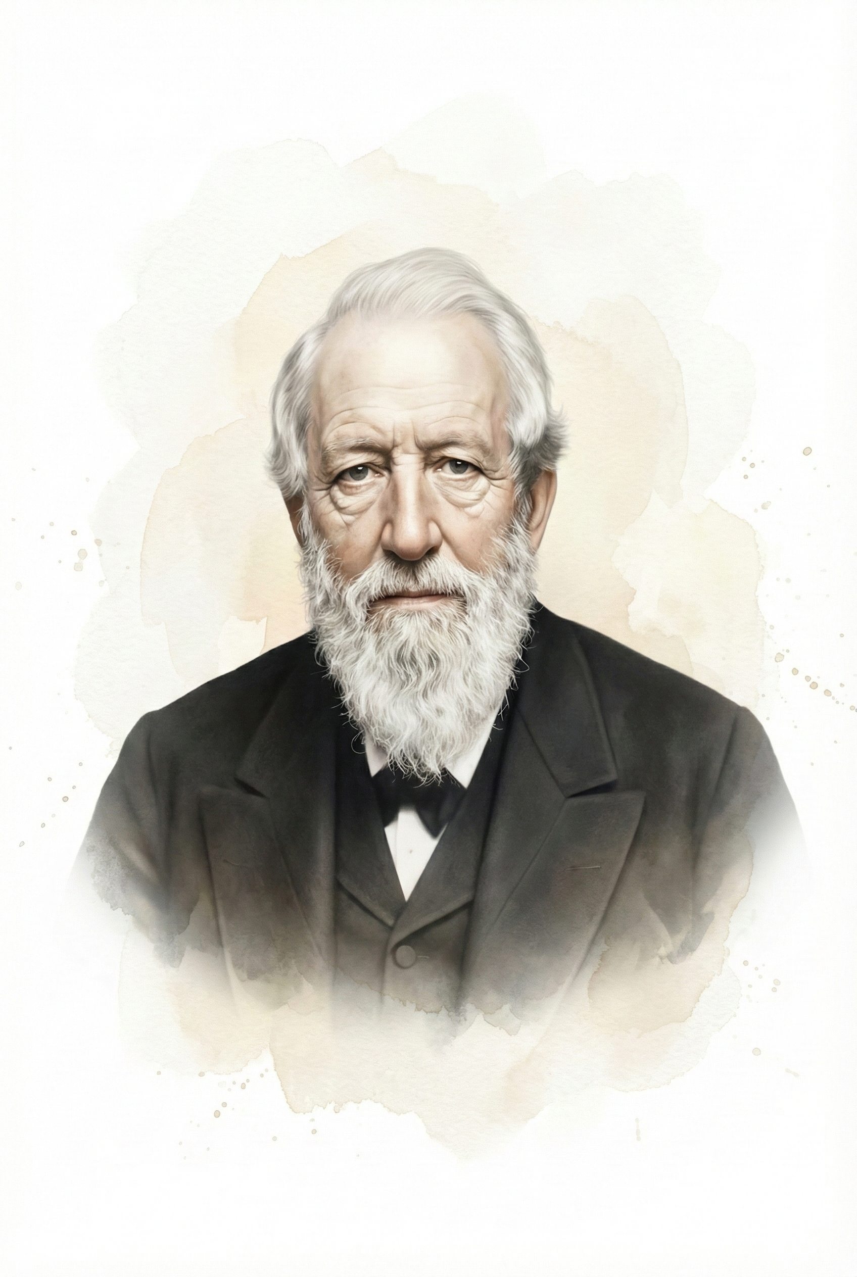
Gustav Fritsch and Eduard Hitzig (1870) discovered that cortical stimulation elicits a localized motor response (Schomer & Lopes da Silva, 2011). Working in Fritsch's Berlin home, they applied weak electrical currents to the exposed cortex of a dog and saw specific muscle groups twitch depending on which region they stimulated. This was the first direct evidence that the cortex is electrically excitable and that motor functions are anatomically localized, a cornerstone of the brain mapping that underlies neurofeedback today.
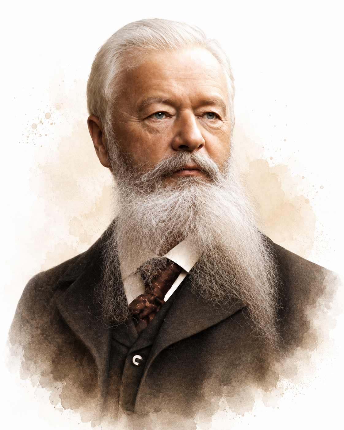
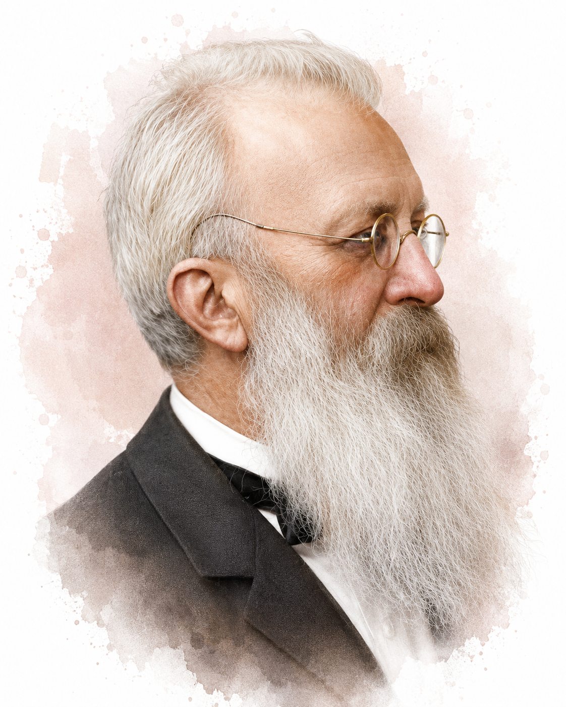
Richard Caton (1875) was an English physician in Liverpool who discovered electrical potentials in vivisected animals and reported his findings to the British Medical Journal. He recorded spontaneous electrical potentials from the exposed cortical surface of monkeys and rabbits. He was the first to measure evoked potentials (EPs), which are EEG responses to stimuli (Schomer & Lopes da Silva, 2011). His instrument was a mirror galvanometer, so basic that he recorded his measurements not in volts but in millimeters of light-beam deflection. Beyond the laboratory, Caton was a civic leader who later served as Lord Mayor of Liverpool.
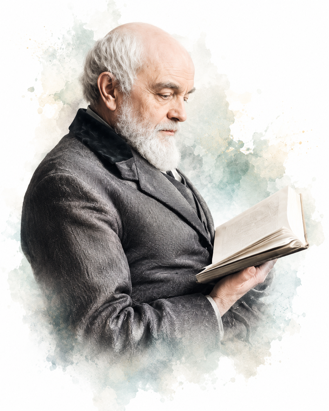
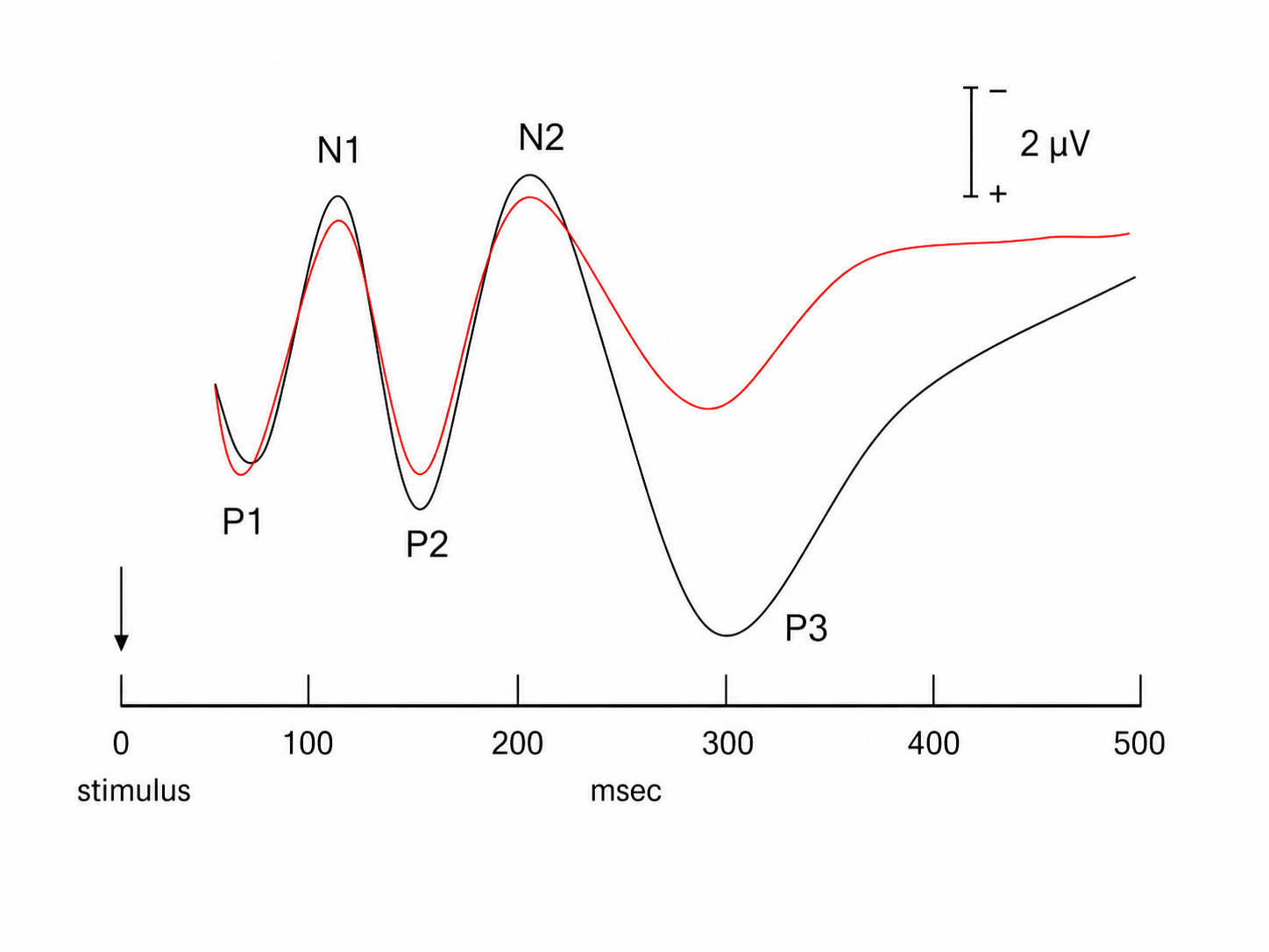
He also reported the first observations of the shift of the cortical gradient to electro-negative during activation.
Vasili Yakovlevic Danilewsky (1877) was a physiologist at Kharkov University who, working independently of Caton, also detected electrical activity in the animal brain. His doctoral thesis, Investigations in the Physiology of the Brain, explored the relationship between the EEG and states of consciousness (Brazier, 1959).
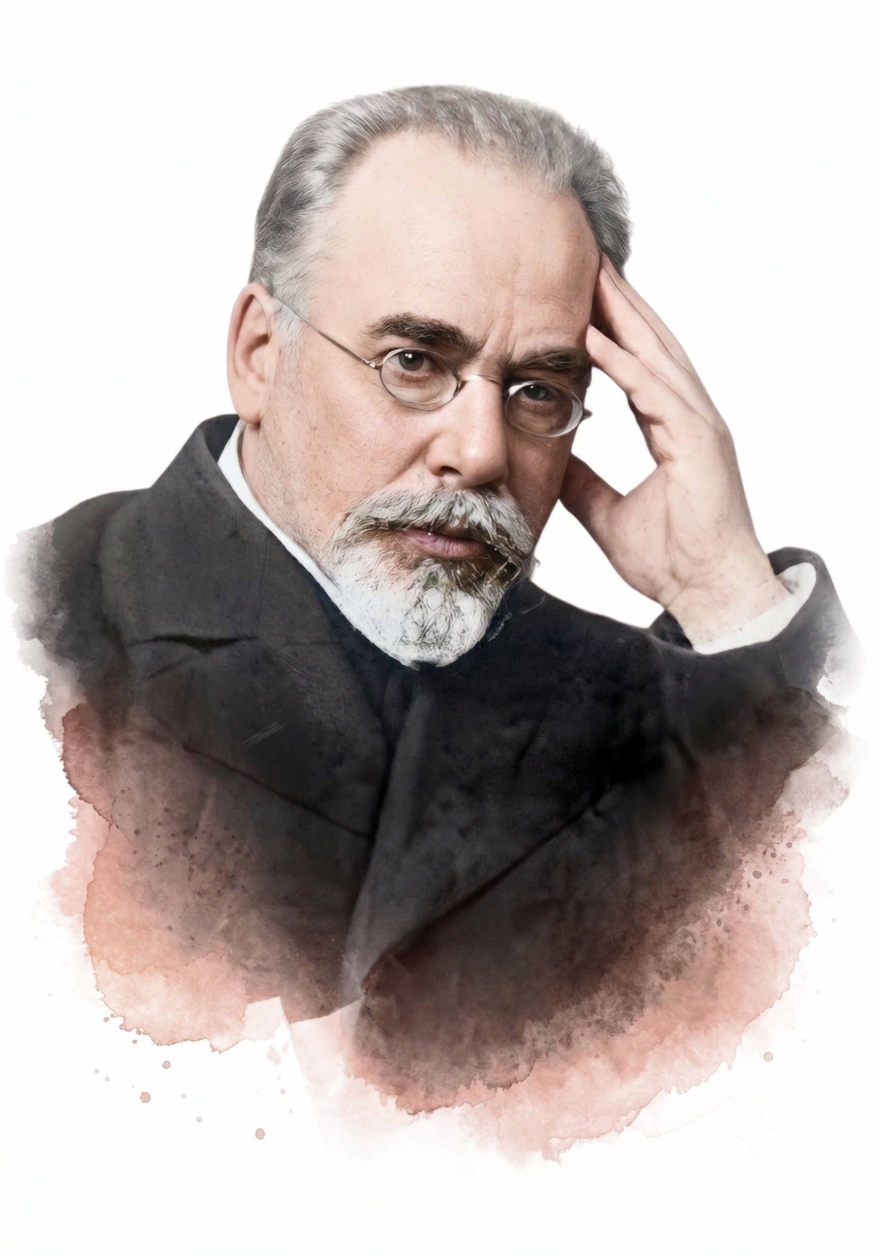
Adolf Beck (1891) was a Polish physiologist at the Jagiellonian University in Kraków and an assistant to Napoleon Cybulski. He published studies of spontaneous electrical potentials detected from the brains of dogs and rabbits. He was the first to document alpha blocking, the desynchronization in which sensory stimulation replaces rhythmic oscillations with faster, lower-amplitude activity (Coenen et al., 1998).
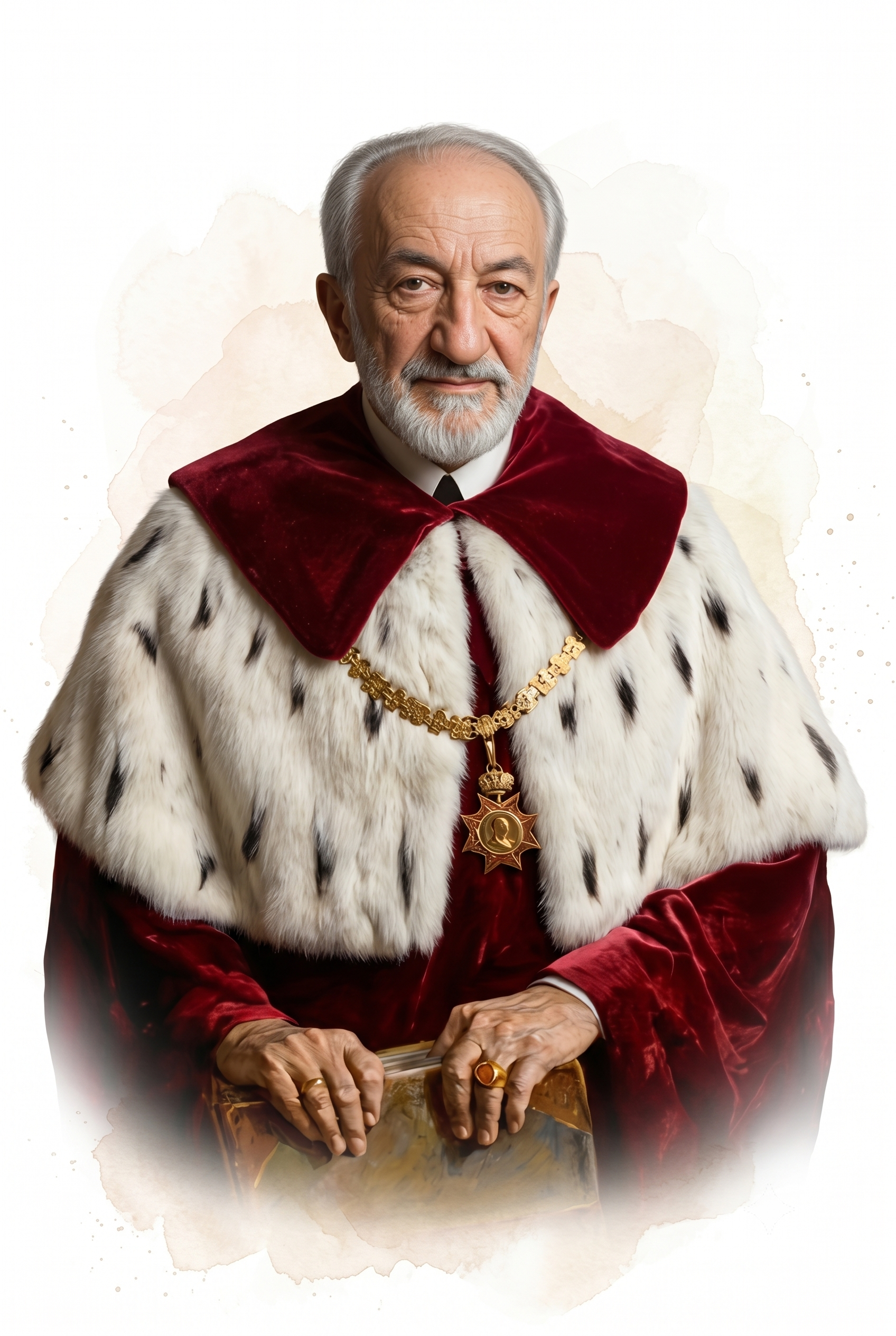
Sir Charles Sherrington (1906) proposed the concept of a synapse to describe the junction across which neurons communicate and published The Integrative Action of the Nervous System. The Cambridge classicist, Arthur Woollgar Verrall, proposed the Greek-derived term (Tansey, 1997). He made significant contributions to understanding muscle action, movement, proprioception, reflexes, and spinal nerves (Schomer & Lopes da Silva, 2011). He shared the 1932 Nobel Prize in Physiology or Medicine with Edgar Adrian for their discoveries on the function of neurons.
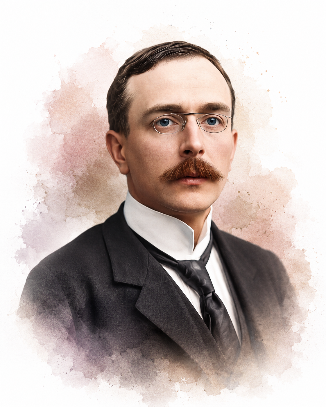
Sherrington proposed the "enchanted loom" metaphor for the human brain in a passage in Man on His Nature, first published in 1940 and quoted here from the 1942 printing, in which he poetically described the change in cortical activity as we awaken:
The great topmost sheet of the mass, that where hardly a light had twinkled or moved, becomes now a sparkling field of rhythmic flashing points with trains of traveling sparks hurrying hither and thither. The brain is waking and with it the mind is returning. It is as if the Milky Way entered upon some cosmic dance. Swiftly the head mass becomes an enchanted loom where millions of flashing shuttles weave a dissolving pattern, always a meaningful pattern though never an abiding one; a shifting harmony of subpatterns.
(p. 178)
Current sleep research shows that the cortical networks are considerably more active during sleep than Sherrington imagined.
Vladimir Pravdich-Neminsky (1912) was a Ukrainian physiologist who, using Einthoven's string galvanometer, produced the first photographic recordings of brain electrical activity in a mammal. He photographed dogs' EEG and event-related potentials, demonstrated a 12-14 Hz rhythm that slowed during asphyxiation, and introduced the term electrocerebrogram (Schomer & Lopes da Silva, 2011).
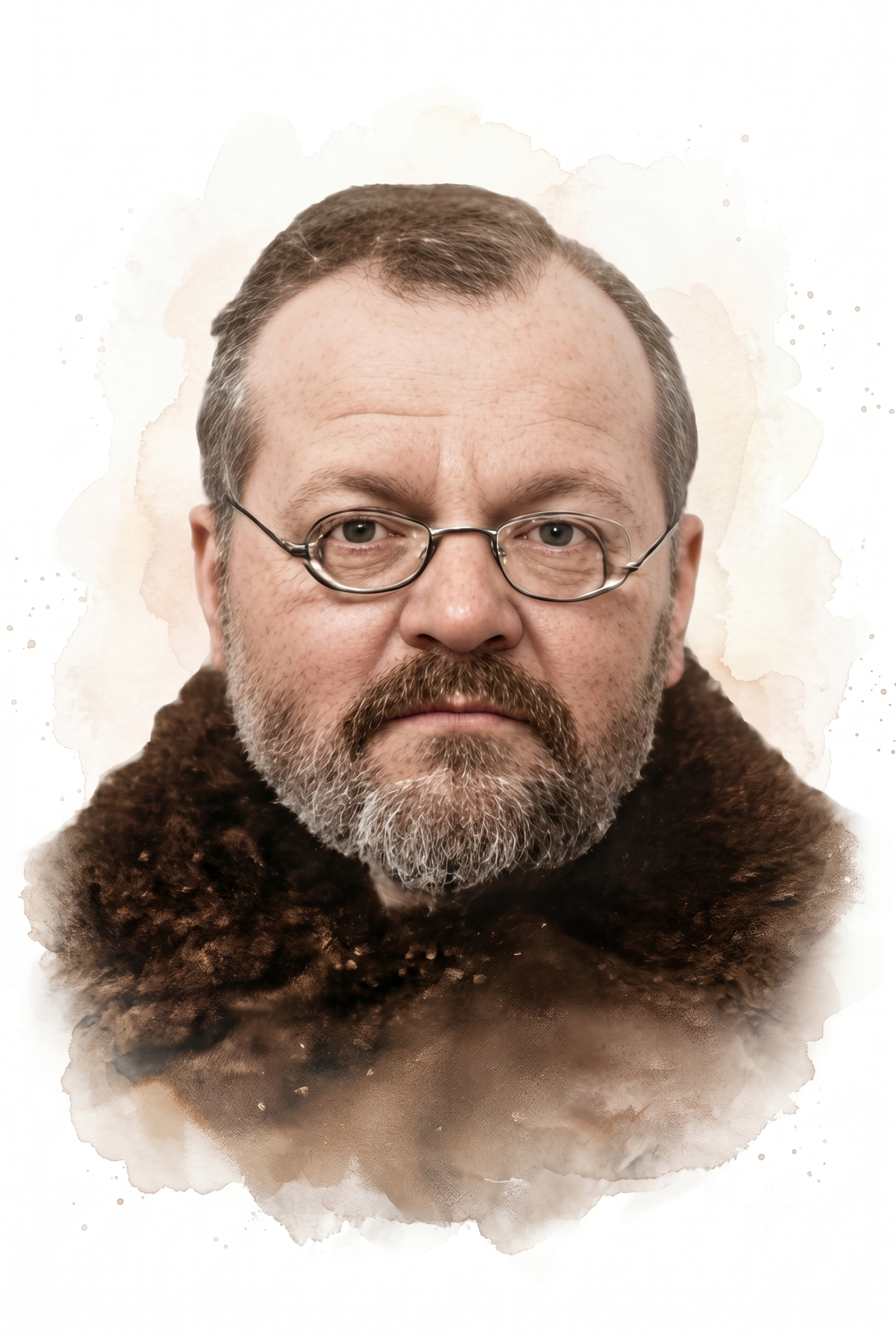
Napoleon Cybulski and Jelenska-Macieszyna (1914) recorded experimental seizures (Swartz & Goldensohn, 1998). Cybulski, a Polish physiologist at the Jagiellonian University and a co-discoverer of adrenaline, mentored Adolf Beck and captured some of the earliest photographic records of seizure activity in the cortex.
Alexander Forbes and Catharine Thacher (1920) reported using a vacuum tube to amplify the faint currents recorded by the string galvanometer. Forbes was an American physiologist at Harvard whose vacuum-tube amplification increased sensitivity roughly fiftyfold, making it possible to display fast electrical potentials that earlier instruments missed. Forbes and Thacher amplified nerve action currents rather than the EEG, which did not yet exist as a human technique, but electronic amplification became the foundation on which EEG instrumentation was built over the following two decades (Swartz & Goldensohn, 1998).
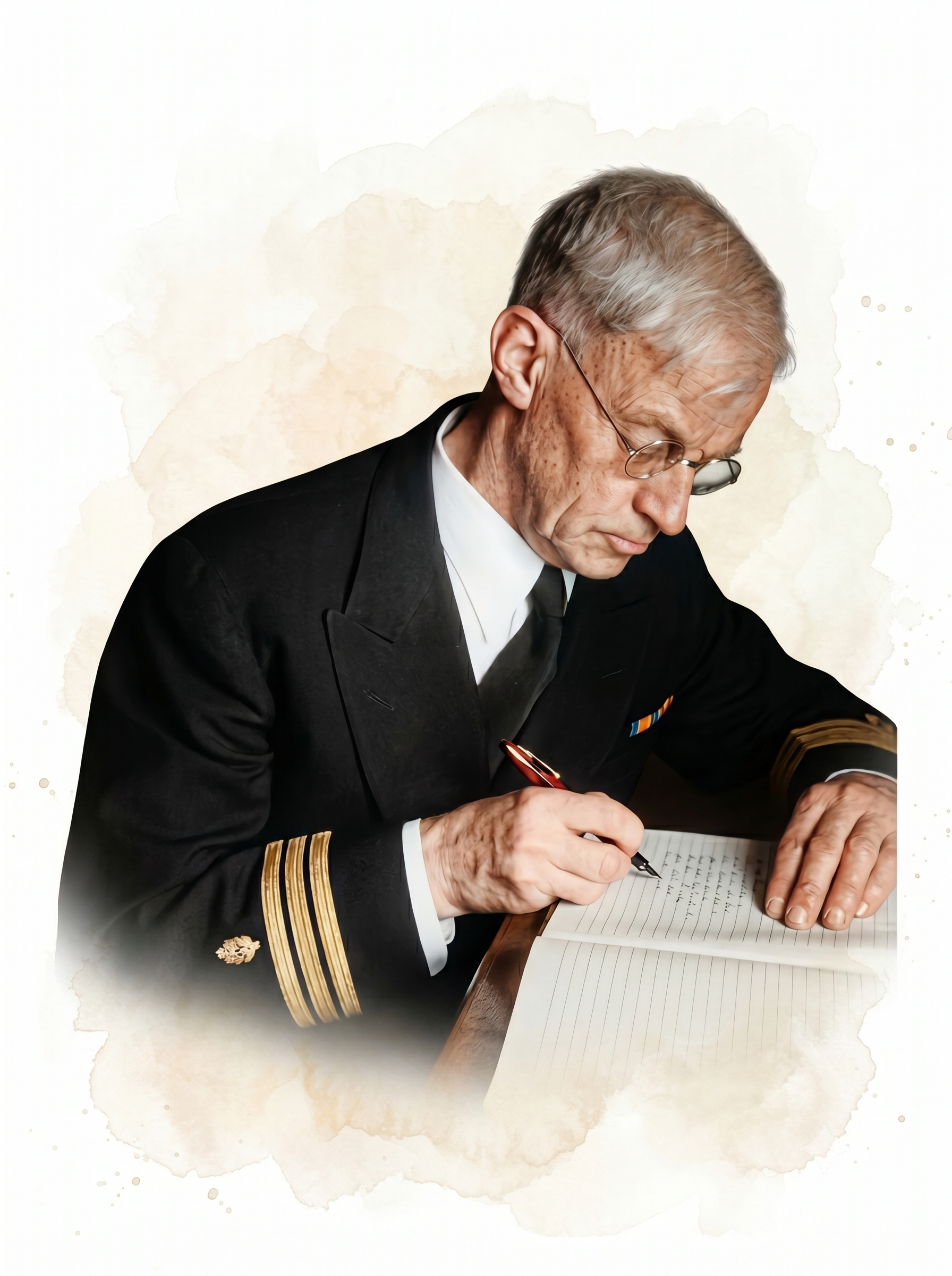
Hans Berger (1929) was a German neurologist and psychiatrist at the University of Jena who published the first human EEG recording. He made that recording in 1924 but did not publish it until 1929. Driven by a lifelong quest to find the physical basis of "psychic energy" and mental phenomena, he worked for years in relative obscurity before publishing fourteen scientific papers from 1929 to 1938 (Schomer & Lopes da Silva, 2011). His conduct under the Third Reich has been substantially revised by recent historical scholarship, as the reappraisal at the end of this section describes (Fields, 2024; Zeidman et al., 2014).
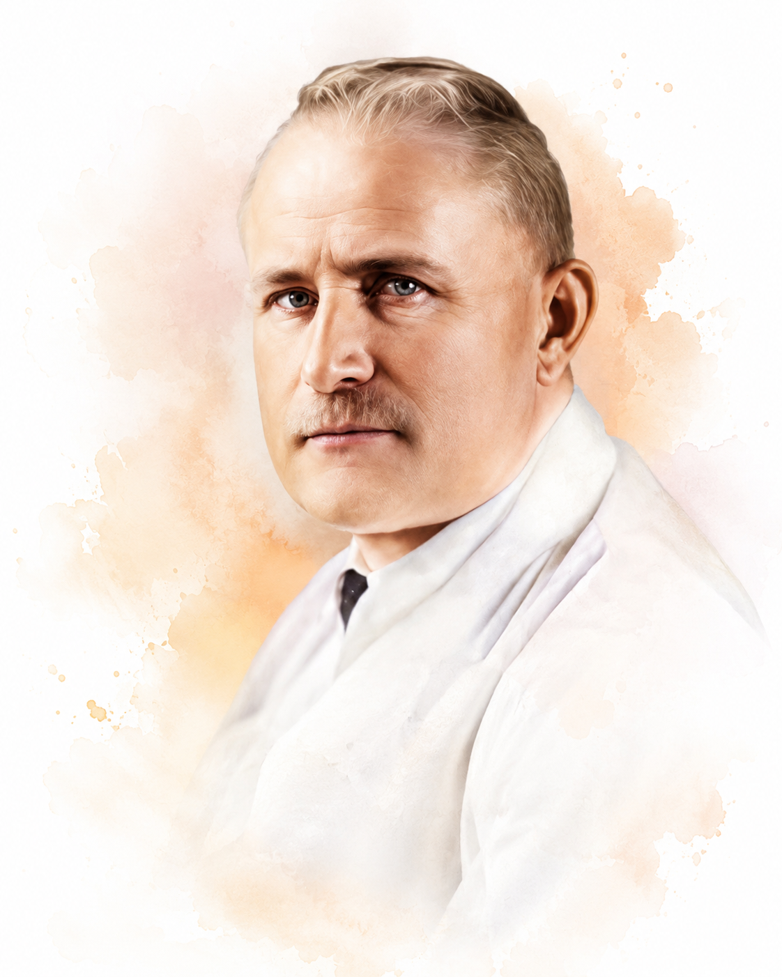
Berger's first human recording, on July 6, 1924, came from a 17-year-old patient with a skull defect left by a neurosurgical procedure. His son Klaus became a frequent subject of the later scalp recordings, and Klaus's tracings appear among the published samples below.
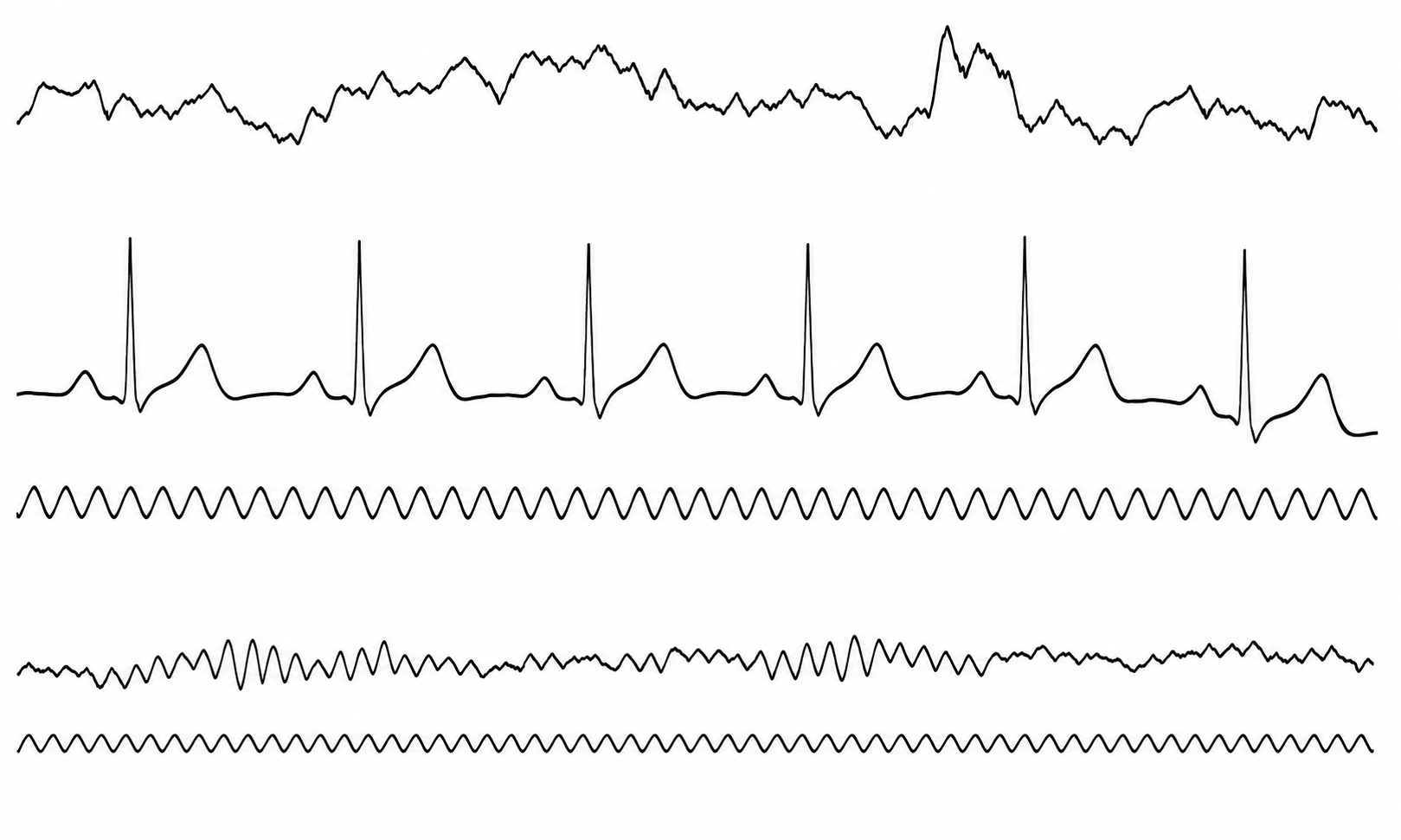
Berger showed that these potentials were not due to scalp muscle contractions. Berger discovered the alpha rhythm, the first EEG rhythm, and named it the alpha wave. The eponym Berger rhythm was applied by Adrian and Matthews, who titled their 1934 confirmation paper "The Berger Rhythm." He identified sleep spindles, which are short bursts of 12-15 Hz activity during stage 2 sleep.
Berger viewed the EEG as analogous to the ECG because it generates an electrical signal that can be amplified and displayed. He introduced the German term Elektrenkephalogramm. He believed that the EEG had diagnostic and therapeutic promise in measuring the impact of clinical interventions.
He demonstrated that alterations in state (sleep/wake, eyes open/closed, drug/no drug, and illness/health) are associated with changes in the EEG. He associated the beta rhythm with alertness. He described interictal activity (EEG potentials between seizures) and recorded a complex partial seizure (impaired awareness and repetitive behaviors called automatisms) in 1933. Finally, he performed the first qEEG, which measures the signal strength of component EEG frequencies (Hassett, 1978; Robbins, 2000; Swartz & Goldensohn, 1998).
Edgar Douglas Adrian and Bryan Matthews (1934), British physiologists at Cambridge, confirmed Berger's findings by recording their own EEGs using a cathode-ray oscilloscope. Because Adrian had just received the 1932 Nobel Prize for his work on the nerve impulse, his endorsement carried great weight, and their demonstration to the Physiological Society at Cambridge in May 1934, at which Adrian displayed his own alpha rhythm, turned skepticism into widespread acceptance (Schomer & Lopes da Silva, 2011).
Adrian provided evidence supporting the all-or-none law: action potentials initiated at the axon hillock either occur or not. When they occur, they have the same amplitude and speed. He used himself as a subject and demonstrated the phenomenon of alpha blocking, where opening his eyes suppressed alpha rhythms.
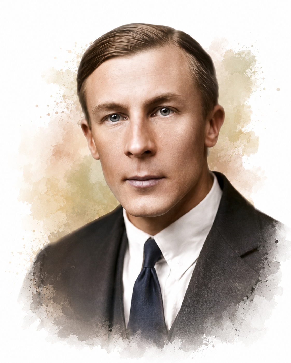
Bryan Matthews was both a physiologist and a gifted instrument engineer whose oscillograph and differential amplifier designs became foundational hardware for recording bioelectric signals. The differential amplifier he refined still lies at the heart of modern biofeedback and neurofeedback systems, where it cancels shared noise and isolates the small signals of interest.
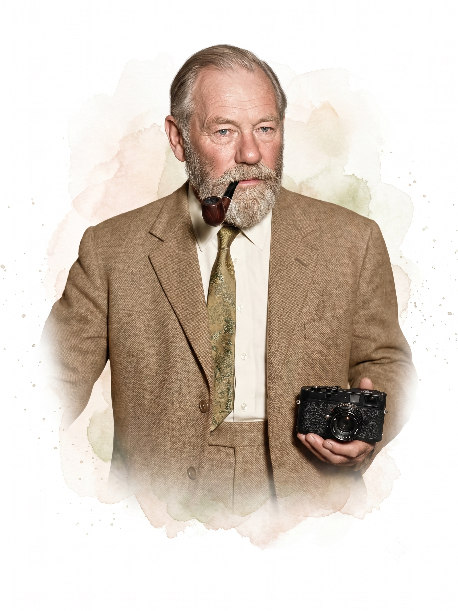
M. H. Fischer and H. Löwenbach (1933/1934) provided the first demonstration of epileptiform spikes, the sharp EEG transients with a duration of less than 70 ms that signal abnormal, hypersynchronous neuronal firing (Swartz & Goldensohn, 1998). Their work helped establish the link between distinctive EEG waveforms and seizure activity that made the EEG a diagnostic tool for epilepsy.
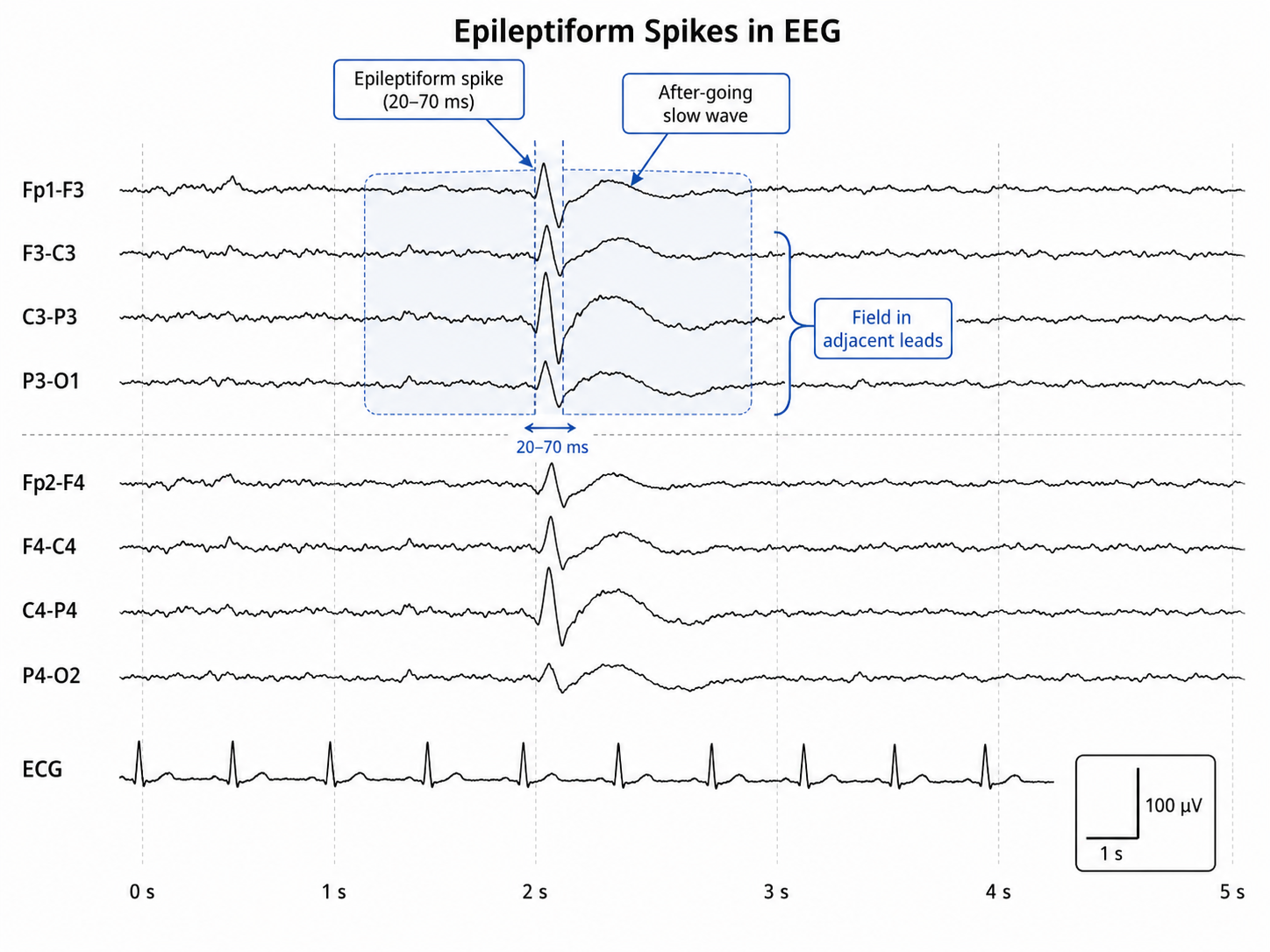
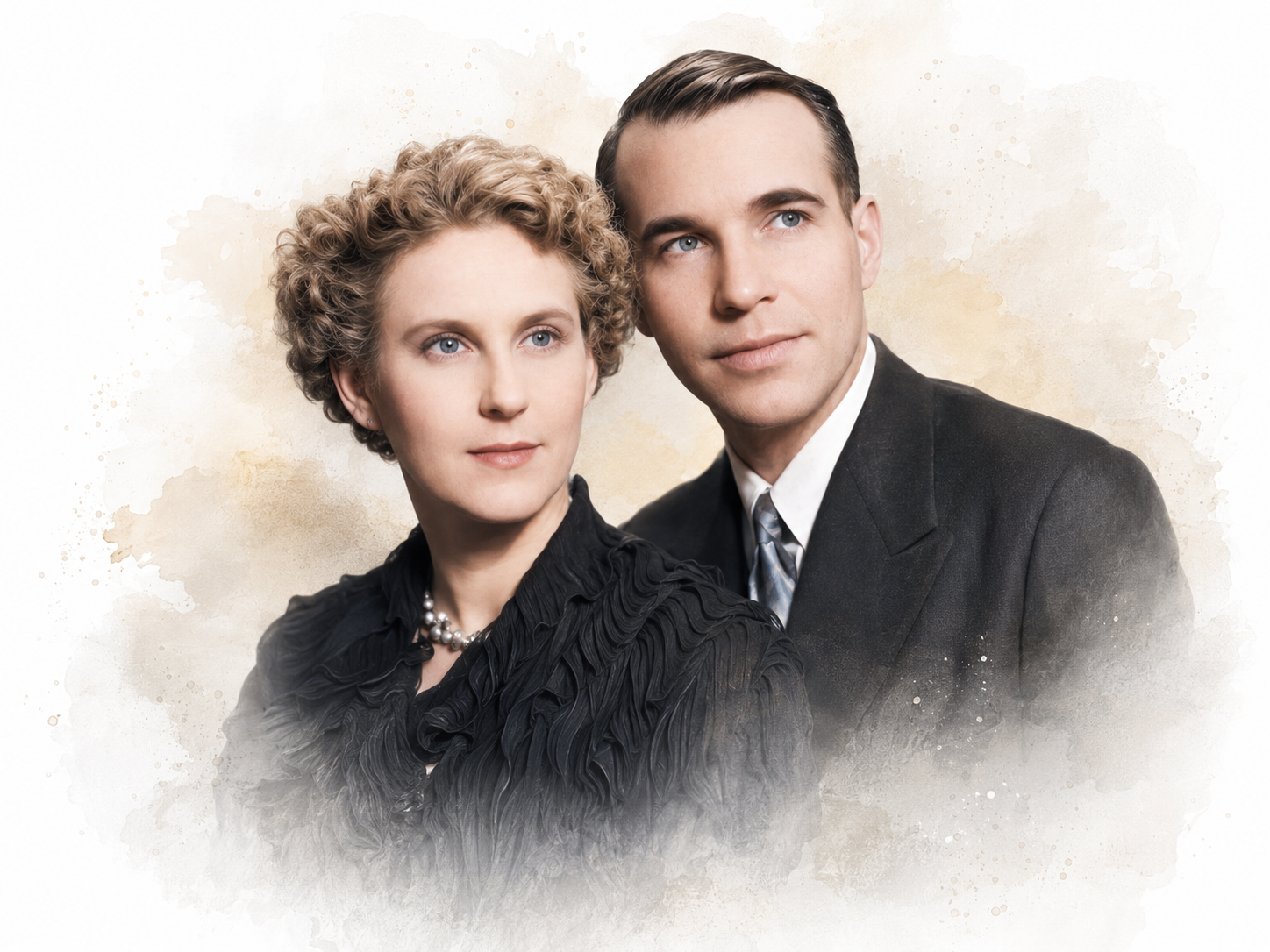
Frederic A. Gibbs, Hallowell Davis, and William G. Lennox, working at Harvard Medical School, inaugurated clinical electroencephalography in 1935 by identifying abnormal EEG rhythms associated with epilepsy, including interictal spike waves and the 3-Hz spike-and-wave activity in absence seizures, which involve periods of less than 15 s during which a client blanks out (Brazier, 1959). This 3-Hz pattern became a reliable diagnostic signature, helping move the EEG from the research laboratory into the hospital. Erna and Frederic Gibbs were married and closely collaborated on their research, including developing a scale for rating beta spindles.
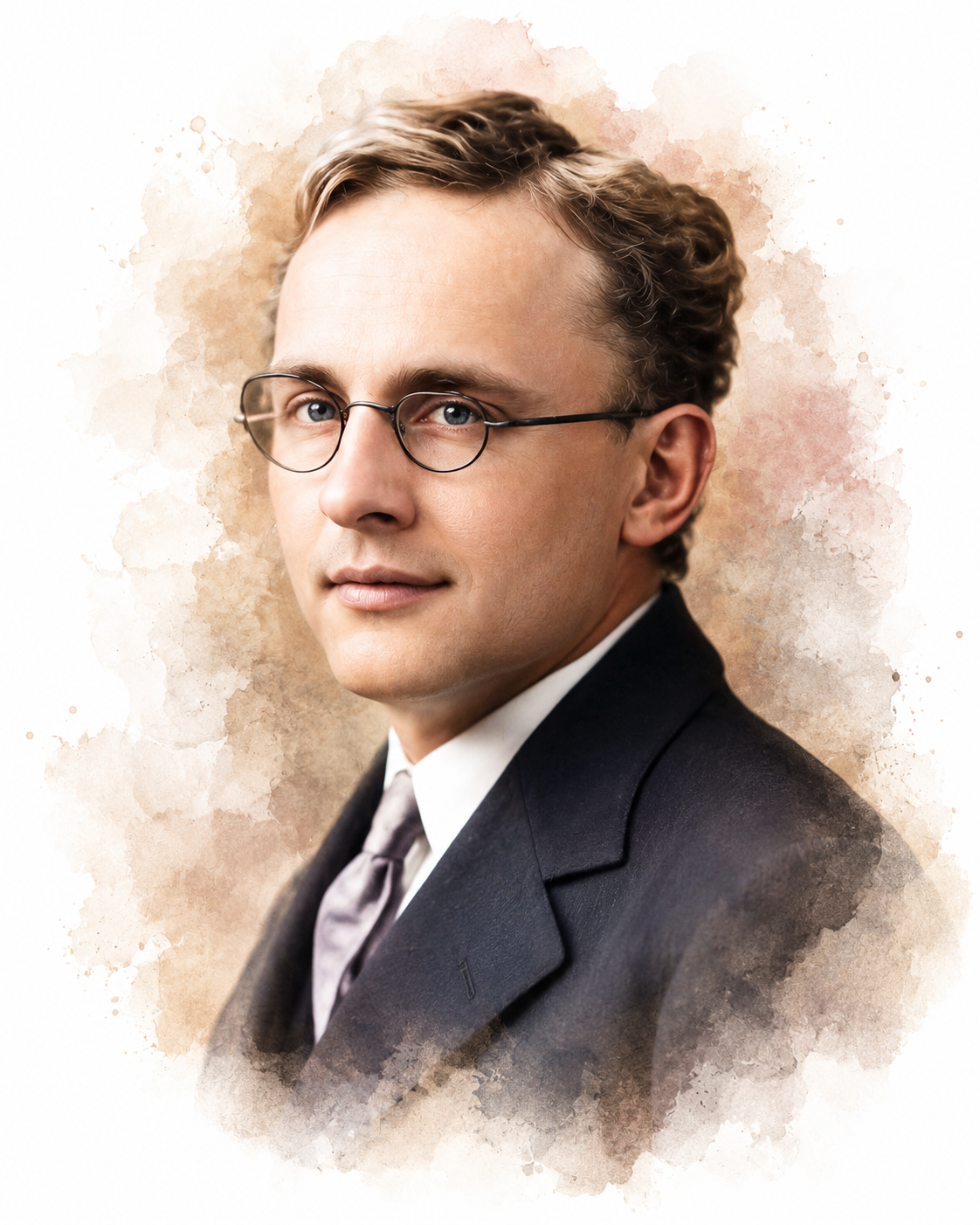
Frederic Bremer (1935) was a Belgian neurophysiologist who used the EEG to show how sensory signals affect vigilance and studied the sleep-wake cycle. Using his cerveau isolé and encéphale isolé brainstem preparations, he showed that lower brainstem structures help sustain cortical arousal. His work is sometimes credited with dividing the EEG into standard bandwidths, but the band names came from others: Berger named alpha and beta in 1929, and Walter named delta in 1936 and theta, with Vivian Dovey, in 1944.
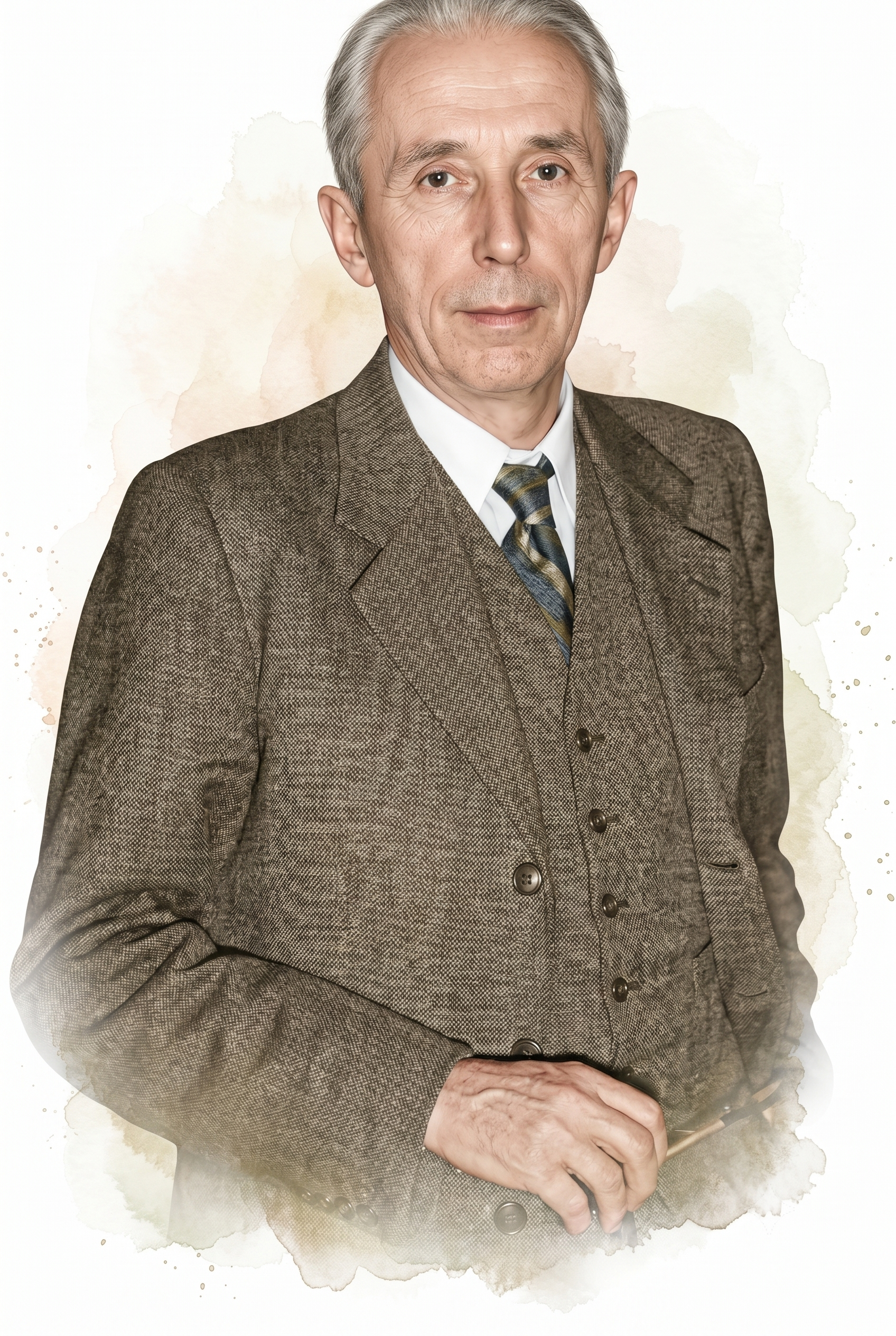
Frederic A. Gibbs and Herbert H. Jasper (1936) showed that the interictal spike was the defining indicator of epilepsy (Schomer & Lopes da Silva, 2011). Jasper went on to standardize electrode placement, work that led to the International 10-20 System and allowed recordings to be compared across laboratories worldwide.
The International 10-20 System positions electrodes at distances of 10% and 20% along measured lines that run between bony landmarks on the skull, such as the nasion at the bridge of the nose and the inion at the back of the head (Jasper, 1958). Because the spacing is proportional rather than fixed, the same sites land over comparable brain regions on heads of different sizes. Each location carries a letter for the underlying region, such as F for frontal or O for occipital, with odd numbers marking the left hemisphere and even numbers the right. This shared map is why a recording made in one laboratory can be meaningfully compared with one made anywhere else.
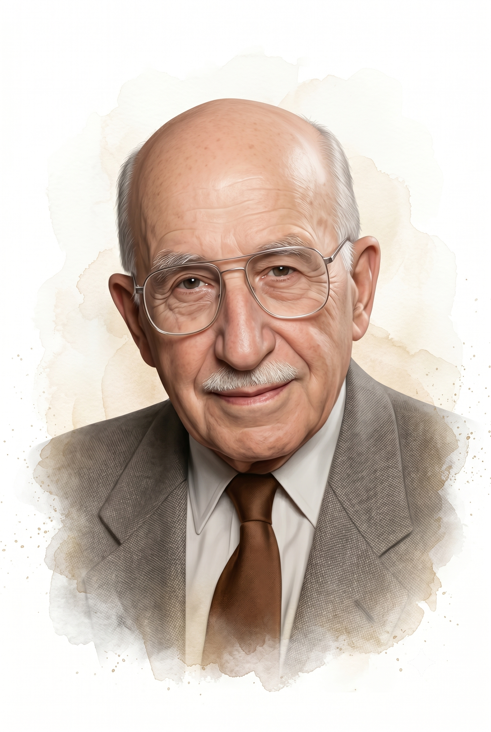
W. G. Walter (1940s) was an American-born British neurophysiologist in Bristol who named the delta and theta waves and discovered the contingent negative variation (CNV). This slow cortical potential may reflect expectancy, motivation, intention to act, or attention (Schomer & Lopes da Silva, 2011). He also built some of the first autonomous robots, his "tortoises," to show that lifelike behavior could emerge from simple feedback circuits.
Walter located an occipital lobe source for alpha waves and demonstrated that delta waves could help find brain lesions like tumors. In 1946, he described electrical responses to photic stimulation (visual stimulation at a specific flash frequency).
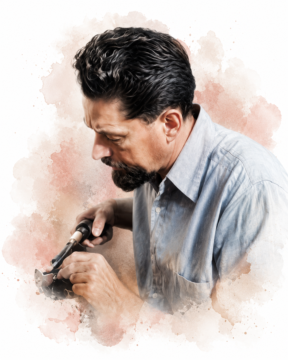
He improved Berger's electroencephalograph and pioneered EEG topography (Bladin, 2006). EEG topography maps electrical activity across the brain surface.
The EEG Society in Britain was established in 1942, and the American EEG Society began in 1947.
Nathaniel Kleitman (1953) was a Russian-born physiologist at the University of Chicago who has been recognized as the "Father of American sleep research" for his seminal work on sleep-wake cycle regulation, circadian rhythms, the sleep patterns of different age groups, and the effects of sleep deprivation. To test whether human rhythms could be reset, he famously spent a month deep in Kentucky's Mammoth Cave in 1938. Kleitman described the basic patterns of sleep cycles and the transitional states that connect them (Schomer & Lopes da Silva, 2011).
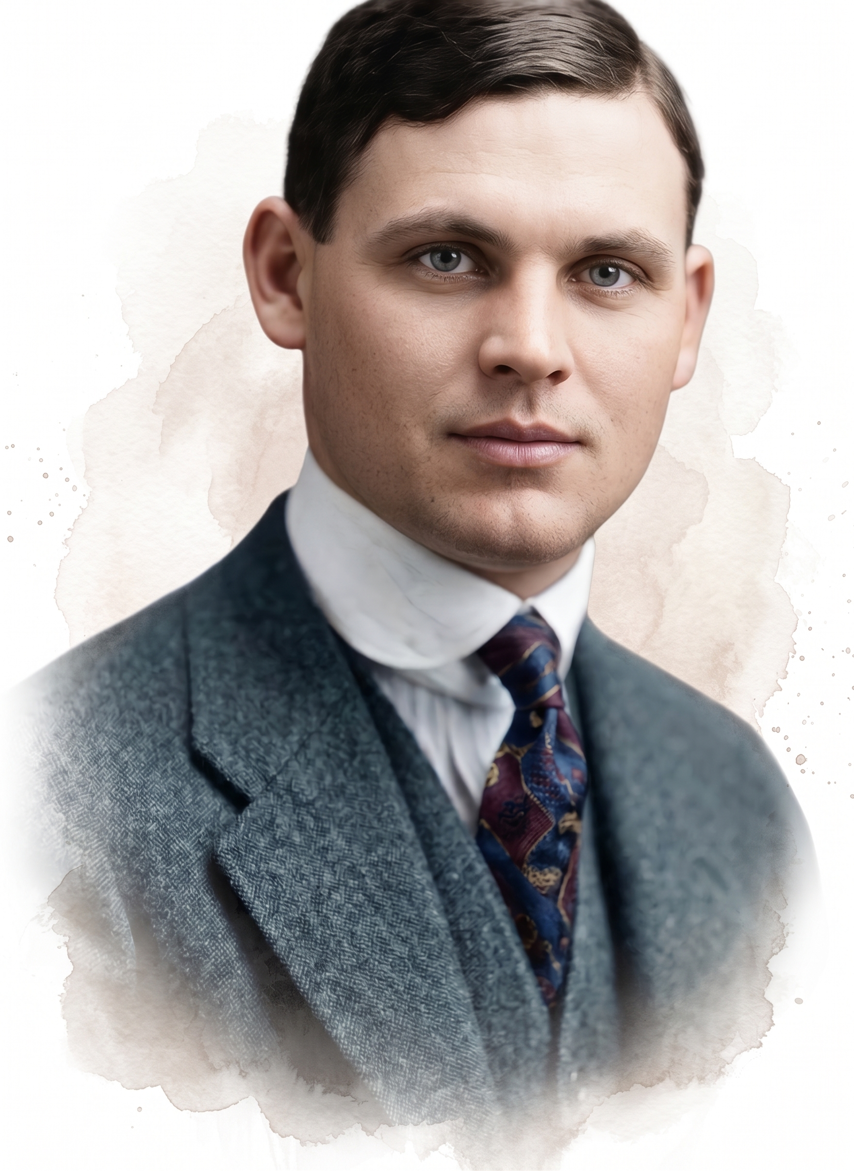
He discovered the phenomenon of rapid eye movement (REM) sleep with his graduate student Eugene Aserinsky. Below is a sample of brainwave activity during REM sleep.
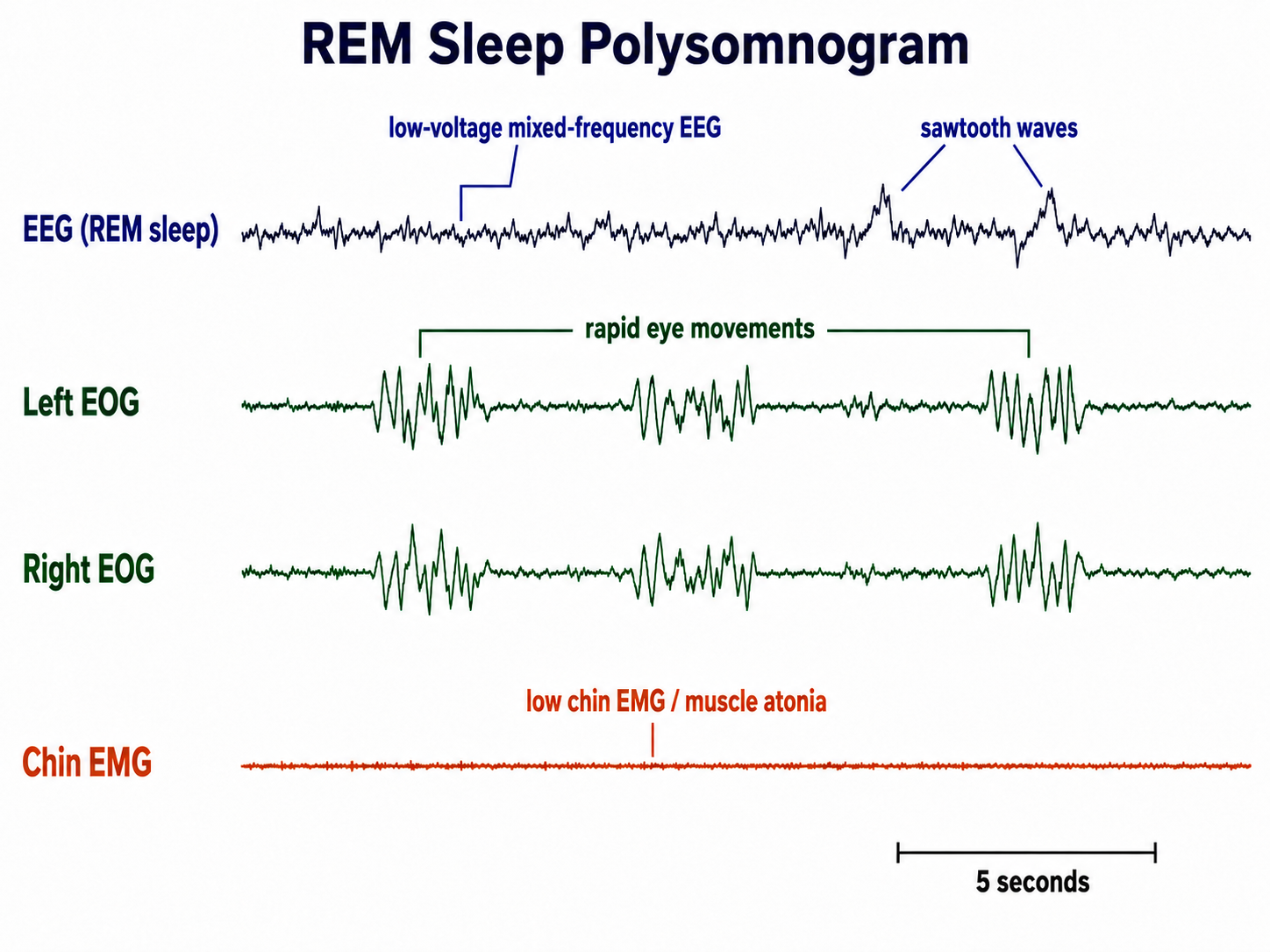
William C. Dement (1950s), another of Kleitman's students, described the EEG architecture and phenomenology of sleep stages and the transitions between them in 1955, associated REM sleep with dreaming in 1957, and documented sleep cycles in another species, cats, in 1958, which stimulated basic sleep research. He established the Stanford University Sleep Research Center, the first international sleep laboratory, in 1970. He has contributed to research on sleep deprivation and the diagnosis and treatment of sleep disorders like apnea and narcolepsy (Schomer & Lopes da Silva, 2011). Widely regarded as the father of sleep medicine, Dement spent decades raising public and clinical awareness of how untreated sleep disorders harm health.
Kleitman and Dement advanced polysomnography by incorporating eye movement and the EEG during an entire night's sleep. These measurements enabled the study of sleep stages and behaviors like dreaming.
Per Andersen and S. A. Andersson (1968) proposed that thalamic pacemaker circuits project synchronous rhythms in the classical alpha band, 8 to 13 Hz, to the cortex through thalamocortical connections. Andersen was a Norwegian neuroscientist at the University of Oslo whose laboratory became the birthplace of long-term potentiation (LTP), the lasting strengthening of synapses that underlies learning and memory. Terje Lømo first observed the effect there in 1966, and Bliss and Lømo characterized it fully in a landmark 1973 paper (Bliss & Lømo, 1973).
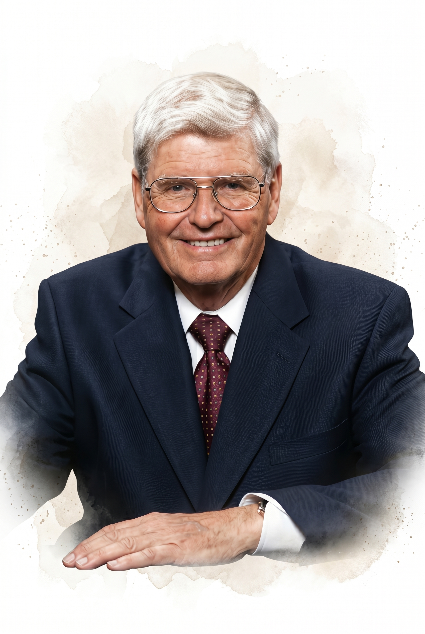
Mary A. B. Brazier (1904-1995) was a British-American neurophysiologist who became one of the foremost authorities on the electrical activity of the brain. After moving from London to Boston on a Rockefeller Fellowship in 1940, she directed the EEG laboratory at Massachusetts General Hospital and, beginning in 1948, pioneered the use of correlation techniques and early digital computers to analyze EEG signals, work that anticipated modern quantitative EEG. She later joined the Brain Research Institute at the University of California, Los Angeles, and served as president of the International Federation of Societies for Electroencephalography and Clinical Neurophysiology. Brazier also became the field's preeminent historian, and her account of the first half-century of brain electrical recording remains a standard reference (Brazier, 1961). She arranged the English translation of Adolf Beck's pioneering thesis, helping secure the place in this history that Beck holds today.
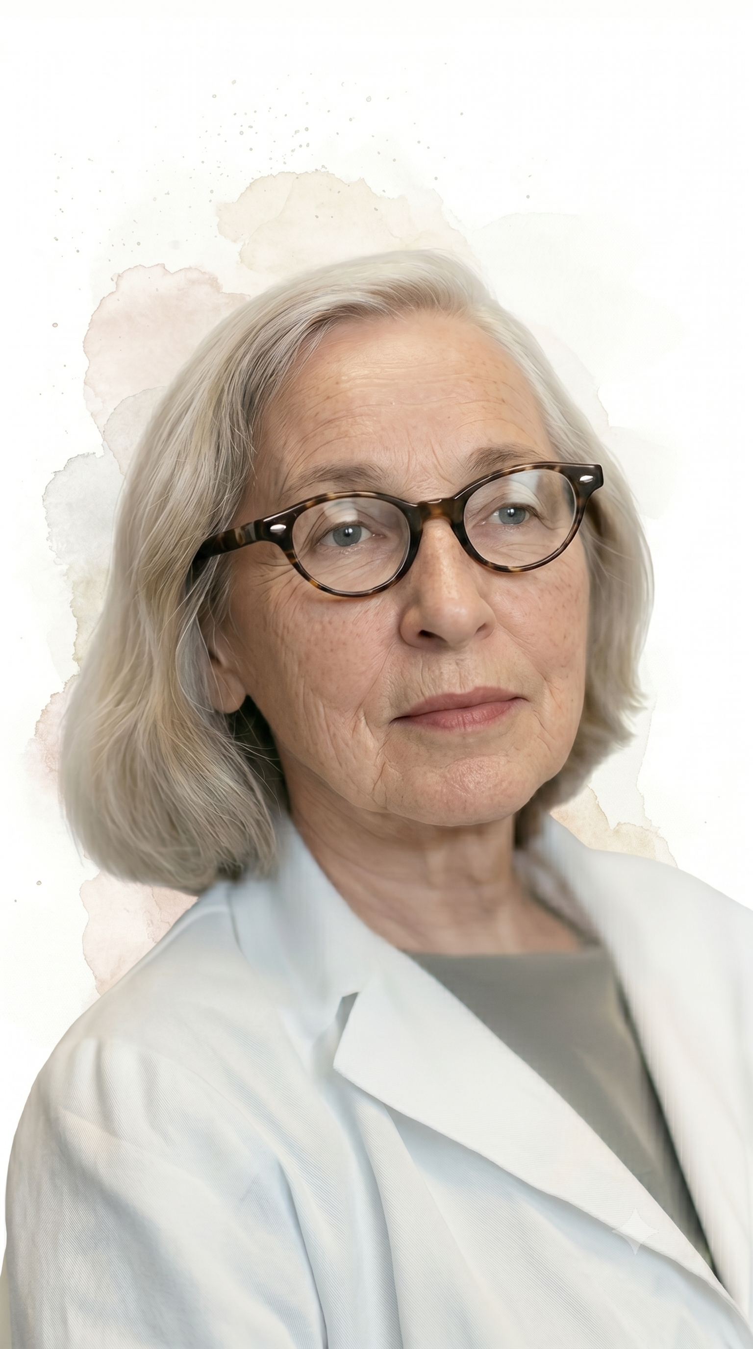
Historical Reappraisal: Caton, Berger, and Beck Reconsidered
The centennial of Berger's first human recording has drawn fresh attention to a revision of this history that scholars have been building since the archives of the former East Germany opened. What emerges is a story more complex, and considerably darker, than the tidy chronology textbooks usually present. Three of the contributors described above anchor it: one pioneer whose priority was forgotten, one revealed as a collaborator, and one destroyed by the regime the second one served (Fields, 2024).
Caton's Forgotten Priority
The story begins not in Jena in the 1920s but in Liverpool in 1875, with the mirror galvanometer described above. When Caton presented his findings at the British Medical Association meeting in Edinburgh, the brain remained largely an enigma and the world still ran on steam power. His insights were too advanced for his contemporaries to absorb, as he reportedly noted after presenting the work again in Washington, D.C., at the Ninth Session of the International Medical Congress in 1887:
Read my paper on the electrical currents of the brain. It was well received but not understood by most of the audience.
(Fields, 2024)
Caton's finding was extended by Danilewsky, Beck, Cybulski, and Pravdich-Neminsky, as the entries above describe, but it never entered mainstream neurology, and his priority was largely forgotten by the time Berger published half a century later. That gap may have delayed advances in brain science and medicine by decades (Fields, 2024).
Berger Reconsidered
Berger made that 1924 recording at the psychiatric clinic in Jena, approaching the problem from a unique if questionable perspective. Initially motivated by a belief in telepathy, he later developed his theories about "psychic energy" and pursued them through experiments that would not survive modern ethical review, including placing rectal thermometers against the exposed brains of patients undergoing surgery to detect temperature changes during mental effort (Fields, 2024).
The more consequential revision concerns Berger's conduct under the Third Reich. For decades the field held that he was forced into retirement in 1938 because the Nazis opposed him and that he took his own life in 1941 as a victim of persecution. Documents recovered after the fall of the Berlin Wall tell a different story. Zeidman and colleagues (2014) document that Berger was a willing participant on the Nazi genetic health higher courts that heard appeals against compulsory sterilization, that he contributed financially to the Schutzstaffel (SS), and that he helped select his successor, Berthold Kihn, who was complicit in the regime's "euthanasia" killings. Drawing on the archival work of medical historian Susanne Zimmermann, Fields (2024) adds that Berger rejected the sterilization appeals that came before him and that his laboratory notebooks carry antisemitic marginalia alongside his scientific observations. His suicide is now attributed to depression rather than persecution, mirroring the fate of many who were implicated in Nazi atrocities as the weight of their actions became clear.
Beck's Tragedy in Eastern Europe
Adolf Beck, whose discovery of alpha blocking is described above, trained under Cybulski in Kraków and then joined the university in Lwów, now Lviv, Ukraine, in 1895, where he later served as rector. His research was repeatedly disrupted by war. After Russian forces occupied the city in 1914, Beck was arrested in 1915 and imprisoned in Kiev, now Kyiv, and returned to Lwów in 1916 following the intervention of Ivan Pavlov (Fields, 2024).
The rise of Nazi Germany brought his life to a tragic end. On August 7, 1942, facing deportation to a Nazi death camp as both a Jew and an intellectual, Beck took cyanide rather than submit to his captors (Fields, 2024).
Why This History Matters
This record is a reminder that scientific progress is inextricably intertwined with broader historical and social forces. Caton's priority went unacknowledged for fifty years. The contrasting fates of Berger and Beck, one a collaborator who participated in the regime's machinery, the other a victim who died escaping it, illustrate that science can never be cleanly separated from the moral choices of the people who practice it. The point is not to diminish Berger's scientific contribution, which remains foundational to neurofeedback, but to hold both facts at once. That tension resonates today, as conflict continues to disrupt scientific work in the very places where this history unfolded, Lviv among them.
Neurofeedback Research
This section highlights the pioneers who transformed EEG biofeedback from laboratory demonstration into clinical practice. We will explore how researchers developed specific training protocols for conditions ranging from epilepsy to ADHD, documented the subjective experiences associated with different EEG states, and established the scientific foundations that legitimized neurofeedback as an evidence-based intervention.
Joseph Kamiya (1960s) is an American psychologist, considered the "Father of Neurofeedback," who began this work in the sleep laboratory at the University of Chicago and later continued it at the Langley Porter Psychiatric Institute in San Francisco. He demonstrated that the alpha rhythm in humans could be operantly conditioned. He published an influential article in Psychology Today showing that subjects could learn to discriminate when alpha was present or absent and could use feedback to shift features of their alpha activity, such as the balance of long and short alpha cycles.
Almost half of his subjects reported experiencing a pleasant "alpha state" characterized as an "alert calmness." These reports may have contributed to the perception of alpha biofeedback as a shortcut to a meditative state. He also studied EEG activity during meditative states, and his work demonstrated a clinical application of alpha-theta neurofeedback.
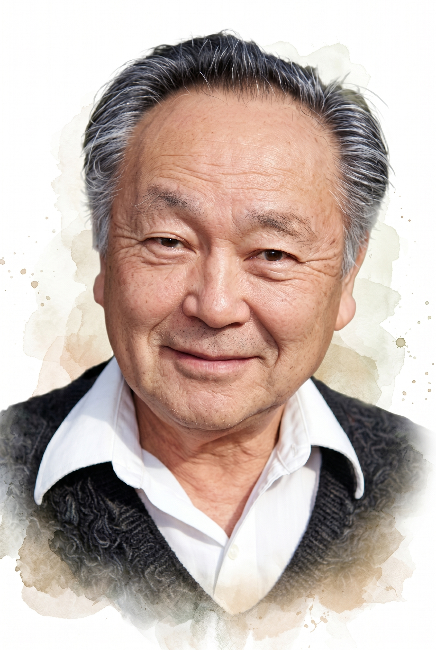
Both the public and academic worlds recognize Joe Kamiya as the father of biofeedback. In 1966, while monitoring subjects' EEGs in his sleep lab at the University of Chicago, he performed a novel experiment by ringing a bell whenever an alpha burst occurred. He discovered that some subjects could discriminate when they produced alpha activity. His 1968 publication of 'Conscious Control of Brain Waves' in Psychology Today summarized research that showed that subjects could learn to discriminate when alpha was present or absent and that they could use feedback to shift the dominant alpha frequency about 1 Hz. Almost half of his subjects experienced a pleasant alpha state, which they characterized as an 'alert calmness.' Kamiya's article made biofeedback accessible to the public and made it exciting because it suggested that individuals can learn to control their own consciousness.
Alpha biofeedback fit an emerging zeitgeist of self-exploration. American culture in the 1960s and 1970s was shaped by a confluence of forces: exploration of consciousness through drugs such as LSD (Timothy Leary and Richard Alpert) and Eastern meditative practices such as transcendental meditation (TM). Harvard physician Herbert Benson repackaged TM as the relaxation response without an overt spiritual dimension. Kamiya's work implied that a language of consciousness was possible and resulted in neurofeedback, one of the most promising areas of biofeedback.
(Peper & Shaffer, 2010, p. 143)
Barbara Brown (1970s) was a pharmacologist at the Veterans Administration who demonstrated the clinical use of alpha-theta biofeedback. In research designed to identify the subjective states associated with EEG rhythms, she trained subjects to increase the abundance of alpha, beta, and theta activity using visual feedback, and she recorded their subjective experiences as the amplitude of these bands increased. She helped popularize biofeedback by publishing a series of books, including New Mind, New Body (1974), Stress and the Art of Biofeedback (1977), and Supermind (1980). Brown was a co-founder and first president of the Biofeedback Research Society, which became the Biofeedback Society of America and then the Association for Applied Psychophysiology and Biofeedback.
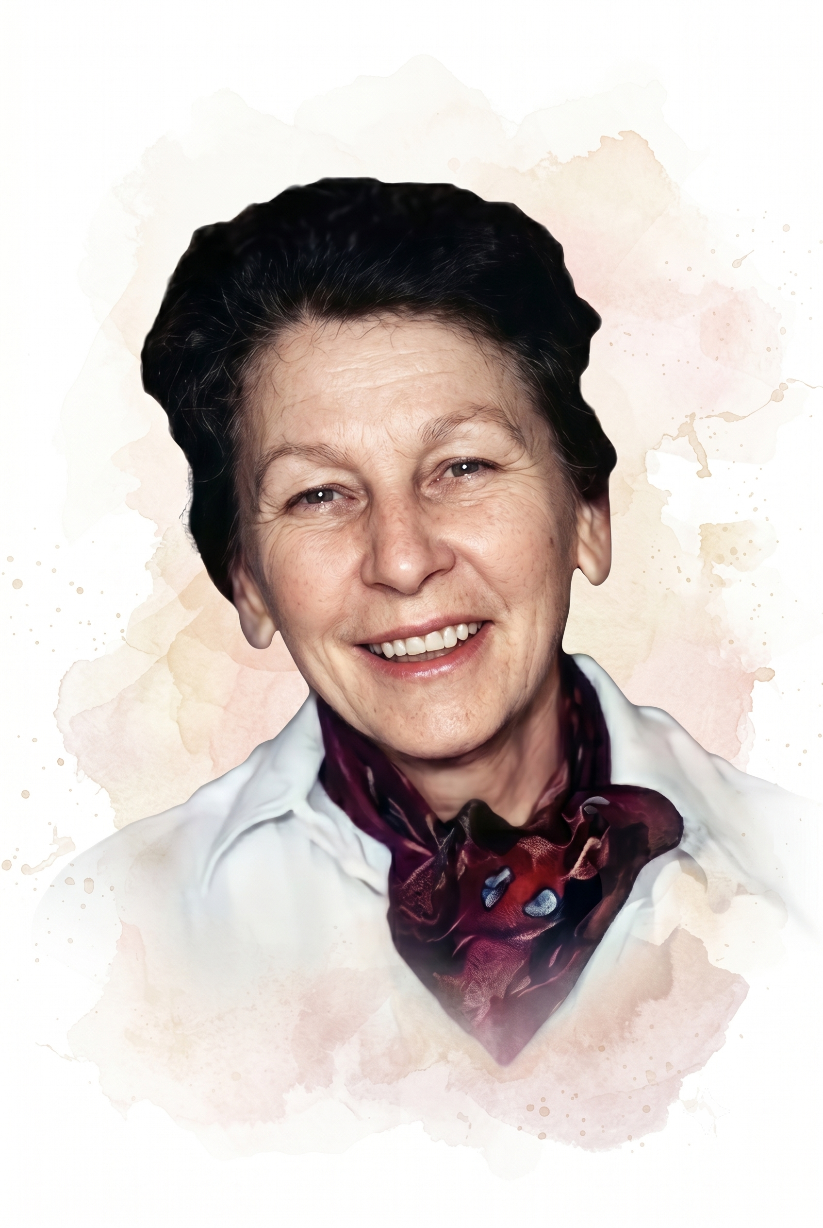
Thomas Mulholland and Erik Peper (1971) showed that occipital alpha increases with eyes open and not focused and is disrupted by visual focusing, a rediscovery of alpha blocking. Mulholland was a Veterans Administration researcher who used feedback loops to study the link between attention and the alpha rhythm.
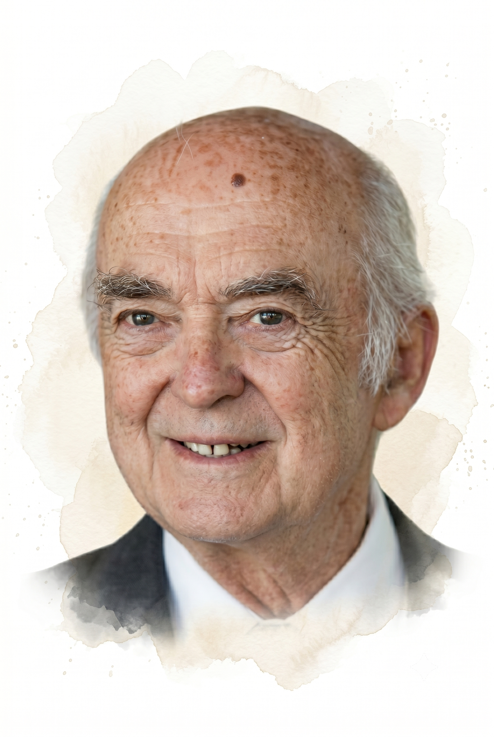
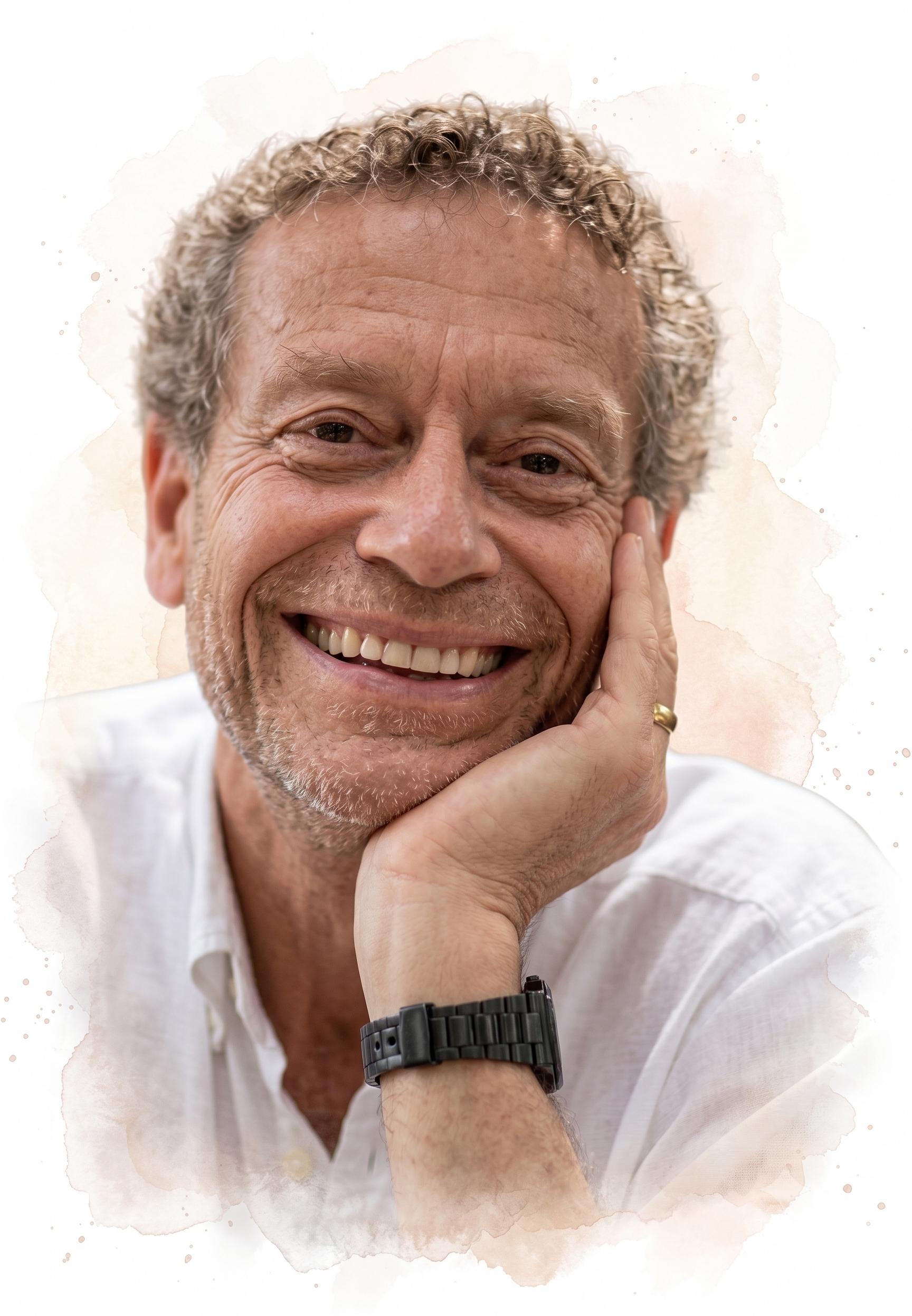
Peper went on to a long career at San Francisco State University and became one of the field's most active educators and leaders.
Elmer Green and Alyce Green (1969, 1970, 1977) were a husband-and-wife research team who led the psychophysiology program at the Menninger Foundation in Topeka, Kansas. They investigated the voluntary control of internal states by individuals like Swami Rama and the American Indian medicine man Rolling Thunder, both in India and at Menninger. They brought portable biofeedback equipment to India and monitored practitioners as they demonstrated self-regulation. A film containing footage from their investigations was released as Biofeedback: The Yoga of the West (1974).
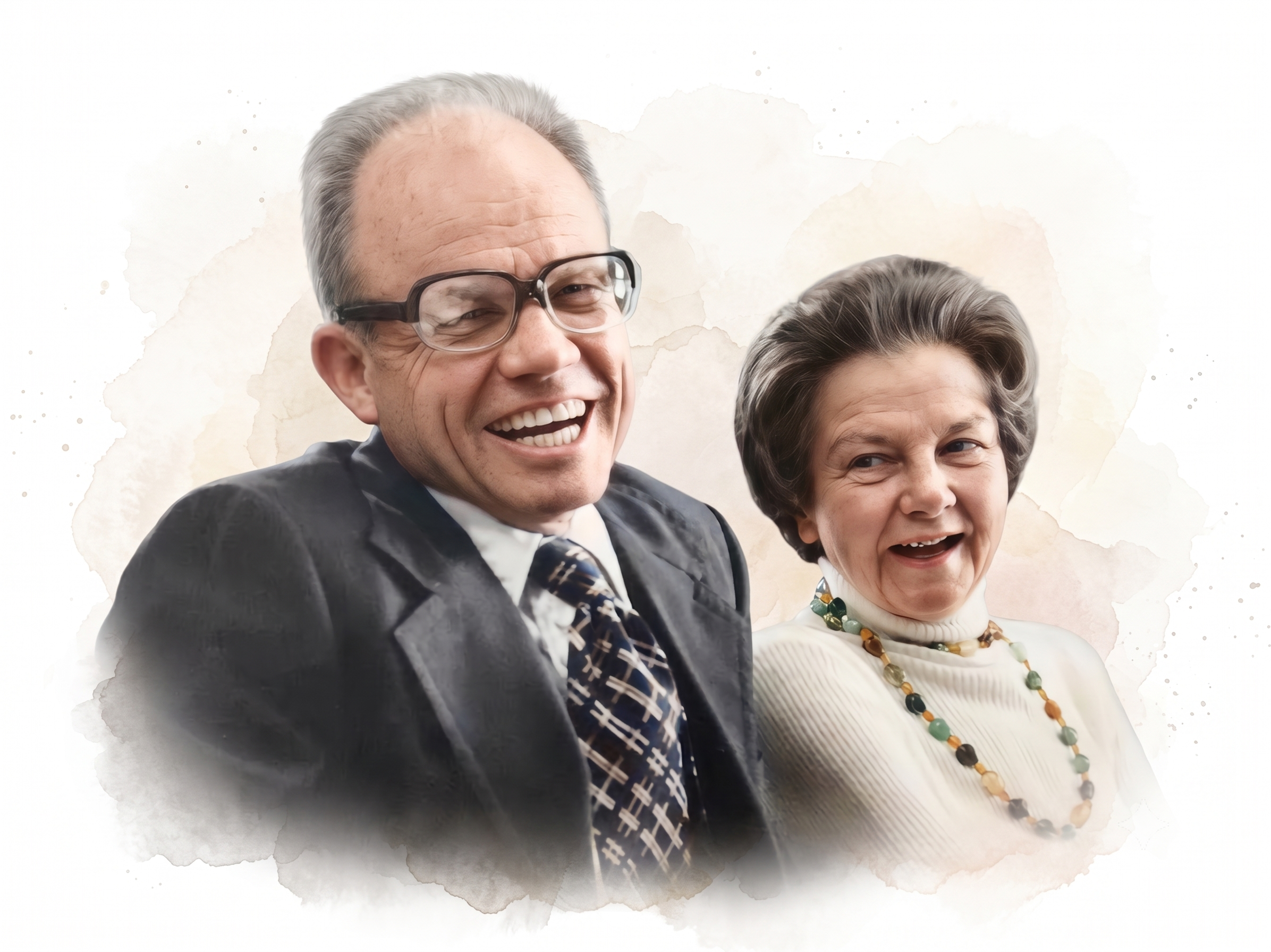
The Greens showed that, like the yogis they studied, ordinary individuals could learn self-regulation of "involuntary" physiological functions like blood pressure, the EEG, heart rate, skeletal muscle activity, and skin temperature.
They developed alpha-theta training at the Menninger Foundation from the 1960s to the 1990s. They hypothesized that theta states allow access to unconscious memories and increase the impact of prepared images or suggestions. Their alpha-theta research fostered Peniston's development of an alpha-theta addiction protocol.
They pioneered temperature biofeedback training for Raynaud's, migraine, and hypertension and wrote the classic Beyond Biofeedback (1977). They developed clinical biofeedback by applying these modalities to various disorders like hypertension and migraine.
Thomas Budzynski (1969, 1973) was a psychologist who, with Johann Stoyva, co-developed EMG biofeedback for tension headache before turning to EEG work. He developed a twilight learning device that monitored left hemisphere EEG while a patient sleeps and played recorded educational content like language lessons and affirmations (positive statements) when theta was present. The premise of twilight learning is that affirmations have a greater impact when presented in a transitional state in which theta waves replace the alpha rhythm.

He studied the lateralization of brain function across the two cerebral hemispheres and the use of neurofeedback and audio-visual stimulation (brain brightening) to correct age-related cognitive decline.
M. Barry Sterman (1973), a physiologist at UCLA and the Veterans Administration, showed that cats and human subjects could be operantly trained to increase the amplitude of the sensorimotor rhythm (SMR) between 12-15 Hz recorded from the sensorimotor cortex. He made his key discovery almost by accident: cats he had earlier trained to boost SMR proved unusually resistant to seizures when later exposed to a toxic rocket fuel in a study for NASA.
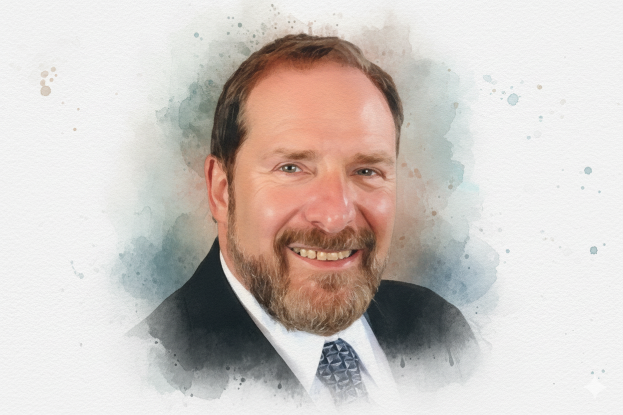
He demonstrated that SMR production protects cats against drug-induced generalized seizures (tonic-clonic seizures involving loss of consciousness) and reduces the frequency of seizures in humans diagnosed with epilepsy. He found that his SMR protocol, which uses visual and auditory EEG biofeedback, normalizes their EEGs (SMR increases while theta and beta decrease toward normal values) even during sleep.
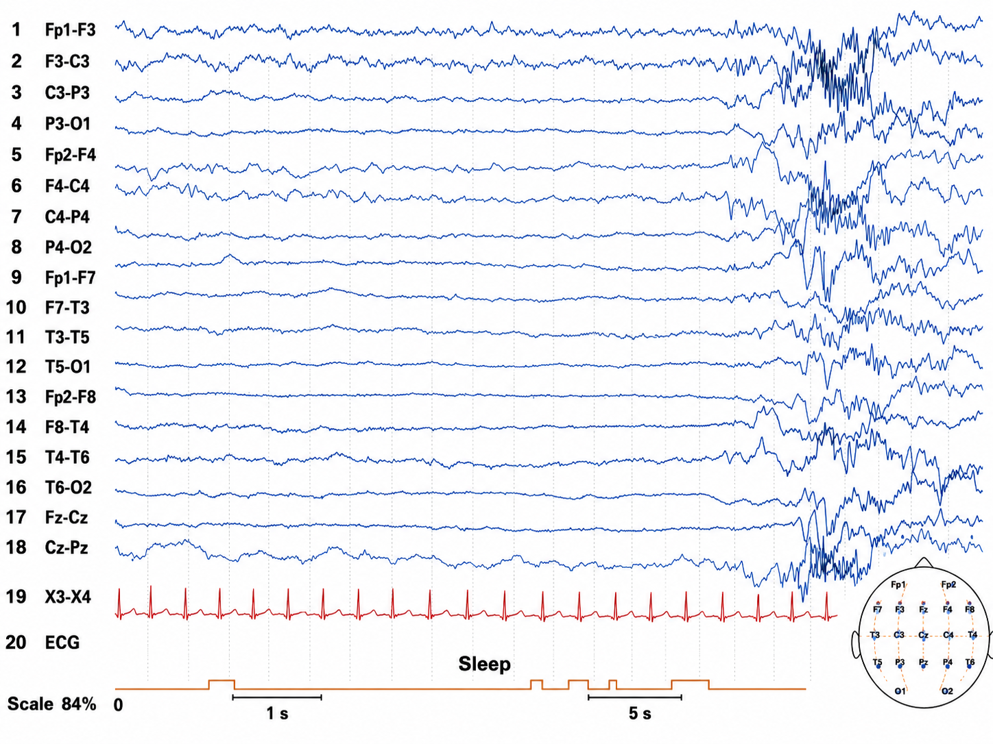
Sterman conducted excellent research published in mainstream journals, which should have propelled SMR training into the forefront of seizure disorder treatment. Sterman also co-developed the Sterman-Kaiser (SKIL) qEEG database.
Joel Lubar (1970s) was a psychologist who collaborated with Sterman to extend SMR biofeedback from epilepsy to attention disorders. He demonstrated that SMR training could improve attention and academic performance in children diagnosed with Attention Deficit Disorder with Hyperactivity (ADHD) at the University of Tennessee.
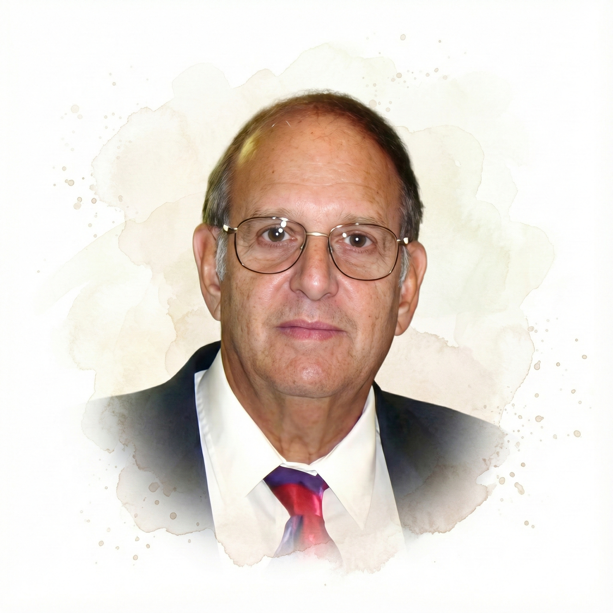
Lubar documented the importance of theta-to-beta ratios (the ratio of power in the theta and beta bands) in ADHD and developed theta suppression-beta enhancement protocols to decrease these ratios and improve student performance.
Judith O. Lubar was a psychologist at the University of Tennessee who, working alongside Joel Lubar, helped turn the laboratory findings on attention into a workable clinical treatment. As first author of a landmark 1984 study, she reported that enhancing SMR or beta activity while suppressing theta in a clinical setting improved school grades and achievement scores in six children with attention deficit disorders trained twice weekly for 10 to 27 months (Lubar & Lubar, 1984). The study reported no follow-up data, so its gains cannot be described as lasting beyond the training period. Their protocol paired neurofeedback with academic tasks and showed changes in the EEG power spectrum that matched the behavioral improvements. This work moved EEG biofeedback for ADHD beyond isolated case studies and helped establish the clinical model that thousands of practitioners later adopted.
Erwin Roy John and Frank H. Duffy (1970s) collaborated in developing quantitative electroencephalography (qEEG) and its use in the assessment of disorders. John, a neuroscientist at New York University, created neurometrics, in which a client's EEG is compared to a normative database, and he contributed to the study of memory by proposing that it is distributed across the brain rather than stored in one place. Duffy, at Harvard, developed Brain Electrical Activity Mapping (BEAM), a method for displaying qEEG data as color topographic maps of the cortex.
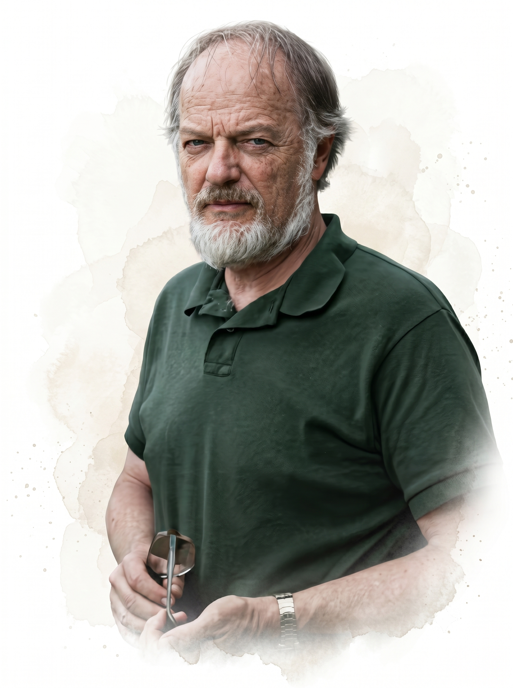
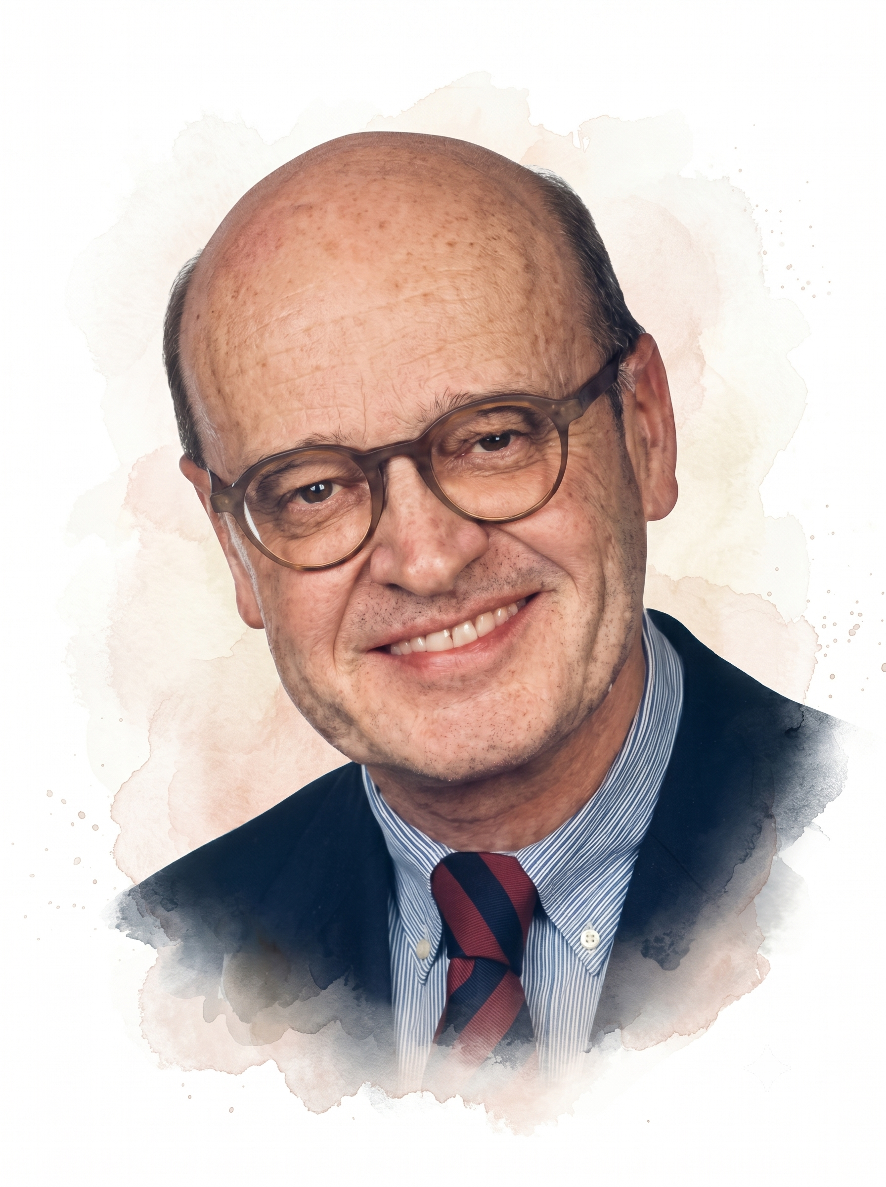
Leslie S. Prichep was E. Roy John's principal collaborator at New York University and a central figure in the development of neurometrics, the comparison of an individual's EEG to an age-regressed normative database. With John and their colleagues, she helped build and validate the reference data and the statistical methods that let clinicians express a client's brain activity as deviations from expected values (John et al., 1988). Her research extended quantitative EEG to the differential diagnosis of dementia, depression, and other disorders, and later to traumatic brain injury and the assessment of concussion. The normative-database approach she helped pioneer underlies the z-score and qEEG-guided neurofeedback described elsewhere in this unit.
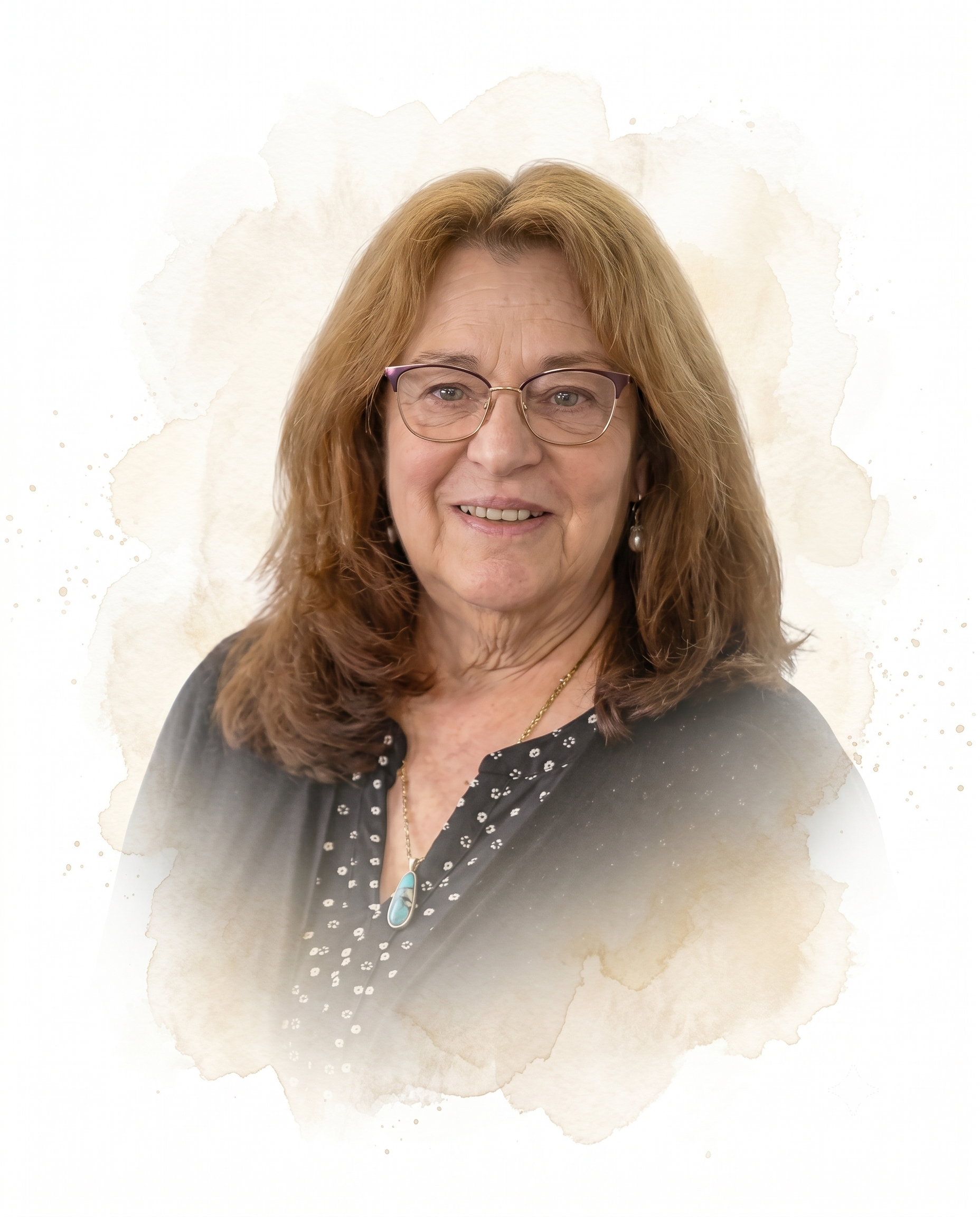
Jay Gunkelman's (Moss & Gunkelman, 2002) involvement in applied psychophysiology and biofeedback dates back to 1972 with the grant funding of the first state hospital-based biofeedback laboratory. Since the mid-1970s, he has specialized in the classical clinical EEG and is one of the world's most experienced EEG/qEEG specialists. Over his career, he has personally reviewed an extraordinary volume of clinical EEG and qEEG records, an experience base that informs his teaching and consultation. He was instrumental in developing biofeedback and neurofeedback efficacy standards that have served as the foundation for the AAPB reference Evidence-Based Practice in Biofeedback and Neurofeedback. Jay has been one of the field's most influential educators and prolific authors.
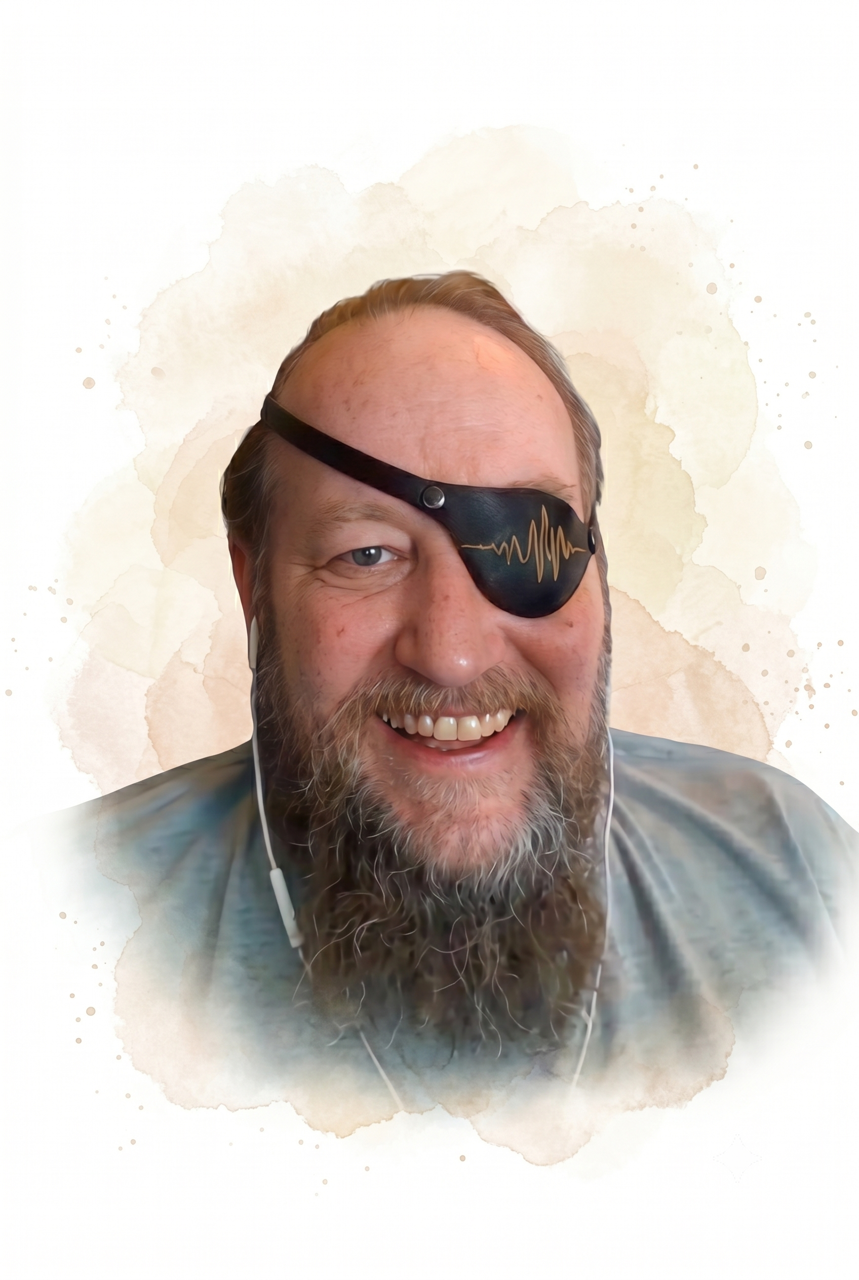
Michael and Lynda Thompson (2015) founded the ADD Centre and the Biofeedback Institute of Toronto, where they have trained thousands of clients and clinicians. Thomas Budzynski described them as "master teachers as well as superb clinicians and clinical researchers. Over the years, their clinic has acquired a much deserved international reputation with clients from other countries seeking their expertise. Somehow they also manage to teach workshops on biofeedback and neurofeedback to other professionals in the field."
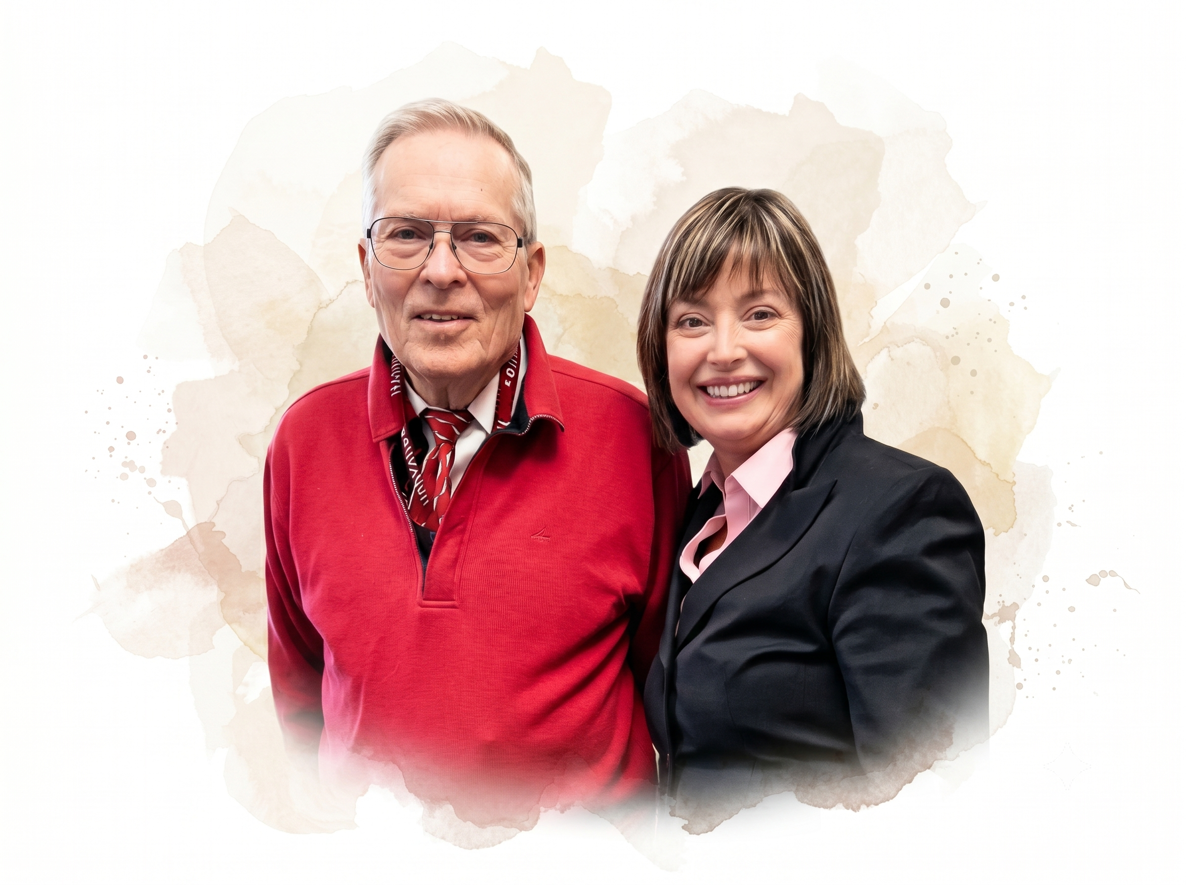
They wrote The Neurofeedback Book: An Introduction to Basic Concepts in Applied Psychophysiology, which has been the "bible" of neurofeedback didactic education. In 2016, AAPB awarded Michael and Lynda its Distinguished Scientist award.
Margaret Ayers (1975) was an early neurofeedback pioneer who established the first private clinical practice devoted exclusively to neurofeedback, based in Beverly Hills, and published the first papers on neurofeedback for treating traumatic brain injury. She developed digital real-time EEG monitoring technology and investigated seizure control and the treatment of coma patients with open head wounds. Her neurofeedback protocol down-trained slow-wave activity and uptrained 15-18 Hz activity. Among the clinicians she trained were Sue and Siegfried Othmer, who went on to shape much of the modern field.
Niels Birbaumer and colleagues (1970s), based at the University of Tübingen, have studied operant conditioning of slow cortical potentials (SCPs; gradual changes in the membrane potentials of cortical dendrites that last from 300 ms to several seconds) since the late 1970s. They have demonstrated that subjects can learn to control these DC potentials and have studied the efficacy of SCP biofeedback in treating ADHD, epilepsy, and schizophrenia. They have shown that negative SCPs improve cognitive performance while positive SCPs increase the seizure threshold. Finally, they pioneered brain-computer interfaces that let completely locked-in patients communicate by regulating their own brain signals.

William N. Kuhlman (1978) mapped the functional topography of the human mu rhythm, peaking near 9-11 Hz, and distinguished it from the SMR. He and B. J. Kaplan reviewed the clinical applications of EEG feedback training the following year. By carefully distinguishing the mu rhythm from the SMR that Sterman had identified, they sharpened the field's understanding of which rhythm a protocol actually trains. Their independent replication of Sterman's SMR work to inhibit generalized seizures added important weight to the evidence for neurofeedback in epilepsy.
Sue and Siegfried Othmer (1985), both trained as physicists, worked with Margaret Ayers to address their son's epilepsy and later founded the EEG Institute. They proposed that protocols be chosen based on a client's symptom patterns and pioneered computer games designed to modify the EEG. They investigated infra-low frequency (ILF) training, which targets the very slow fluctuations of the slow cortical potentials.

Eugene Peniston (1989) was a Veterans Administration psychologist who studied with the Greens at the Menninger Foundation. He developed the Peniston-Kulkosky Protocol, which uses alpha-theta training to treat alcoholism and post-traumatic stress disorder, and he applied it especially to combat veterans whose addiction and PTSD had resisted conventional care.
This multimodal protocol incorporated alpha-theta neurofeedback that uptrained alpha (8-13 Hz) and theta (4-8 Hz), autogenic training, constructed visualizations, diaphragmatic breathing, guided imagery, systematic desensitization, and temperature biofeedback. The diverse suggestion techniques utilized to potentiate alpha-theta training have influenced the use of guided imagery and behavior change scenarios in current alpha-theta neurofeedback.
Peniston and Kulkosky reported that 8 of 10 experimental patients who received their alpha-theta protocol for alcoholism maintained abstinence from alcohol at the 13-month follow-up, compared with none of 10 traditionally treated controls. The two experimental patients who relapsed showed significantly reduced tolerance for alcohol. Peniston published seminal articles concerning alpha-theta training.
Vincent Monastra published early qEEG studies concerning the theta/beta ratio in ADHD and the neurofeedback treatment of ADHD. Along with Lubar and Linden, he developed the statistical basis for using theta/beta power ratio measurement as a marker for ADHD. His large-sample studies brought statistical rigor to the measure and helped establish neurofeedback for attention disorders as a credible clinical approach.
Robert Thatcher (2010) developed a quantitative EEG database known as the lifespan database (NeuroGuide) and published the Handbook of QEEG and EEG Biofeedback. His key insight was to express each client's EEG as a z-score relative to an age-matched normative database, so that training could target the measures that deviate most from typical values. Building on this idea, he developed 2- and 4-channel z-score training, 19-channel surface and LORETA z-score neurofeedback training, and BrainSurfer.
Juri Kropotov, who holds doctoral degrees in theoretical physics, philosophy, and neurophysiology, studied event-related potentials, developed EEG amplifiers (Mitsar 201 and 202), and is a prolific author, including his 2009 book Quantitative EEG: Event-Related Potentials and Neurotherapy. He is affiliated with the Human Brain Institute (HBI) and developed WinEEG software, using the HBI normative database for EEG and ERP analysis. Working in St. Petersburg, he has been an important bridge between Russian and Western traditions of quantitative EEG research. He received the USSR State Prize in 1985 and the Copernicus Prize from the Polish Neuropsychological Society in 2009.
Roberto Pascual-Marqui has contributed to functional brain mapping using EEG and magnetoencephalography, and to the study of functional connectivity in brain networks. He developed three-dimensional Low Resolution Electromagnetic Tomography (LORETA), a method that estimates the deep cortical sources of scalp EEG. LORETA made it possible to localize EEG activity in three dimensions and opened the door to source-based qEEG analysis and neurofeedback.
Neurofeedback Timeline
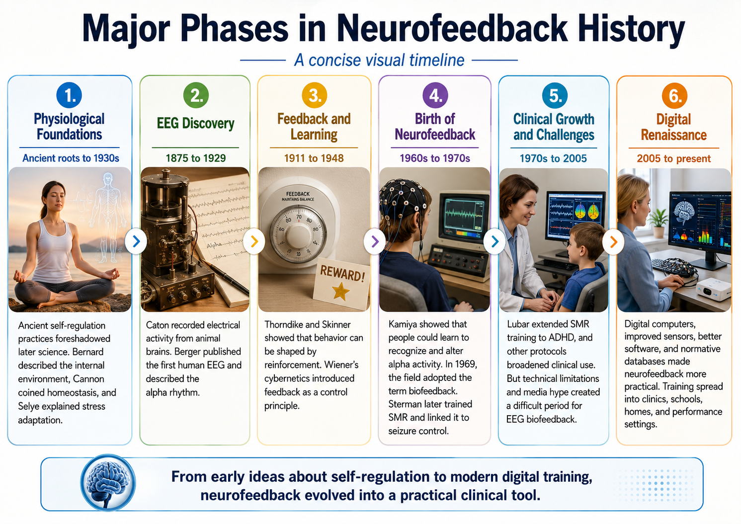
Neurofeedback did not arrive all at once. It was assembled from four separate lines of work: the physiology of homeostasis and stress, the cybernetic idea of feedback, the learning theory of operant conditioning, and the long technical effort to make brain electrical activity visible. The entries below place the contributors described in this chapter in sequence, so that you can see where those threads converge and how the field moved from laboratory demonstration through its Dark Ages to the present renaissance.
Glossary
absence seizure: a seizure usually lasting less than 15 s in which a client blanks out; formerly called petit mal seizures, a label now considered obsolete.
affirmations: positive statements like "Every day I am getting better in every way."
all-or-none law: action potentials either occur or not. When they occur, they have the same amplitude and speed.
alpha blocking: replacement of the alpha rhythm by low-amplitude desynchronized beta activity during movement, attention, mental effort like complex problem-solving, and visual processing.
alpha rhythm: the first EEG rhythm discovered by Berger that ranges from 8 to 12 Hz which appears in three-quarters of adults when they are calm, awake, and not actively processing information.
alpha-theta training: training to progressively slow the EEG, increasing alpha and then theta abundance, developed by Brown and the Menninger Foundation and then adopted by Peniston and Kulkosky in treating alcoholics.
animal electricity: Galvani's theory that living tissue generates and stores its own electricity, demonstrated by the contraction of a frog's leg when touched by metal.
autonomic viscera: the internal organs (such as the heart, blood vessels, lungs, stomach, and intestines) whose functions are regulated involuntarily by the autonomic nervous system but which can be consciously controlled through biofeedback training.
beta rhythm: the second EEG rhythm described by Berger, who defined it simply as activity faster than alpha. This curriculum uses 12-38 Hz.
biofeedback: information about psychophysiological performance obtained by noninvasive monitoring and used to help individuals achieve self-regulation through a learning process that resembles motor skill learning.
comparator: in cybernetic theory, the component that measures the difference between a regulated variable's actual value and its setpoint and then passes the resulting error signal to the effectors.
complex partial seizure: a seizure in which awareness is impaired and repetitive behaviors called automatisms (e.g., lip smacking) may occur.
contingent negative variation (CNV): a steady, negative shift in potential (15 microvolts in young adults) detected at the vertex discovered by Walter. This slow cortical potential may reflect expectancy, motivation, intention to act, or attention. The CNV appears 200-400 ms after a warning signal (S1), peaks within 400-900 ms, and sharply declines after a second stimulus that requires the performance of a response (S2).
cortical gradient: the standing difference in electrical potential across the depth of the cortex. Caton observed that this gradient shifts toward negativity at the cortical surface when a region becomes active, the observation later formalized as the negative slow cortical potential.
curare: a paralytic drug used by Miller and DiCara to prevent rats from "cheating" by using skeletal muscles during operant conditioning of heart rate, blood pressure, kidney blood flow, skin blood flow, and intestinal contraction.
cybernetic theory: Wiener proposed that living systems are controlled by monitoring their results.
cybernetics: the study of control and communication in both living organisms and machines, founded by Norbert Wiener and named from the Greek kybernetes, meaning steersman.
delta rhythm: EEG rhythm named by Walter that ranges from 0.5 to 3.5 Hz and is increased in adult Stage 3 sleep, brain injury, brain tumor, and developmental disability.
desynchronization: the replacement of large, synchronized EEG rhythms by faster, lower-amplitude activity when the brain engages with a stimulus or task.
dysponesis: Whatmore and Kohli's concept of misplaced effort. For example, bracing your shoulders when you hear a loud sound.
EEG topography: the display that maps EEG activity across the brain surface in color.
effectors: in cybernetic theory, the control mechanisms that use the error signal to adjust the system variable and drive the error toward zero.
electrocerebrogram: Pravdich-Neminsky's early term for a recording of the brain's electrical activity.
epileptiform spikes: sharp EEG transients lasting less than 70 ms that signal abnormal, hypersynchronous neuronal firing associated with seizure activity.
error signal: in cybernetic theory, the difference between a regulated variable's actual value and its target, or setpoint.
evoked potentials: brain electrical potentials that are elicited by sensory stimulation.
feedback: in cybernetic theory, orders to correct small errors to prevent larger future errors in a closed system.
feedforward: in cybernetic theory, orders to perform an action based on anticipated conditions in an open system.
fight-or-flight response: Cannon's term for the pattern of sympathetic and adrenal arousal that prepares the body to confront or flee a threat.
galvanism: the contraction of muscle produced by an electric current, named for Luigi Galvani.
general adaptation syndrome (GAS): Selye's three-stage model of the body's response to prolonged stress, consisting of alarm, resistance, and exhaustion.
generalized seizures: seizures characterized by a peculiar cry, loss of consciousness, falling, tonic-clonic convulsions of all extremities, incontinence, and amnesia for the episode. These were previously called grand mal seizures.
genetic health higher courts: the Nazi appellate tribunals (Erbgesundheitsobergerichte) that reviewed appeals against orders for compulsory sterilization issued under the Third Reich's racial hygiene laws. Hans Berger served on one of these courts and rejected the appeals that came before him.
homeostasis: the dynamic maintenance of a steady internal state. Bernard described the underlying idea as the milieu interieur in 1865; Cannon coined the term homeostasis in 1926.
instrumental learning: operant conditioning, an unconscious associative learning process that modifies the form and occurrence of voluntary behavior by manipulating its consequences.
interictal activity: EEG potentials between seizures.
International 10-20 System: Jasper's standardized scheme for naming and placing EEG electrodes at proportional distances (10% and 20%) between skull landmarks, allowing recordings to be compared across laboratories.
law of effect: Thorndike's principle that responses followed by satisfying consequences become more likely to recur.
long-term potentiation (LTP): a long-lasting increase in synaptic strength following repeated stimulation, widely regarded as a cellular basis of learning and memory.
manometric flame: a device developed by Koenig and studied by Alexander Graham Bell for teaching the deaf to speak that displayed sounds as patterns of light.
milieu interieur: Bernard's concept of the stable internal environment that an organism actively maintains.
mirror galvanometer: an early galvanometer in which a small mirror suspended between magnets deflects a beam of light across a scale in proportion to the current passing through the instrument. Richard Caton used one to detect brain potentials in 1875, recording his measurements in millimeters of light-beam deflection rather than in volts.
motor unit: alpha motor neuron and the skeletal muscle fibers it controls.
mu rhythm: an arch-shaped rhythm recorded over the sensorimotor strip at C3 and C4, peaking near 9-11 Hz within a broader 8-13-Hz range; Kuhlman distinguished it from the sensorimotor rhythm (SMR).
neuron: an excitable nervous system cell that processes and distributes information, chemically and electrically, and usually contains a soma (cell body), dendrites, and axon.
normative database: means and standard deviations for EEG variables such as amplitude, power, coherence, and phase that are calculated for single hertz bins, frequency bands, or band ratios based on the EEG data collected from healthy normal subjects who are grouped by age, eyes open or eyes closed conditions, and sometimes gender and task, which also allows for the specification of z-scores with a mean of 0 and standard deviation of 1 for the various combinations of EEG variables, frequency ranges, subject ages, eyes-open or eyes-closed conditions and other variables.
operant conditioning: an unconscious associative learning process that modifies the form and occurrence of an operant behavior (emitted behavior) by manipulating its consequences.
Peniston-Kulkosky addiction protocol: a multimodal approach that incorporates visualization training, temperature biofeedback, rhythmic breathing techniques, autogenic training exercises, construction of personalized imagery, guided imagery, systematic desensitization, and 30 alpha-theta sessions. The 1989 trial itself used 15 sessions; the 30-session figure belongs to the later generalized protocol.
phonautograph: a device developed by Leon Scott and studied by Alexander Graham Bell to teach the deaf to speak, translating sound vibrations into tracings on smoked glass to show their acoustic waveforms.
photic stimulation: visual stimulation at a specific flash frequency.
punisher: a consequence that weakens an operant behavior. For example, shoulder pain (punisher) following excessive exercise (operant behavior) may reduce exercise intensity.
punishment: in operant conditioning, the consequence of an operant behavior reduces its probability.
quantitative EEG (qEEG): digitized statistical brain mapping using at least a 19-channel montage to measure EEG amplitude within specific frequency bins.
rapid eye movement (REM) sleep: a sleep stage marked by darting eye movements and dreaming, discovered by Aserinsky and Kleitman, who published the finding in 1953.
reinforcement: in operant conditioning, the consequence of an operant behavior increases its probability.
reinforcer: a consequence that strengthens an operant behavior. For example, weight loss (reinforcer) following daily walks (operant) may increase the probability of walking.
self-regulation: the control of your behavior (voluntary hand-warming).
sensorimotor rhythm (SMR): EEG rhythm that ranges from 12-15 Hz and appears when you inhibit movement and relax your muscles.
setpoint: the system goal, for example, a thermostat setting of 75 degrees F (23.9 degrees C).
sleep spindles: short bursts of 12-15 Hz activity during stage 2 sleep.
slow cortical potentials (SCPs): gradual changes in the membrane potentials of cortical dendrites that last from 300 ms to several seconds. These potentials include the contingent negative variation (CNV), the readiness potential, and movement-related potentials (MRPs). The boundary between a long-latency event-related potential such as the P300 or N400 and a slow cortical potential is conventional rather than sharp. SCPs modulate the firing rate of cortical pyramidal neurons by exciting or inhibiting their apical dendrites. They group the classical EEG rhythms using these synchronizing mechanisms.
spike: a sharp EEG transient with a duration of less than 70 ms.
stress: Selye's concept of a nonspecific response to stimuli called stressors.
synapse: specialized chemical and electrical junctions across which neurons communicate with each other and non-neural cells.
system variable: the variable that is controlled. For example, room temperature is the system variable for a thermostat.
thalamus: a forebrain structure above the hypothalamus that receives, filters, and distributes most sensory information. The thalamus contains neurons that can block or relay ascending sensory information. When these thalamic neurons rhythmically fire, this blocks the transmission of information to the cortex. When they depolarize in response to sensory information, this integrates and transmits this information to the cortex. Inputs to the thalamus determine whether these neurons block or relay sensory information.
theta/beta training: a protocol that decreases theta amplitude and increases beta amplitude.
theta rhythm: the EEG rhythm discovered by Walter that ranges from 4 to 7 Hz and is associated with drowsiness, the transition from wakefulness to sleep, rapid eye movement (REM) sleep, and the processing of information.
theta-to-beta ratio: the ratio of power in the theta band to power in the beta band.
twilight learning: Budzynski's training paradigm in which recorded material is played when theta activity replaces alpha activity.
visceral learning: the operant conditioning of autonomic responses such as heart rate, blood pressure, and intestinal contraction, studied by Neal Miller and Leo DiCara in curarized animals.
visual cortical potentials: brain electrical potentials evoked by visual stimuli like flashing lights.
z-score training: a strategy that attempts to normalize brain function with respect to mean values in a clinical database. EEG amplitudes that are 2 or more standard deviations above or below the database means are down-trained or uptrained to treat symptoms and improve performance.
Test Yourself on ClassMarker
Click on the ClassMarker logo below to take a 10-question exam over this entire unit. There is no password.

Review Flashcards on Quizlet
Click on the Quizlet logo to review our chapter flashcards.

Visit the BioSource Software Website
BioSource Software offers Physiological Psychology, which satisfies BCIA's Neuroanatomy requirement, and the Neurofeedback100 Testing Service, which provides extensive multiple-choice testing over the Neurofeedback Blueprint.

Assignment
From your perspective, which EEG researcher contributed most to the development of neurofeedback? Why?
References
Adrian, E. D., & Matthews, B. H. C. (1934). The Berger rhythm: Potential changes from the occipital lobes in man. Brain, 57(4), 355-385. https://doi.org/10.1093/brain/57.4.355
Andersen, P., & Andersson, S. A. (1968). Physiological basis of the alpha rhythm. Appleton-Century-Crofts.
Andreassi, J. L. (2007). Psychophysiology: Human behavior and physiological response (5th ed.). Lawrence Erlbaum and Associates, Inc.
Ayers, M. E. (1995a). EEG neurofeedback to bring individuals out of level 2 coma [Abstract]. Biofeedback and Self-Regulation, 20(3), 304-305.
Ayers, M. E. (1995b). Long-term follow-up of EEG neurofeedback with absence seizures. Biofeedback and Self-Regulation, 20(3), 309-310.
Berger, H. (1929). Ueber das elektroenkephalogramm des menschen. Archiv für Psychiatrie und Nervenkrankheiten, 87(1), 527-570. https://doi.org/10.1007/BF01797193
Bernard, C. (1957). An introduction to the study of experimental medicine. Dover. (Originally published, 1865.)
Birbaumer, N., Elbert, T., Lutzenberger, W., Rockstroh, B., & Schwarz, J. (1981). EEG and slow cortical potentials in anticipation of mental tasks with different hemispheric involvement. Biol Psychol, 13, 251-260. https://doi.org/10.1016/0301-0511(81)90040-5
Bladin, P. F. (2006). W. Grey Walter, pioneer in the electroencephalogram, robotics, cybernetics, artificial intelligence. Journal of Clinical Neuroscience, 13(2), 170-177. https://doi.org/10.1016/j.jocn.2005.04.010
Bliss, T. V. P., & Lømo, T. (1973). Long-lasting potentiation of synaptic transmission in the dentate area of the anaesthetized rabbit following stimulation of the perforant path. Journal of Physiology, 232(2), 331-356. https://doi.org/10.1113/jphysiol.1973.sp010273
Bokser, I. O. (1999). History of creation of the doctrine, equipment and methods of formation of biological feedback. Med Tekh, 4, 44-46. PMID: 10464764
Brazier, M. A. B. (1959). The EEG in epilepsy: A historical note. Epilepsia, 1(1-5), 328-336. https://doi.org/10.1111/j.1528-1157.1959.tb04270.x
Brazier, M.A.B. (1961). A history of the electrical activity of the brain. The first half-century. Pitman.
Bremer, F. (1935). Cerveau isole et physiologie du sommeil. Com Ren Soc Bio (Paris), 118, 1235-1241.
Brown, B. (1974). New mind, new body. Harper & Row.
Brown, B. (1977). Stress and the art of biofeedback. Harper & Row.
Brown, B. (1980). Supermind: The ultimate energy. Harper & Row.
Bruce, R. V. (1973). Bell: Alexander Graham Bell and the conquest of solitude. Gollancz.
Cannon, W. B. (1914). The emergency function of the adrenal medulla in pain and the major emotions. American Journal of Physiology, 33(2), 356-372. https://doi.org/10.1152/ajplegacy.1914.33.2.356
Cannon, W. B. (1932). The wisdom of the body. W. W. Norton.
Caton, R. (1875). The electric currents of the brain. British Medical Journal, 2, 278.
Coenen, A. M. L., Zajachkivsky, O., & Bilski, R. (1998). Scientific priority of A. Beck in the neurophysiology. Experimental and Clinical Physiology and Biochemistry, 1, 105-109.
Cybulski, N., & Jelenska-Macieszyna, X. (1914). Action currents of the cerebral cortex [in Polish]. Bull Acad Sci Cracov, B, 776-781.
Dement, W. (2000). The promise of Sleep: A pioneer in sleep medicine explores the vital connection between health, happiness, and a good night's sleep. Random House.
Evans, J. R., & Abarbanel, A. (1999). Introduction to quantitative EEG and neurofeedback. Academic Press.
Fields, R. D. (2024, December 20). The human brainwaves centennial exposes a darker science history. Scientific American.
Green, E., & Green, A. (1977). Beyond biofeedback. Delacorte Press.
Green, E., Green, A. M., & Walters, E. D. (1970a). Self-regulation of internal states. In J. Rose (Ed.), Progress of cybernetics: Proceedings of the First International Congress of Cybernetics, London, September 1969 (pp. 1299-1318). Gordon and Breach Science Publishers.
Green, E., Green, A. M., & Walters, E. D. (1970b). Voluntary control of internal states: Psychological and physiological. Journal of Transpersonal Psychology, 2, 1-26.
Hassett, J. (1978). A primer of psychophysiology. W. H. Freeman.
Hilgard, E. R., & Marquis, D. G. (1940). Conditioning and learning. Appleton-Century.
Jasper, H. H. (1958). The ten-twenty electrode system of the International Federation. Electroencephalography and Clinical Neurophysiology, 10, 371-375. https://doi.org/10.1016/0013-4694(58)90053-1
John, E. R., Prichep, L. S., Fridman, J., & Easton, P. (1988). Neurometrics: Computer-assisted differential diagnosis of brain dysfunctions. Science, 239(4836), 162-169. https://doi.org/10.1126/science.3336779
Kamiya, J. (1968). Conscious control of brain waves. Psychology Today, 1, 56-61.
Kamiya, J. (1969). Operant control of the EEG alpha rhythm. In C. Tart (Ed.), Altered states of consciousness. Wiley.
Kleitman, N. (1960). Patterns of dreaming. Scientific American, 203(5), 82-88. https://doi.org/10.1038/scientificamerican1160-82
Kuhlman, W. N. (1978). Functional topography of the human mu rhythm. Electroencephalography and Clinical Neurophysiology, 44(1), 83-93. https://doi.org/10.1016/0013-4694(78)90107-4
Lubar, J. F. (1989). Electroencephalographic biofeedback and neurological applications. In J. V. Basmajian (Ed.), Biofeedback: Principles and practice for clinicians (3rd ed.), pp. 67-90. Williams and Wilkins.
Lubar, J. F. (1991). Discourse on the development of EEG diagnostics and biofeedback treatment for attention-deficit/hyperactivity disorders. Biofeedback and Self-regulation, 16, 201-225. https://doi.org/10.1007/bf01000016
Lubar, J. F., & Shouse, M. N. (1977). Use of biofeedback in the treatment of seizure disorders and hyperactivity. In B. B. Lahey & A. E. Kazdin (Eds.), Advances in clinical child psychology (pp. 203-265). NY: Plenum Press.
Lubar, J. O., & Lubar, J. F. (1984). Electroencephalographic biofeedback of SMR and beta for treatment of attention deficit disorders in a clinical setting. Biofeedback and Self-Regulation, 9(1), 1-23. https://doi.org/10.1007/BF00998842
McKnight, J. T., & Fehmi, L. G. (2001). Attention and neurofeedback synchrony training: Clinical results and their significance. Journal of Neurotherapy, 5. https://doi.org/10.1300/J184v05n01_05
Miller, N. E. (1969). Learning of visceral and glandular responses. Science, 163, 434-445. https://doi.org/10.1126/science.163.3866.434
Miller, N. E. (1978). Biofeedback and visceral learning. Annual review of psychology, 29, 373-404. https://doi.org/10.1146/annurev.ps.29.020178.002105
Miller, N. E., & DiCara, L. (1967). Instrumental learning of heart rate changes in curarized rats: Shaping and specificity to discriminative stimulus. Journal of Comparative and Physiological Psychology, 63, 12-19. https://psycnet.apa.org/doi/10.1037/h0024160
Miller, N. E., & Dworkin, B. (1974). Visceral learning: Recent difficulties with curarized rats and significant problems for human research. In P. A. Obrist; A. H. Black, J. Brener, & L. V. DiCara (Eds.), Cardiovascular psychophysiology (pp. 312-331). Aldine.
Moss, D. (Ed.). (1998). Humanistic and transpersonal psychology: A historical and biographical sourcebook. Greenwood Press.
Moss, D., & Gunkelman, J. (2002). Task force report on methodology and empirically supported treatments: Introduction and summary. Applied Psychophysiology and Biofeedback, 27 (4), 261-262. https://doi.org/10.1023/a:1021009301517
Mulholland, T., & Peper, E. (1971). Occipital alpha and accommodative vergence, pursuit tracking, and fast eye movements. Psychophysiology, 8, 556-575. https://doi.org/10.1111/j.1469-8986.1971.tb00491.x
Niedermeyer, E. (1993). Historical aspects. In E. Niedermeyer & F. H. Lopes da Silva (Eds.), Electroencephalography: Basic principles, clinical applications, and related fields (3rd ed., pp. 1-14). Williams & Wilkins.
O'Leary, J. L., & Goldring, S. (1976). Science and epilepsy. Raven Press.
Peper, E., & Shaffer, F. (2010). Biofeedback history: An alternative view. Biofeedback, 38(4), 142-147. https://doi.org/10.5298/1081-5937-38.4.03
Pravdich-Neminsky, V. V. (1913). Ein versuch der registrierung der elektrischen gehirnerscheinungen. Zbl Physiol 27, 951-960.
Robbins, J. (2000). A symphony in the brain. Atlantic Monthly Press.
Schomer, D. L., & Lopes da Silva, F. H. (Eds.). (2011). Niedermeyer's electroencephalography: Basic principles, clinical applications, and related fields (6th ed.). Lippincott Williams & Wilkins.
Selye, H. (1936). A syndrome produced by diverse nocuous agents. Nature, 138(3479), 32. https://doi.org/10.1038/138032a0
Sherrington, C. S. (1906). The integrative action of the nervous system. Yale University Press.
Sherrington, C. S. (1942). Man on his nature (2nd printing; originally published 1940). Cambridge University Press.
Skinner, B. F. (1938). The behavior of organisms: An experimental analysis. Appleton-Century.
Sterman, M. B. (1973). Neurophysiologic and clinical studies of sensorimotor EEG biofeedback training: Some effects on epilepsy. Seminars in Psychiatry, 5, 507-524.
Swartz, B. E., & Goldensohn, E. S. (1998). Timeline of the history of EEG and associated fields. Electroencephalography and Clinical Neurophysiology, 106(2), 173-176. https://doi.org/10.1016/s0013-4694(97)00113-2
Tansey, E. M. (1997). Not committing barbarisms: Sherrington and the synapse, 1897. Brain Research Bulletin, 44(3), 211-212. https://doi.org/10.1016/S0361-9230(97)00312-2
Thompson, M., & Thompson, L. (2015). The neurofeedback book: An introduction to basic concepts in applied psychophysiology (2nd ed.). The Association for Applied Psychophysiology and Biofeedback.
Thorndike, E. (1911). Animal intelligence: Experimental studies. The Macmillan Company.
Walter, W. G. (1937). Electroencephalogram in cases of cerebral tumour. Proceedings of the Royal Society of Medicine, 30(6), 579-598. https://doi.org/10.1177/003591573703000526
Walter, W. G. (1953). The living brain. Norton.
Walter, W. G., Cooper, R., Aldridge, V. J., McCallum, W. C., & Winter, A. L. (1964). Contingent negative variation: An electric sign of sensorimotor association and expectancy in the human brain. Nature, 203, 380-384. https://doi.org/10.1038/203380a0
Wiener, N. (1948). Cybernetics: Or control and communication in the animal and the machine. MIT Press.
Zeidman, L. A., Stone, J., & Kondziella, D. (2014). New revelations about Hans Berger, father of the electroencephalogram (EEG), and his ties to the Third Reich. Journal of Child Neurology, 29(7), 1002-1010. https://doi.org/10.1177/0883073813486558
Return to Top