Assessment Demonstration
What You Will Learn in This Chapter
This unit provides hands-on demonstrations of EEG assessment, from streamlined abbreviated Q recordings to comprehensive 19-channel evaluations. You will observe how clinicians collect, process, and interpret EEG data using a variety of tools and systems. Whether you work in a VA medical center, a hospital neurology department, or a private clinic, these skills form the foundation of effective neurofeedback practice.
Whether you are beginning your neurofeedback training or refining your assessment expertise, these demonstrations will guide you through the step-by-step process of transforming raw EEG recordings into clinically actionable information. By the end, you will understand how different assessment approaches serve different clinical needs.
BCIA Blueprint Coverage: This unit addresses VI. Patient/Client Assessment - D. Assessment Demonstration: Perform a basic EEG assessment, an abbreviated Q recording and/or attaching electrode cap and completing an abbreviated Q or 19-channel QEEG recording.
Learning Objectives
After completing this section, you will be able to:
Describe the purpose and procedure of an abbreviated Q recording.
Explain the difference between clinical databases and normative databases for EEG assessment.
Identify the key steps in conducting a 19-channel EEG recording with normative database comparison.
Describe the process of visual inspection, artifact rejection, and data extraction from EEG recordings.
Overview
This section covers the practical side of EEG assessment, building on the foundational concepts introduced in earlier units of Neurofeedback Tutor. You will encounter two broad approaches: abbreviated Q recordings that sample a subset of scalp sites, and full 19-channel evaluations that provide a comprehensive picture of brain electrical activity. Understanding when to use each approach, and what each can and cannot tell you, is a core clinical skill.
Dr. Ronald Swatzyna has generously permitted the authors to share the Houston Neuroscience Brain Center's client qEEG cap orientation video.
As discussed in previous units, every EEG assessment begins with a visual inspection of the raw recording. Clinicians use various montages, specific groupings of electrode comparisons (Thomas, 2007), to identify the EEG's basic characteristics and flag any abnormal patterns that may warrant a neurological referral. Once visual inspection is complete, the clinician may pursue a quantitative analysis, and this section demonstrates several examples of that process. This progression from visual inspection to quantitative analysis mirrors the way experienced clinicians think: first look at the data with your own eyes, then let the numbers refine your interpretation.
Neurofeedback Tutor describes and demonstrates an example of a 2-channel assessment that uses a series of three pairs of 10-20 system sites, with specific tasks assigned to each pair. This should not be confused with an EEG assessment that uses two channels at only two sites, which would have limited clinical usefulness; any particular complaint may be associated with deviations at different locations depending on the individual. Since Neurofeedback Tutor is equipment, software, and database agnostic, this unit illustrates its concepts with products from several manufacturers.

EEG assessment follows a logical progression: visual inspection first, then quantitative analysis. This unit demonstrates both abbreviated Q recordings, which sample selected scalp sites, and full 19-channel assessments, which provide comprehensive coverage. You will see examples from multiple manufacturers, reflecting the equipment-agnostic approach of Neurofeedback Tutor.
Abbreviated Q Recordings
NewQ
This section introduces abbreviated Q recordings, streamlined EEG assessments that sample a subset of scalp locations rather than the full 19-channel array. These tools offer clinicians a faster, more automated path from data collection to clinical interpretation. We will examine how they work, what distinguishes their databases from normative approaches, and why they have become popular in diverse practice settings.
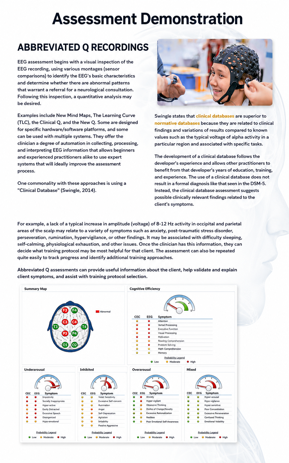
The first example is the NewQ, a six-location assessment that samples selected sites from the International 10-20 system, the standardized electrode placement grid used across clinical EEG practice. Similar tools include New Mind Maps, The Learning Curve (TLC), the Clinical Q, and others. Some are designed for specific hardware and software platforms, while others work across multiple systems. As a class, these tools offer a degree of automation in collecting, processing, and interpreting EEG information that benefits both beginners and experienced practitioners by incorporating expert decision-support systems into the assessment process.
A shared feature of these abbreviated approaches is their reliance on a clinical database, a collection of EEG metrics linked to known clinical findings rather than to the statistical norms of a healthy population (Swingle, 2014). Swingle argues that clinical databases are superior to normative databases (for a survey of database use in qEEG evaluation, see Johnstone & Gunkelman, 2003) because they connect EEG patterns directly to clinical observations, such as the typical voltage of alpha activity (8–12 Hz rhythms associated with relaxed wakefulness) in a particular region during specific tasks. Each clinical database reflects its developer's years of education, training, and clinical experience, allowing other practitioners to leverage that accumulated expertise.
Importantly, a clinical database assessment does not produce a formal diagnosis like those found in the Diagnostic and Statistical Manual of Mental Disorders (5th ed., text rev.; DSM-5-TR). Instead, it flags possible clinically relevant findings related to the client's presenting symptoms. This distinction matters in practice: the assessment guides your clinical reasoning and protocol selection without overstepping the boundaries of what the EEG data alone can establish.
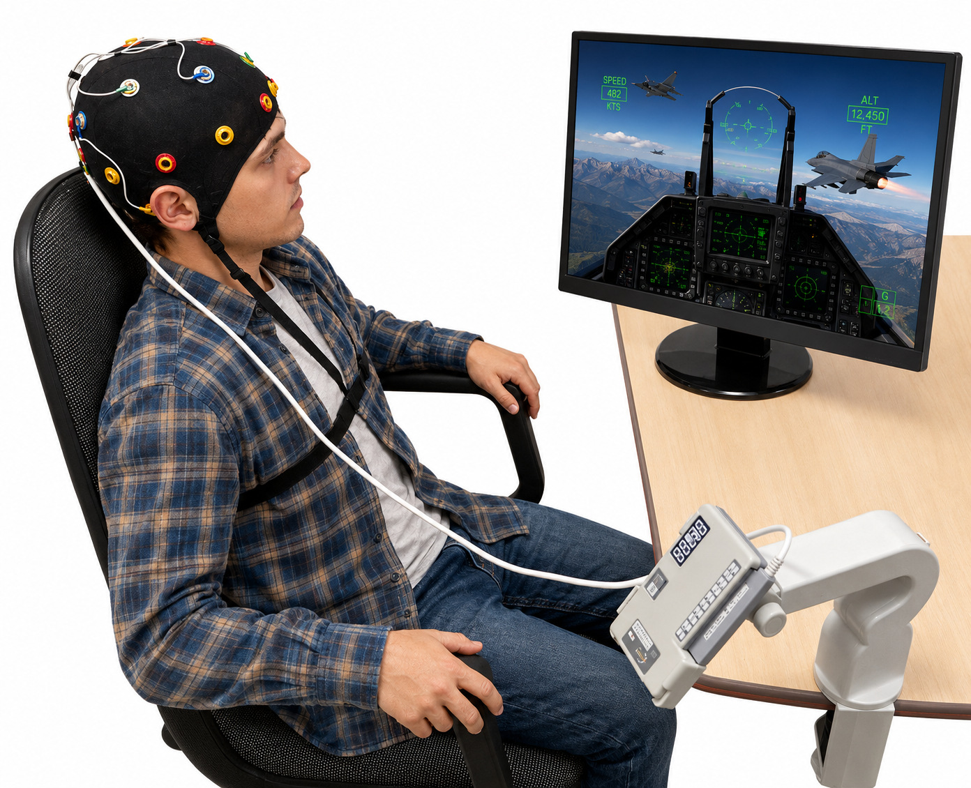
The following video describes the NewQ as an example of an abbreviated Q assessment. A subsequent demonstration walks through the complete process of collecting, processing, and interpreting the data. While this video features one specific tool, the workflow is representative of the broader category. Some tools assess additional sites, some collect data simultaneously rather than sequentially, and some use individual sensors while others employ an EEG "cap," an elastic fabric cap that holds sensors at fixed 10-20 positions, as shown in the image below.
Video © J. S. Anderson.
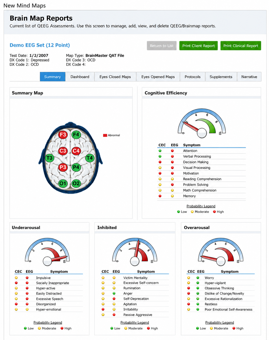
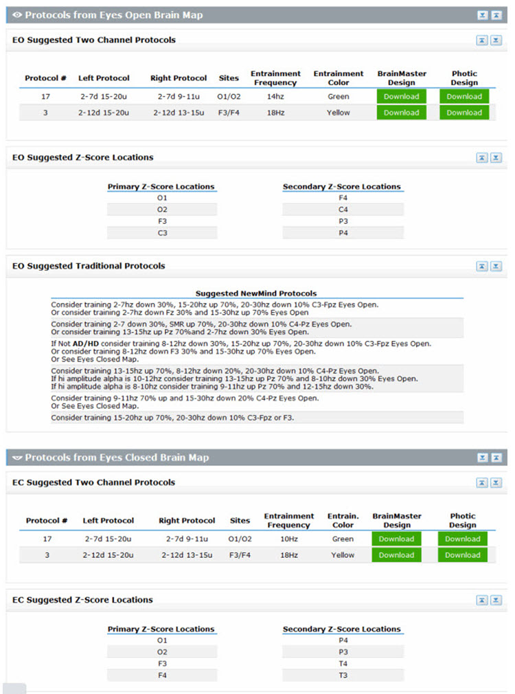
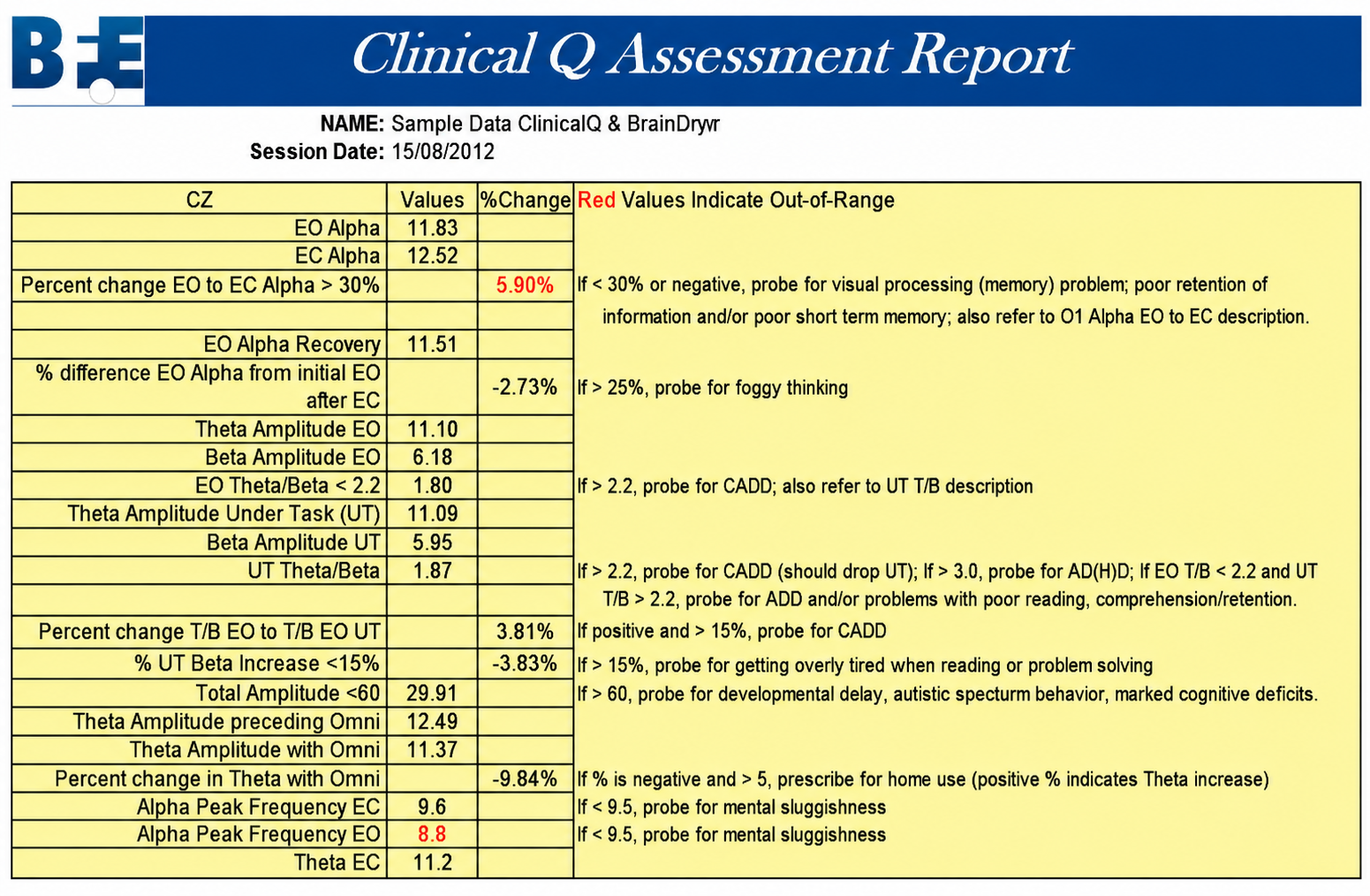
In summary, abbreviated Q assessments provide clinically useful information in a time-efficient format. They help validate and explain client symptoms, support training protocol selection, and can be readily repeated to monitor progress, making them practical tools for busy clinical settings. Two additional report examples appear below.
Clinical Q Assessment Report
Abbreviated Q recordings like the NewQ use a subset of 10-20 scalp locations to produce clinically useful analyses in less time than a full 19-channel assessment. Their reliance on clinical databases, which link EEG patterns to observed clinical findings rather than population norms, allows clinicians to identify relevant patterns and select appropriate training protocols. These tools do not produce formal diagnoses but instead guide clinical reasoning and support treatment planning.
19-Channel Recordings
This section explores the full 19-channel EEG recording with normative database comparison, the most comprehensive of the assessment approaches covered in this unit. Where abbreviated Q recordings offer speed and clinical specificity, 19-channel assessments provide a detailed, whole-brain picture of electrical activity. We will walk through the recording process, the critical steps of artifact rejection and visual inspection, and how the resulting data are compared to normative values to guide treatment planning.
The primary alternative to abbreviated Q assessments is the full 19-channel recording, which captures data from all standard International 10-20 system scalp locations simultaneously. The data are then compared to a normative database, a collection of EEG metrics from a representative sample of healthy individuals recorded under standardized resting and active-task conditions. This comparison reveals where a client's brain electrical activity falls relative to age-matched norms, expressed as z-scores (standard deviations from the database mean; Thatcher, 1998; Thatcher et al., 2019). The following video demonstrates the complete recording process. Video © J. S. Anderson.
EEG Recording, Testing, and Protocol Development
We encourage you to view the following two-hour video in stages, as it provides a comprehensive overview of the full assessment-to-treatment workflow. The recording covers 19-channel EEG data collection, protocol selection, treatment implementation, and qEEG-guided neurofeedback. This end-to-end demonstration shows how assessment findings translate directly into clinical decisions, the bridge from data to practice that defines effective neurofeedback. Video © J. S. Anderson.
Clean Data Extraction and Comparison to a Normative Database
Before quantitative analysis can begin, the raw EEG must be carefully cleaned, a process that connects directly to the artifact rejection skills covered in earlier units. Visual inspection starts with the standard 19-channel recording referenced to linked ears. The clinician then extracts segments for each recording condition (for example, eyes open and eyes closed), inspects the data using multiple montages, different electrode comparison configurations that reveal different aspects of brain activity, and selects only clean, artifact-free epochs. These artifact-free segments are the foundation of a reliable quantitative comparison to the normative database. Video © J. S. Anderson.
The rigor of this data-cleaning process directly affects the quality of your assessment results. Artifacts, false signals from sources like muscle tension, eye blinks, or electrical interference, can distort quantitative values and lead to inaccurate comparisons with the normative database. Taking time to carefully reject contaminated epochs is what separates a clinically trustworthy assessment from a misleading one.
Full 19-channel recordings with normative database comparisons offer the most comprehensive of the EEG assessment approaches covered in this unit. The process follows a structured sequence: recording from all standard scalp locations, visual inspection using multiple montages, careful artifact rejection, extraction of clean data, and statistical comparison to age-matched norms. The quality of the final assessment depends directly on the rigor of each preceding step, making meticulous data cleaning essential to accurate clinical interpretation.
Check Your Understanding
- What is the difference between a clinical database and a normative database in EEG assessment?
- Why might a 2-channel assessment using only two sites have limited clinical usefulness?
- What are the key steps involved in processing a 19-channel EEG recording for normative database comparison?
- How can abbreviated Q assessments like the NewQ assist clinicians in selecting training protocols?
- Why is visual inspection of the raw EEG an important first step before quantitative analysis?
Assignment
Now that you have completed this unit, explain why the visual inspection of raw EEG waveforms is important.
Glossary
A (auricular): the International 10-20 system letter designating an earlobe reference placement, as in A1 and A2.
abbreviated Q recording: an EEG assessment that samples a subset of International 10-20 system scalp sites, usually against a clinical database, in place of a full 19-channel recording.
alpha rhythm: an 8 to <13 Hz EEG rhythm dominant posteriorly during relaxed wakefulness with eyes closed and attenuated by eye opening.
amplitude: the magnitude of a signal's excursion from a specified reference level, reported using a stated convention such as peak, peak-to-peak, or RMS.
artifact: an unwanted signal component introduced by movement, environmental interference, hardware, processing, or biological activity outside the signal of interest.
artifact rejection: the removal of epochs contaminated by noncerebral signals before quantitative analysis.
beta rhythm: a 13 to <30 Hz EEG rhythm, usually low amplitude, associated with alert wakefulness and active cognitive or motor processing.
bipolar montage: an EEG montage composed of bipolar derivations, often arranged in longitudinal or transverse chains to emphasize spatial voltage gradients.
bridging artifact: electrical coupling between adjacent electrodes caused by conductive gel, sweat, or moisture, reducing their independence and distorting recorded voltage differences.
C fibers: small unmyelinated afferent fibers conducting slowly, commonly about 0.5 to 2 m/s, and carrying polymodal nociceptive, thermal, pruritic, and autonomic information.
channel: one differential amplifier input, formed by a pair of electrodes whose voltage difference is recorded. A single ground electrode is shared across all channels rather than belonging to any one of them.
clinical database: a reference set built from a clinician's accumulated experience with clinical populations that maps EEG findings onto probable client symptoms and suggests training approaches.
delta rhythm: a 0.5 to <4 Hz EEG rhythm in the tutorial convention, prominent during N3 sleep and potentially abnormal when focal or excessive in awake adults.
drowsiness artifact: the intrusion of drowsiness or light sleep into a recording intended to sample wakefulness, marked by attenuation of the posterior dominant rhythm, slow rolling eye movements of opposite polarity at F7 and F8, and 1-2 Hz alpha slowing.
EEG artifacts: noncerebral electrical activity in an EEG recording, which can be divided into physiological and exogenous artifacts.
EEG power: the squared amplitude of EEG activity quantified over time or frequency, expressed as µV² for band power or µV²/Hz for power spectral density.
electro-ocular artifact: contamination of EEG recordings by potentials generated by eye blinks, eye flutter, and eye movements.
electrode impedance meter: an instrument that applies a small test signal to estimate electrode-skin impedance.
electrode-pop artifact: an abrupt high-amplitude transient caused by a sudden change in electrode-skin contact potential or impedance.
EMG artifact (ECG): false or obscured ECG events caused by skeletal-muscle electrical activity, potentially producing spurious or missed beat detections.
epoch: a defined time segment of a physiological signal selected for recording, averaging, artifact review, or analysis.
exogenous artifact: signal contamination originating outside the organism, such as electromagnetic interference, vibration, or recording-equipment noise.
F (frontal): the International 10-20 system letter designating sites that detect frontal lobe EEG activity.
Fp electrode sites: frontopolar scalp locations designated Fp1, Fpz, and Fp2 in the international 10–10 system; the 10–20 system uses Fp1 and Fp2.
hertz (Hz): the SI unit of frequency equal to one cycle per second.
impedance (Z): frequency-dependent opposition to alternating current, comprising resistance and reactance and measured in ohms.
impedance test: measurement of electrode-skin impedance before or during biopotential recording.
inion: the most prominent point of the external occipital protuberance at the posterior skull.
international 10–20 system: a standardized method for locating EEG electrodes at intervals of 10% or 20% of measured head distances; the original array includes 19 scalp and two auricular sites, with ground specified separately.
low-resolution electromagnetic tomography (LORETA): the inverse solution of Pascual-Marqui and colleagues (1994) that estimates three-dimensional cortical sources of scalp-recorded EEG at low spatial resolution.
mastoid bone: the bony prominence behind the ear.
microvolt (µV): a unit of electric potential equal to 10⁻⁶ volt.
montage: the specified arrangement of electrode derivations or channel pairings used to display a physiological recording.
movement artifact: signal distortion caused by movement of the person, sensor, electrode, cable, or tissue interface; its appearance and effects depend on recording modality.
nasion: the midline depression at the junction of the frontal bone and nasal bones.
normative database: a reference set of EEG measures collected from healthy subjects across age ranges, against which an individual client's values are compared as z-scores.
notch filter: a band-stop filter that strongly attenuates a narrow frequency range, commonly centered on 50 or 60 Hz.
O (occipital): the International 10-20 system letter designating sites that detect occipital lobe EEG activity.
ohm (Ω): the unit of impedance or resistance.
parietal EEG site (P): an electrode location over the parietal scalp designated P in international EEG-placement systems.
physiological artifact: contamination generated by biological activity other than the target signal, such as eye, muscle, cardiac, respiratory, or sweat activity.
posterior dominant rhythm (PDR): the dominant occipital rhythm during relaxed wakefulness with eyes closed, usually in the alpha range in healthy adults and attenuated by eye opening.
preauricular point: a landmark immediately anterior to the tragus used in cranial and EEG measurements.
protocol: a rigorously organized plan for training.
pulse artifacts: noncerebral voltages due to the mechanical movement of an electrode in relation to the skin surface caused by the pressure wave of each heartbeat.
quantitative EEG (qEEG): the numerical analysis of digitized EEG features, such as spectral power, asymmetry, and connectivity, sometimes compared with normative databases; no universal minimum channel count defines it.
reference electrode: an electrode whose potential is used as the comparison for an active recording electrode; it is distinct from the ground electrode.
rhythmic midtemporal theta of drowsiness (RMTD): a benign drowsiness variant, not an epileptiform pattern, consisting of notched or sharply contoured rhythmic theta waveforms localized to the midtemporal regions. It may be seen over either hemisphere, and its asymmetry shifts within and between recordings.
sensorimotor rhythm (SMR): a 12 to 15 Hz rhythm recorded over sensorimotor cortex, enhanced during physical stillness and attenuated by movement; it is defined topographically and behaviorally, so its range overlaps the mu and beta bands.
standardized LORETA (sLORETA): a refinement of LORETA that produces images of standardized current density, partitioning the intracerebral volume into 6,239 voxels at 5-mm spatial resolution, with exact localization for test point sources and no localization bias under noise.
surface Laplacian (SL) analysis: a family of mathematical algorithms that provide two-dimensional images of radial current flow from cortical dipoles to the scalp.
swLORETA: a version of sLORETA that adds a singular-value-decomposition-based lead-field weighting, improving localization under realistic noise and for deep sources (Palmero-Soler et al., 2007).
theta rhythm: a 4 to <8 Hz EEG rhythm associated with drowsiness and sleep transitions and, depending on location and task, memory and cognitive processing.
theta/beta ratio (TBR): EEG theta-band power divided by beta-band power using explicitly stated bands, electrodes, reference, and processing methods; it is not a diagnostic test for ADHD.
tragus: the small cartilaginous flap projecting backward over the opening of the external acoustic meatus; the preauricular point lies immediately anterior to it.
transient (EEG): an isolated waveform or brief sequence that is distinguishable from the ongoing EEG background.
vertex and Cz: the vertex is the highest point of the skull; Cz is the midline EEG position at the intersection of nasion-inion and left-right preauricular measurements.
z score: a standardized value expressing how far a measurement falls from a reference mean in standard-deviation units; it is not the probability of a diagnosis or evidence that a feature requires treatment.
z-score training: a neurofeedback approach using standardized deviations from a reference database to guide feedback; thresholds such as ±2 SD are protocol choices, not universal criteria.
References
Fein, G., Galin, D., Johnstone, J., Yingling, C. D., Marcus, M., & Kiersch, M. E. (1983). EEG power spectra in normal and dyslexic children. I. Reliability during passive conditions. Electroencephalography and Clinical Neurophysiology, 55(4), 399-405. https://doi.org/10.1016/0013-4694(83)90127-x
Hardt, J. V., & Kamiya, J. (1978). Anxiety change through electroencephalographic alpha feedback seen only in high anxiety subjects. Science, 201(4350), 79-81. https://doi.org/10.1126/science.663641
John, E. R., Ahn, H., Prichep, L., Trepetin, M., Brown, D., & Kaye, H. (1980). Developmental equations for the electroencephalogram. Science, 210(4475), 1255-1258. https://doi.org/10.1126/science.7434026
Johnstone, J., & Gunkelman, J. (2003). Use of databases in QEEG evaluation. Journal of Neurotherapy, 7(3-4), 31-52. https://doi.org/10.1300/J184v07n03_02
Kamiya, J. (1968). Conscious control of brain waves. Psychology Today, 1, 56-60.
Nuwer, M. R., & Coutin-Churchman, P. (2014). Brain mapping and quantitative electroencephalogram. In Encyclopedia of the Neurological Sciences (2nd ed.). Elsevier.
Rogala, J., Jurewicz, K., Paluch, K., Kublik, E., Cetnarski, R., & Wróbel, A. (2016). The do's and don'ts of neurofeedback training: A review of the controlled studies using healthy adults. Frontiers in Human Neuroscience, 10, 301. https://doi.org/10.3389/fnhum.2016.00301
Soutar, R., & Longo, R. (2022). Doing neurofeedback: An introduction (2nd ed.). Foundation for Neurofeedback and Neuromodulation Research.
Swingle, P. G. (2014, Spring). Clinical versus normative databases: Case studies of Clinical Q assessments. NeuroConnections.
Thatcher, R. W. (1998). Normative EEG databases and EEG biofeedback. Journal of Neurotherapy, 2(4), 8-39. https://doi.org/10.1300/J184v02n04_02
Thatcher, R. W., Lubar, J. F., & Koberda, J. L. (2019). Z-Score EEG biofeedback: Past, present, and future. Biofeedback, 47(4), 89-103. https://doi.org/10.5298/1081-5937-47.4.04
Thomas, C. (2007). What is a montage? In EEG instrumentation. American Society of Electroneurodiagnostic Technologists, Inc.
Thompson, M., & Thompson, L. (2015). The neurofeedback book: An introduction to basic concepts in applied psychophysiology (2nd ed.). Association for Applied Psychophysiology and Biofeedback.
Return to Top