
Basic Knowledge of the Neurophysiology of the EEG
What You Will Learn in This Chapter
Place an electrode on the scalp and you are eavesdropping on roughly a hundred million neurons at once. What reaches your amplifier is not the chatter of individual cells firing. It is the slow, rhythmic swell of synaptic voltage rising and falling across sheets of pyramidal neurons stacked in cortical columns. Understanding that distinction is the foundation of everything you will do as a qEEG practitioner.
This unit takes you from the single pyramidal neuron to the scalp tracing. You will learn where the EEG comes from, why postsynaptic potentials rather than action potentials dominate the signal, and how sinks, sources, and dipoles determine what your electrodes can and cannot see. You will then examine amplitude and frequency, the two dimensions along which every EEG signal varies, and the oscillatory circuits that organize brain activity into processing windows.
The second half turns to event-related potentials and slow cortical potentials, the very fast and the very slow ends of the electrophysiological spectrum. You will see how SCPs index cortical excitability, why a depolarized cortex produces a surface-negative shift, and what SCP abnormalities look like in ADHD, epilepsy, Parkinson's disease, and depression. The unit closes with neuroplasticity, the long-term depression and long-term potentiation that make neurofeedback learning possible in the first place.
IQCB Blueprint Coverage: This unit addresses IV. EEG, specifically A. Basic Knowledge of Neurophysiology of the EEG.
Learning Objectives
After completing this section, you will be able to:
Describe how the scalp EEG is generated by the summation of excitatory and inhibitory postsynaptic potentials in cortical columns of large pyramidal neurons.
Explain why action potentials contribute little to the scalp EEG while postsynaptic potentials dominate it.
Define sink, source, and dipole, and explain why the EEG is most sensitive to radially oriented dipoles in gyri.
Distinguish amplitude from frequency, and relate each to neuronal synchrony and to the size of the participating neuronal pool.
Compare and contrast event-related potentials and slow cortical potentials, including the CNV, readiness potential, MRPs, P300, and N400.
Explain paradoxical negativity and relate surface-negative and surface-positive SCP shifts to cortical excitability.
Summarize the role of long-term depression and long-term potentiation in the neuroplasticity that makes neurofeedback learning possible.
What is the EEG?
This section explains what the scalp EEG actually measures, how it is generated by pyramidal neurons in cortical columns, and the roles of local field potentials, dipole generators, amplitude, and frequency. This knowledge is foundational for every neurofeedback clinician because it connects the electrical activity displayed on a neurofeedback screen to the underlying neural processes being trained.
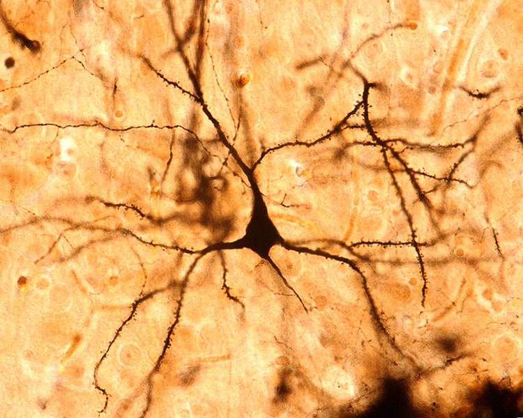
The scalp EEG is the voltage difference between two recording sites recorded over time. The EEG is primarily generated by large pyramidal neurons located in layers 3 and 5 of the cortical gray matter, which ranges from about 1 to 4.5 mm thick and averages about 2.5 mm. Image of a pyramidal neuron revealed using Golgi silver chromate © Jose Luis Calvo/Shutterstock.com. Note that the cell body's apical dendrite, which projects toward the cortical surface, and basilar dendrites, which arise from the base, feature an extensive network of spines.
Local activity is a composite of local and network influences. Network communication systems and local cortical functions show different characteristics across the cortex and produce unique and specific EEG patterns in different regions.
The movie below is a BioTrace+/NeXus-32 display of the raw EEG with voltage shown as μV peak to peak © John S. Anderson.
What Can the EEG Tell Us?
With the EEG, we can follow the progression from stimulus to behavioral response. This allows us to determine whether each step in the processing chain is functioning correctly and identify causal factors in dysfunctional outcomes. For clinicians, this capability makes the EEG an invaluable tool for both assessment and treatment planning.
Source of the Scalp EEG
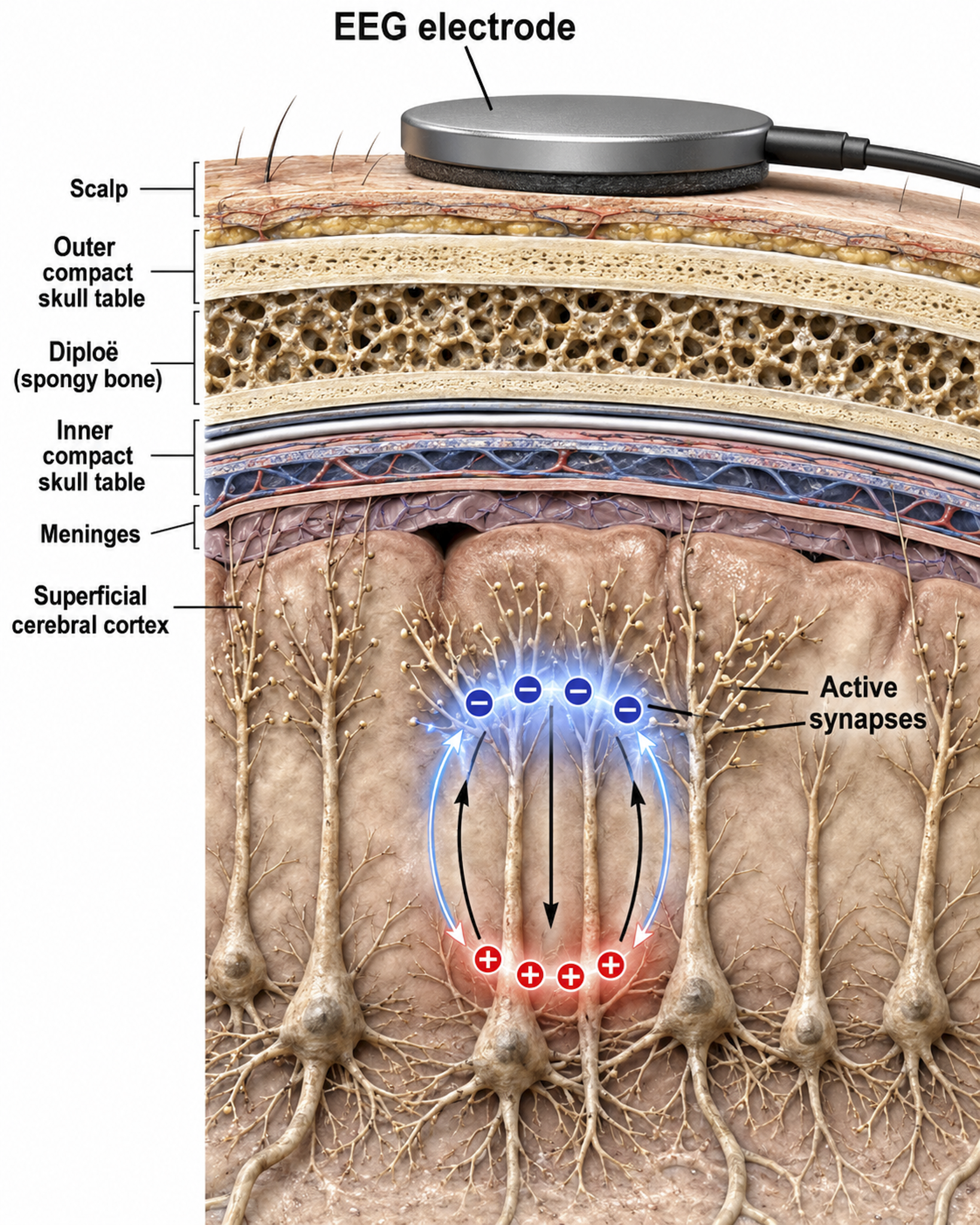
The scalp EEG results from the summation of large areas of gray matter activity. These areas are polarized synchronously due to the input of oscillatory or transient evoked activity, and they comprise thousands of cortical columns containing large pyramidal cells aligned perpendicularly to the cortical surface.
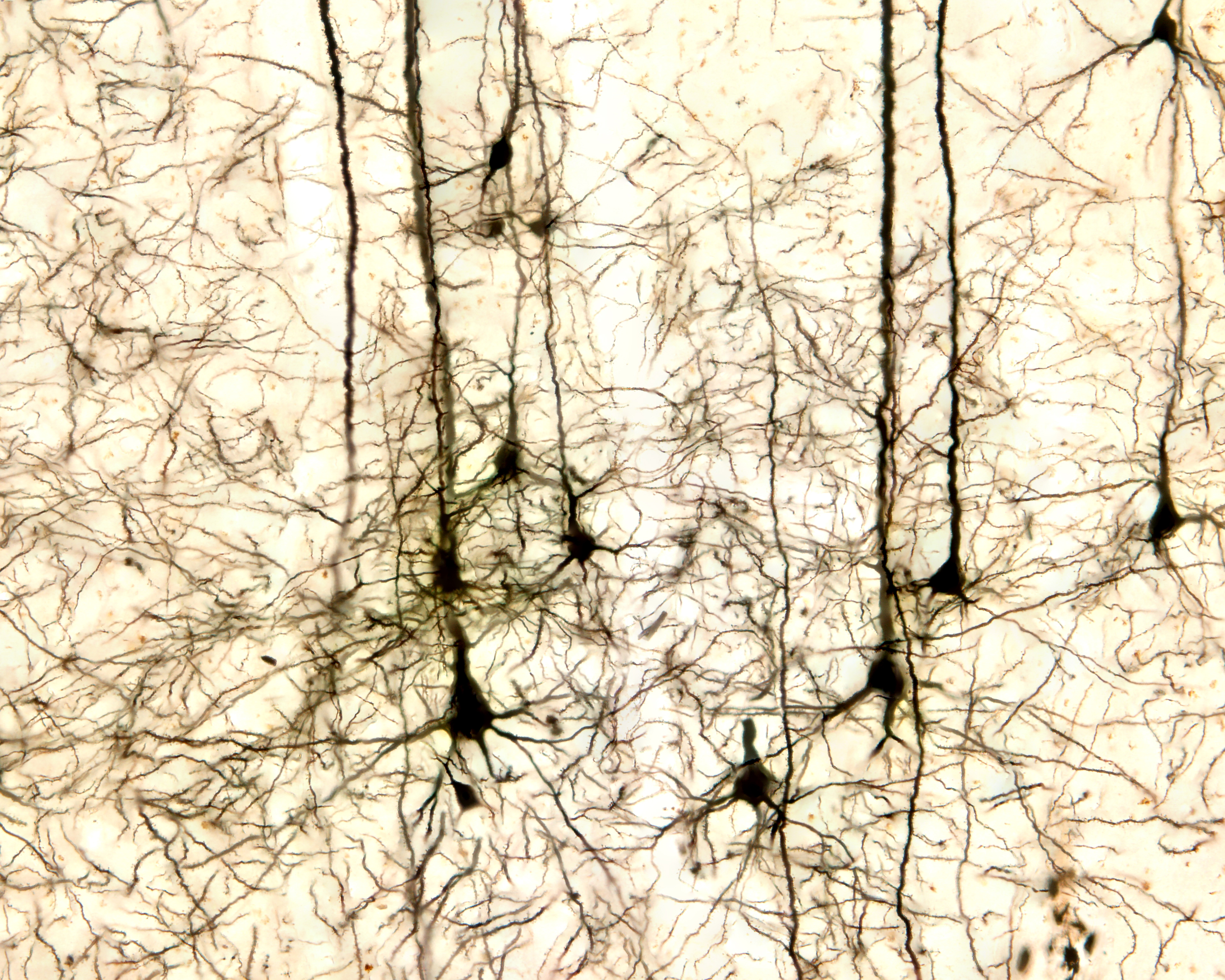
Pyramidal neurons of the cerebral cortex stained with the Golgi silver chromate © Jose Louis Calvo/Shutterstock.com.
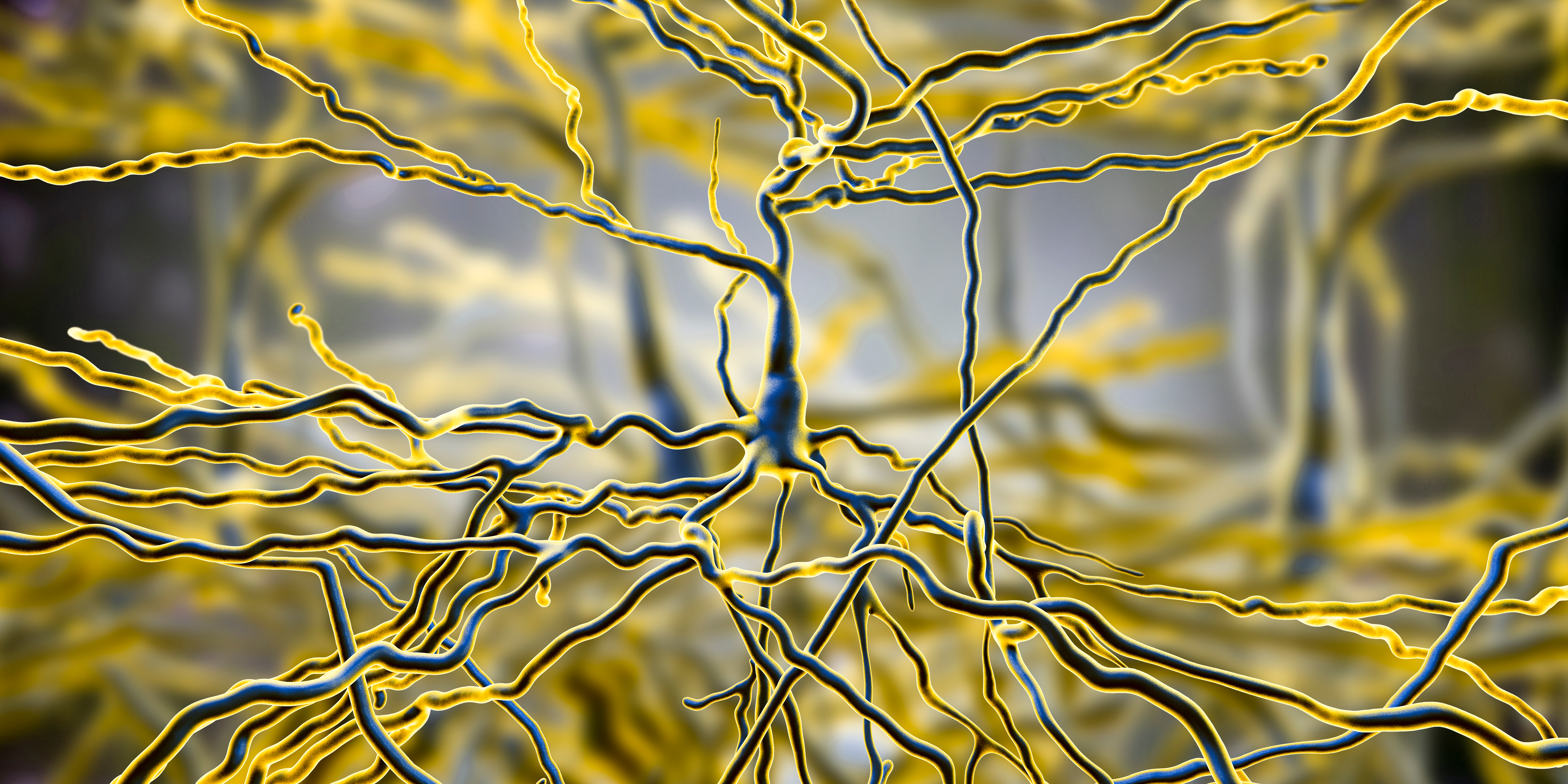
Pyramidal neurons are found in all cortical layers except layer 1 and represent the primary type of output neuron in the cerebral cortex. Pyramidal neuron graphic © Kateryna Kon/Shutterstock.com.
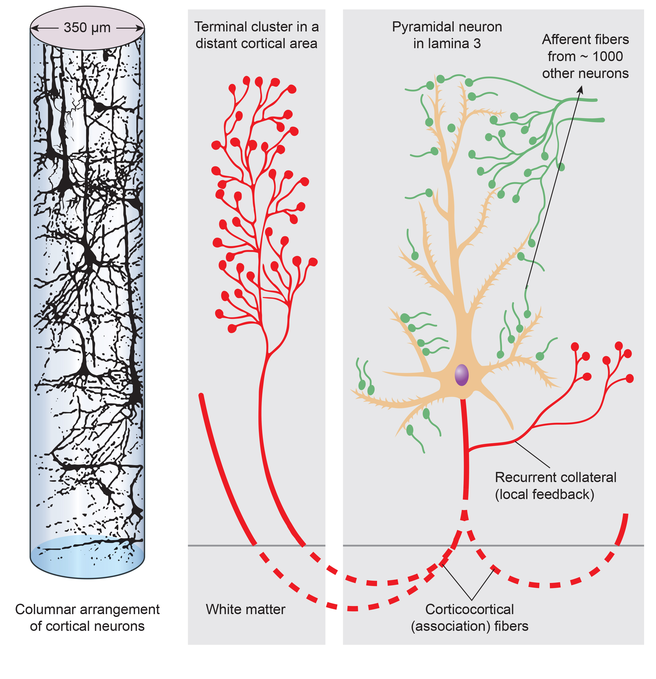
The scalp EEG results from the summation of EPSPs and IPSPs in thousands of cortical columns containing large pyramidal cells perpendicular to the cortical surface. The columns are synchronously polarized (made more negative) and depolarized (made less negative) due to the input of oscillatory or transient evoked activity. Graphic redrawn by minaanandag on Fiverr.com.
Artist: Dani S@unclebelang. This WEBTOON is part of our Real Genius series.

Local Field Potentials
The local field potential (LFP) is the aggregate electrical effect of interconnected pyramidal neurons firing within cortical columns, plus additional mechanisms like glial cell modulation of the cortical electrical gradient. The LFP is the bridge between individual neuron activity and the macroscopic EEG signal; it reflects the collective behavior of local neural populations.
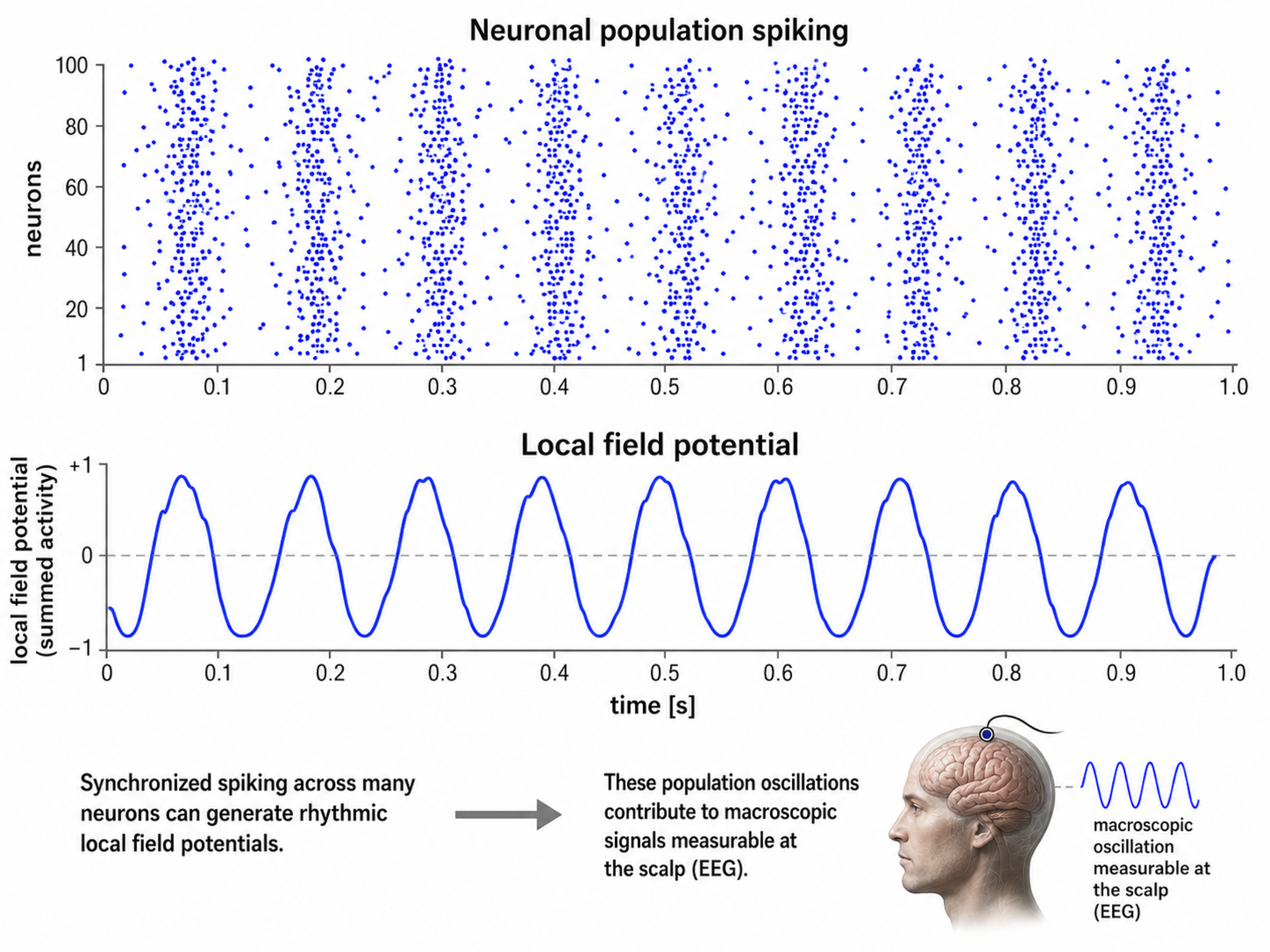
Caption from Wikipedia's article on Neural Oscillation. Simulation of neural oscillations at 10 Hz. The upper panel shows spiking of individual neurons (with each dot representing an individual action potential within the population of neurons). On the lower panel, the local field potential reflects their summed activity. This figure illustrates how synchronized patterns of action potentials may result in macroscopic oscillations that can be measured outside the scalp.
Do not confuse the "spiking" of individual neurons with epileptogenic spikes in the scalp EEG.
Scalp Electrical Potentials
Scalp electrical potentials represent the sum of all available electrical fields. Fields of opposite polarity (+/-) cancel each other out so that scalp potentials are greater when large aggregates of neurons polarize and depolarize synchronously. The scalp EEG represents a weighted sum of all active currents within the brain that generate open fields, including non-cortical sources.
Action potentials reflect neuronal output. In extracellular recordings they appear as brief events lasting less than 2 ms, isolated by high-pass filtering above roughly 300 Hz. The familiar 100-mV swing of an action potential is an intracellular measurement; recorded extracellularly, a spike is only tens to a few hundred microvolts. Action potentials play a minor role in the scalp EEG because their amplitude falls below 60 µV outside a 50-μm (0.050-mm) radius of the cell body. Since scalp electrodes are several centimeters from cortical neurons and are generally aligned away from the scalp, action potentials are unlikely to contribute significant voltages to the scalp EEG. This is a key point: what we measure on the scalp during neurofeedback are postsynaptic potentials, not action potentials.
Local Field Potentials Regulate Neuron Excitability and Firing
Neurons are most likely to fire during the depolarizing phase of the local field potential. Neurons are more excitable when they are "in phase" with the LFP and are inhibited when they are out of phase. Thus, at any instant of time, the amplitude and frequency of the EEG are regulated by the LFP, which in turn is influenced by oscillatory mechanisms such as slow cortical potentials. This rhythmic gating of neural excitability is what makes neurofeedback training possible; by modifying these oscillatory patterns, clients can influence which neural populations fire and when.
The movie is a 19-channel BioTrace+ /NeXus-32 display of SCPs © John S. Anderson. Negative SCPs drift up, and positive SCPs drift down, depending on the software settings. However, the convention in electroencephalography is to show negative up. SCPs represent a global shift in DC voltage across the cortex and reflect a generally higher (negative SCPs) or lower (positive SCPs) state of cortical excitability regulating neural networks.
The EEG is a moment-to-moment measure of the excitability of action potential firing, like gates opening and closing on the half cycle. The synchronous activity of large pyramidal neurons networked in cortical columns creates the EEG.
The Composition of the EEG
The EEG is composed of electrical potentials, varying in two dimensions: frequency and amplitude.
Sources of IPSP and EPSP Inputs
Many sources contribute input that results in IPSP and EPSP activity within cortical neurons. These sources primarily contribute influences such as oscillatory generator input or ascending event-related evoked input.
EEG Sources
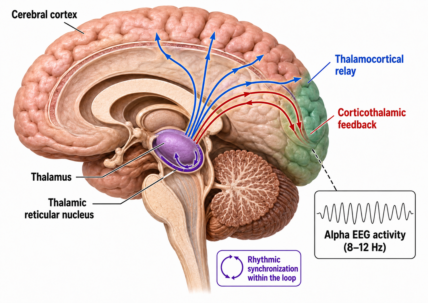
Generators like the thalamus produce oscillatory activity among many interconnected neurons, including EEG patterns like the alpha rhythm. The thalamus functions as the brain's central relay station, and its rhythmic output shapes much of the EEG activity that clinicians observe during recording.
Movie © John S. Anderson. The recording begins with eyes open. The eyes-closed condition starts at 14'01" and clearly shows increased 8-12 Hz voltage (posterior dominant rhythm or PDR) in occipital and parietal locations in the line tracing and topographic maps to the right of the tracing.
The eyes open again at 14'31", and alpha attenuates (alpha blocking). This demonstrates the posterior dominant rhythm (generally known as "alpha") appearing in the eyes-closed condition when visual sensory input is stopped, and the attenuation or blocking of this rhythm as sensory input returns in the eyes-open condition. This eyes-open/eyes-closed comparison is one of the most common assessment procedures in clinical neurofeedback.
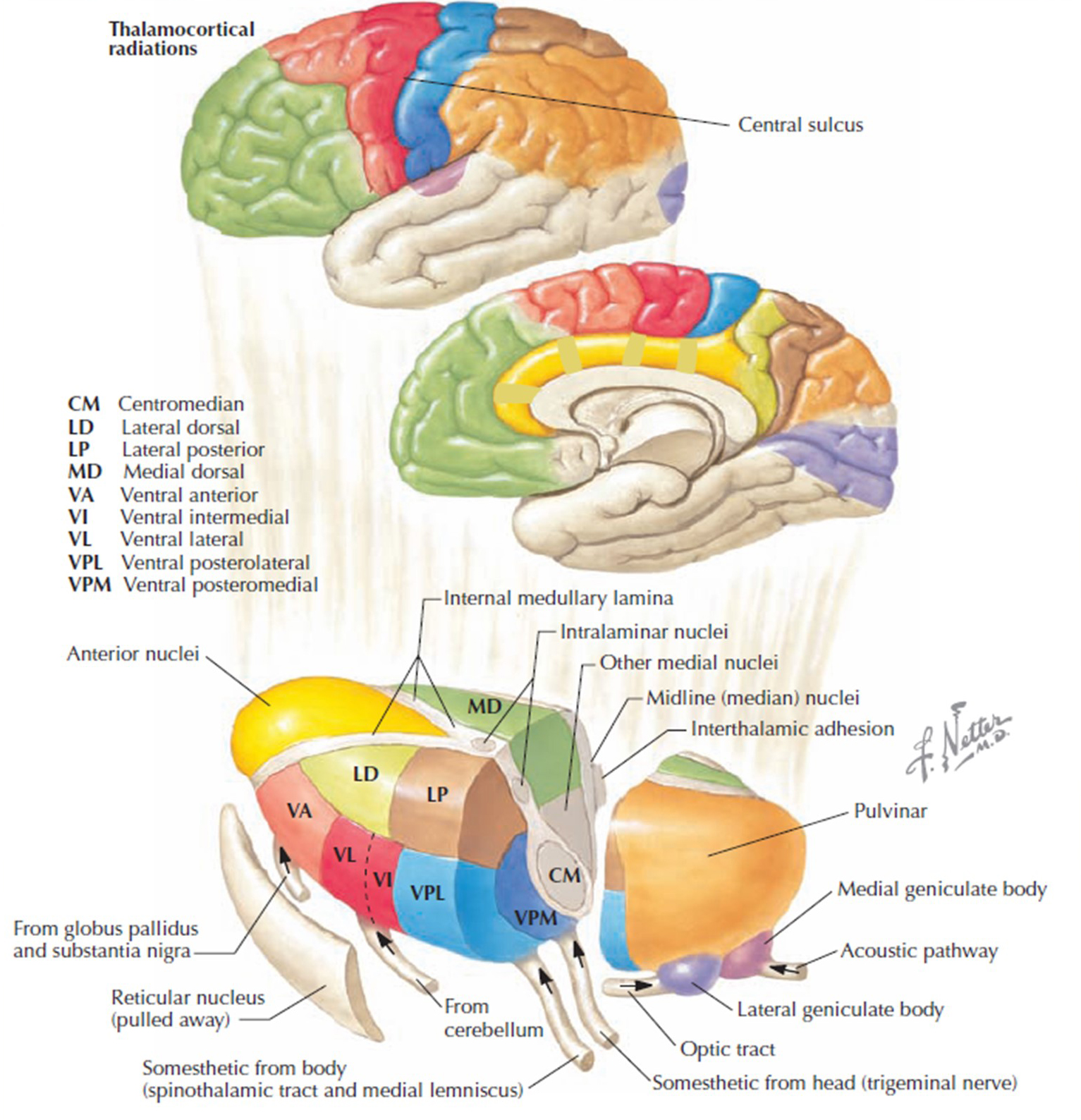
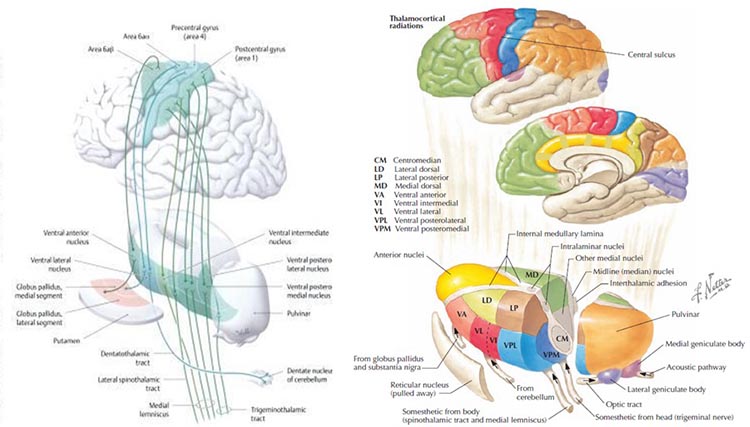
The thalamus contributes to slow cortical potentials, 1-4 Hz delta, 8-12 Hz alpha, and beta activity in the 20-38 Hz range, as well as to 40-Hz gamma activity. The diagram shows the connections between the pulvinar (bottom right) and reticular nuclei (bottom left) of the thalamus and the cortex © Elsevier Inc. - Netterimages.com.
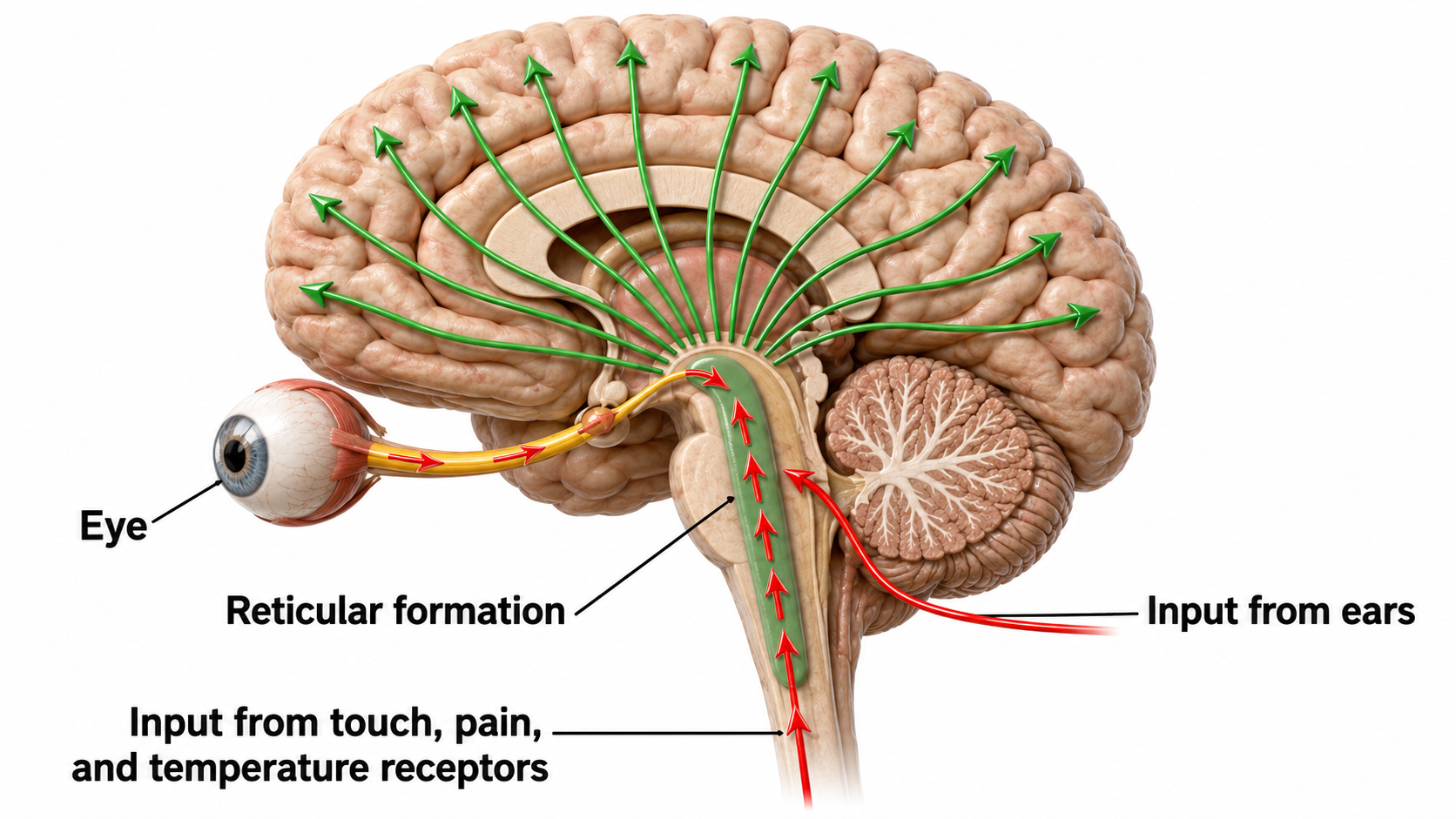
Thalamocortical (signaling from the thalamus to the cortex) cells are subject to excitatory drive from their system afferents, from monosynaptic corticothalamic (signaling from the cortex to the thalamus) fibers, and from the brainstem reticular formation (ascending reticular activating system, ARAS). They receive inhibitory drive from local interneurons and neurons in the reticular nucleus of the thalamus (RNT).
Note that the RNT neurons are excited by activity in thalamocortical cells and corticothalamic cells. The connections are precisely organized; each column in a primary cortical area sends corticothalamic fibers back to the same part of its specific thalamic nucleus that sends its thalamocortical fibers to that cortical column. The corticothalamic fibers also synapse on the RNT cells receiving input from that part of the thalamic nucleus. Each cortical receiving area is said to be "reciprocally connected" with its specific thalamic nucleus. Like the thalamocortical cells, RNT cells and cortical neurons also receive excitatory drive from the ARAS (Jackson & Stoney, 2006).
The EEG is generated by thalamocortical (delta and alpha), medial cortical and subcortical regions (theta), and cortico-cortical (beta) sources.
Ascending reticular activating system input helps drive the cortical responses recorded as event-related potentials following diverse stimuli like a flashing light or a sound. Event-related potentials (ERPs) are the brain's response to externally applied stimuli, events, or cognitive/motor tasks. They are time-locked measures of brain electrical activity.
Dipole Generators
Large cortical pyramidal neurons organized in macrocolumns are oriented with an apical dendrite projecting toward the scalp and an axon descending in the opposite direction. An "Equivalent Dipole Generator" usually represents the sum of all multipolar current sources. Summed generators are modeled as dipoles to aid the conceptual understanding of the electrical fields involved. Graphic adapted from Lipping (2017).
EEG Signals (Brainwaves)
The EEG represents changes in a brain area's electrical activity (potential) compared to a "neutral" site or another brain area. The EEG is displayed as oscillations or voltage fluctuations, which show a "wave" pattern when plotted on a graph.
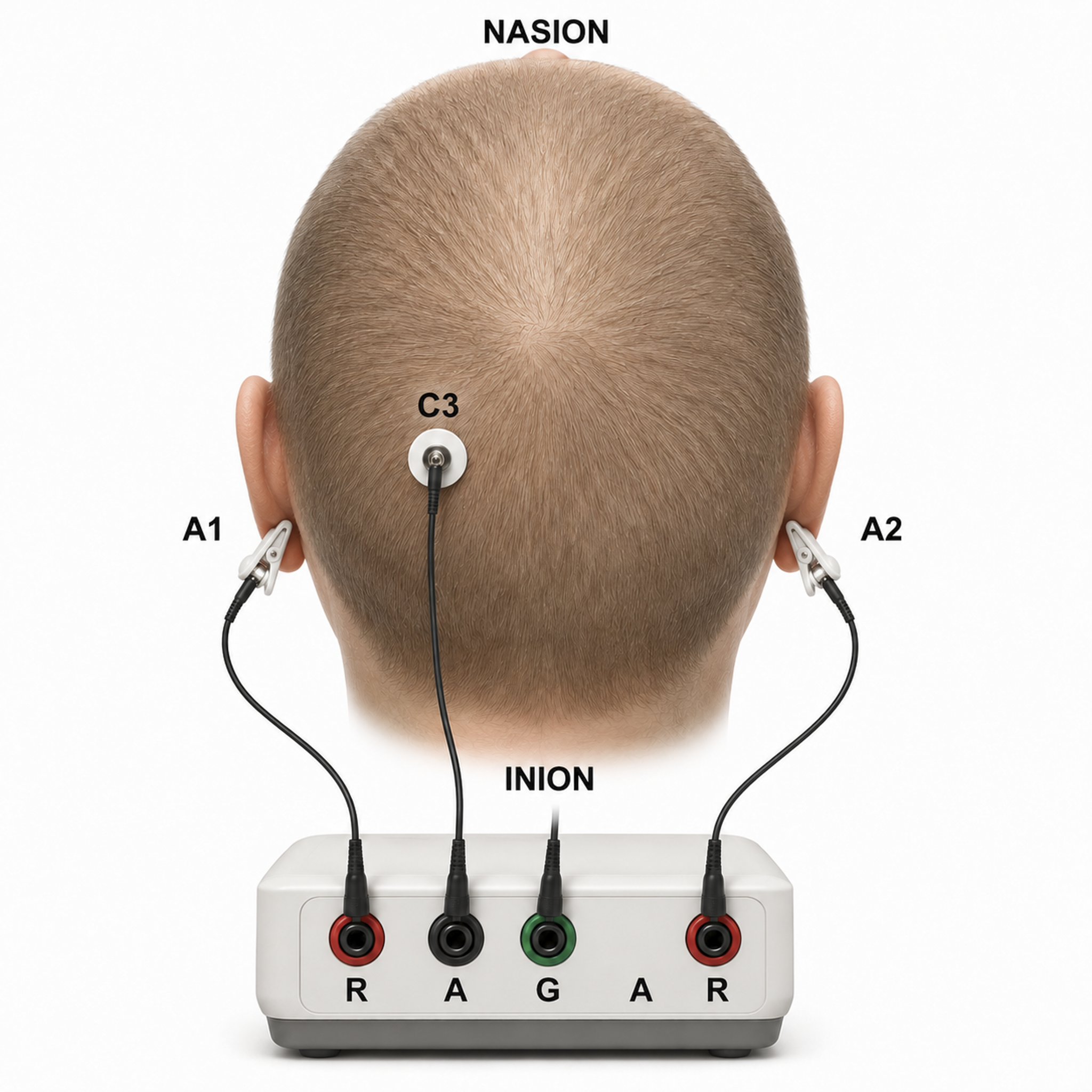
"These oscillations are generated spontaneously in several areas of the cerebral cortex as neuronal networks transiently form assemblies of synchronously firing cells." Klaus Linkenkaer-Hansen.
Sink, Source, and Dipole
We can describe pyramidal cells in terms of their sink, source, and dipole. A sink (-ve), which may be located at the bottom, middle, or top of the apical dendrite, is where positive ions enter the dendrite. Cation (positive ion) entry gives the extracellular space a negative charge. The source (+ve) is where the current exits the cell. Finally, the dipole is the field created between the sink and source (Thompson & Thompson, 2015).
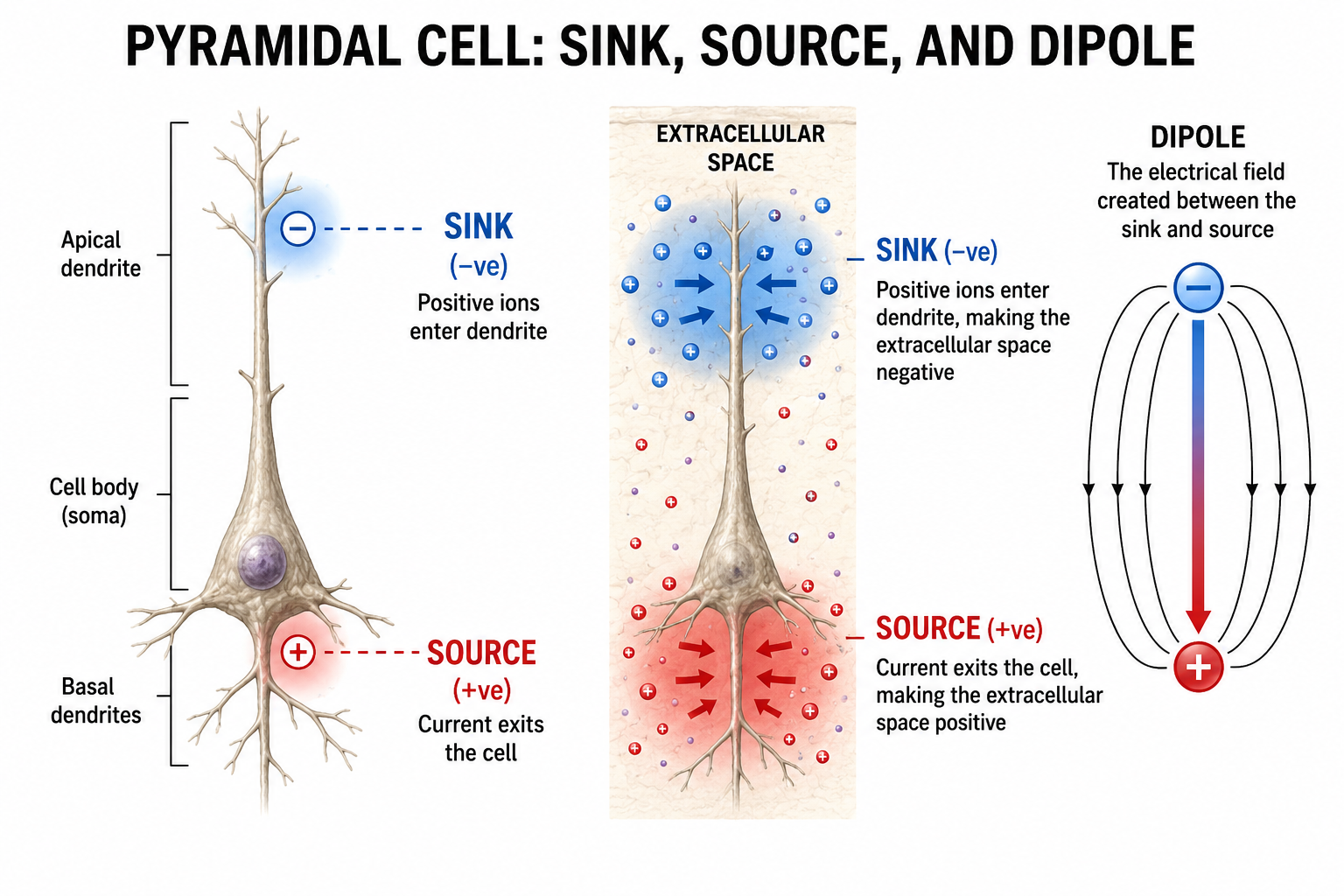
The postsynaptic potentials (EPSPs and IPSPs) propagated by the apical dendrites in layers 2 and 3 create an extracellular dipole layer parallel to the cortical surface. The dipole layer's electrical polarity is the opposite of the deeper cortical layers 4 and 5 (Fisch, 1999).
A cortical dipole is created when pyramidal neurons depolarize simultaneously, a phenomenon called local synchrony. Synchrony matters more than sheer numbers. Because asynchronously active neurons contribute potentials that partly cancel, a comparatively small synchronized population can dominate the recorded signal; roughly 6 to 10 cm2 of contiguous, synchronously active cortex is generally taken as the minimum needed to register at the scalp. A small fraction of these neurons firing in step can produce visible changes in EEG feedback. This creates the potential for operant conditioning to help clients learn to modify EEG activity through neurofeedback.
Cortical dipoles have three properties: site (depends on source), size (oscillation frequency and voltage), and relative position with respect to sulci and gyri (Collura, 2014).
The EEG is Mainly Sensitive to Radially Oriented Dipoles
Evolution has convoluted the human brain to increase its computing power without enlarging the skull. This enfolding has created two easily visible anatomical features: gyri and sulci.
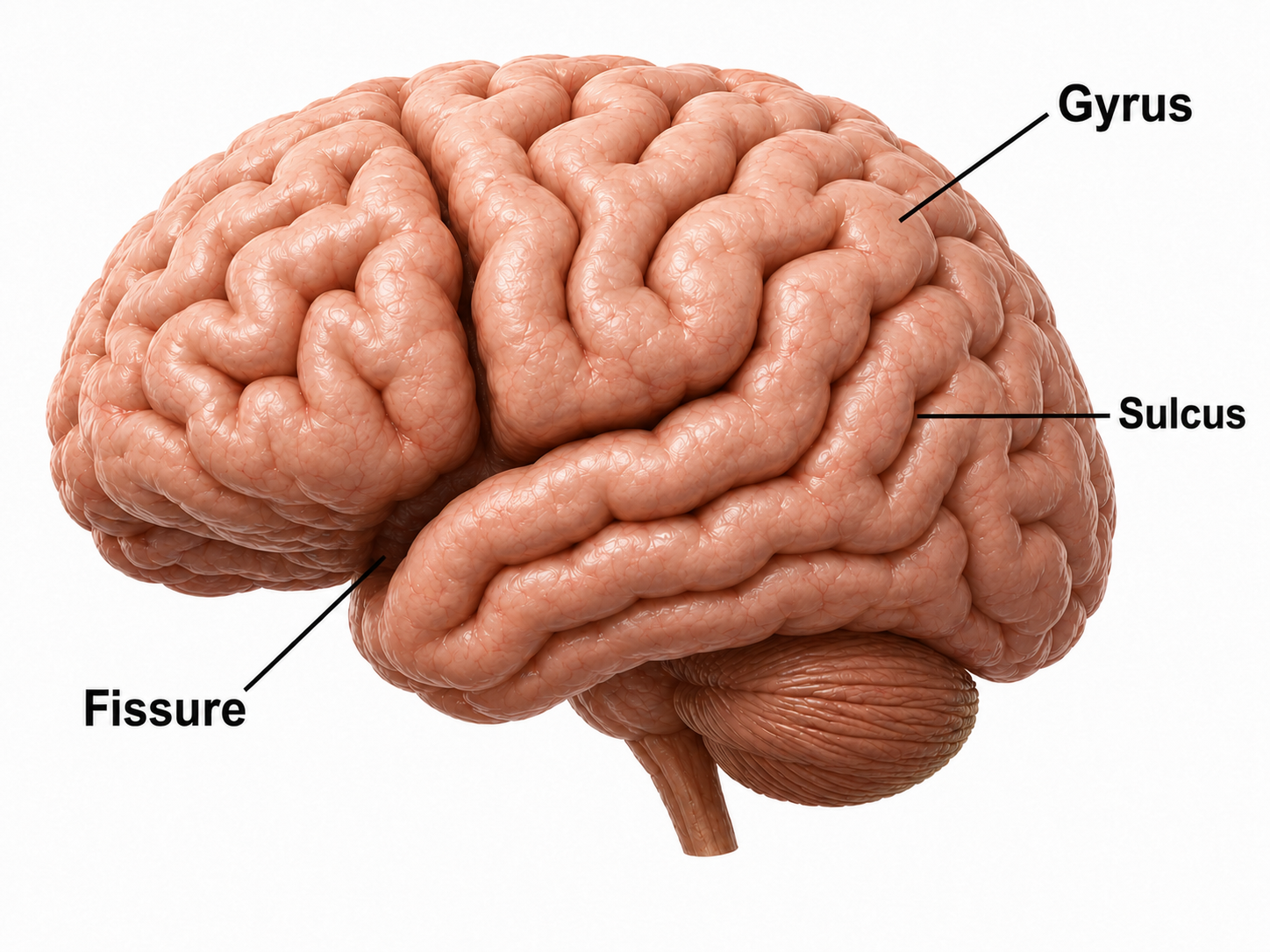
Recall that a gyrus is a ridge of the convoluted cerebral cortex, while a sulcus is a valley.
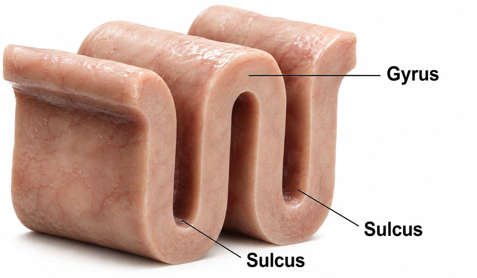
A dipole's orientation relative to the scalp determines how well the EEG detects it. A radial dipole points outward from a gyral crown, roughly perpendicular to the overlying scalp, so its field projects directly toward the recording electrodes and is detected well. A tangential dipole lies in the wall of a sulcus, oriented parallel to the scalp, so much of its field runs sideways beneath the electrodes rather than toward them. When the two facing walls of a sulcus generate opposing tangential dipoles, their fields largely cancel at the surface. This geometry is why the scalp EEG favors activity from gyral crowns and can underrepresent sources buried in sulci (Bear, Connors, & Paradiso, 2026).
The EEG is most sensitive to a correlated dipole layer in gyri. The EEG is less sensitive to a correlated dipole layer in sulci, valleys within the cortex. Finally, the EEG is insensitive to an opposing dipole layer in sulci. This sensitivity pattern is a key limitation of scalp EEG that neurofeedback clinicians should keep in mind: the EEG preferentially detects activity from gyral surfaces and may miss activity originating deep within sulci.
The signal also spreads as it travels from cortex to scalp, a process called volume conduction. The brain, cerebrospinal fluid, skull, and scalp act as a passive conductor that smears each cortical source across a broad area of the scalp, so a single electrode records a blend of contributions from many underlying generators. Volume conduction is the main reason scalp EEG has limited spatial resolution and why an electrode's reading depends heavily on the choice of reference. It should not be confused with volume transmission, the chemical process described earlier (Collura, 2014).
The EEG is composed of electrical potentials that vary along the dimensions of amplitude and frequency.
EEG Amplitude
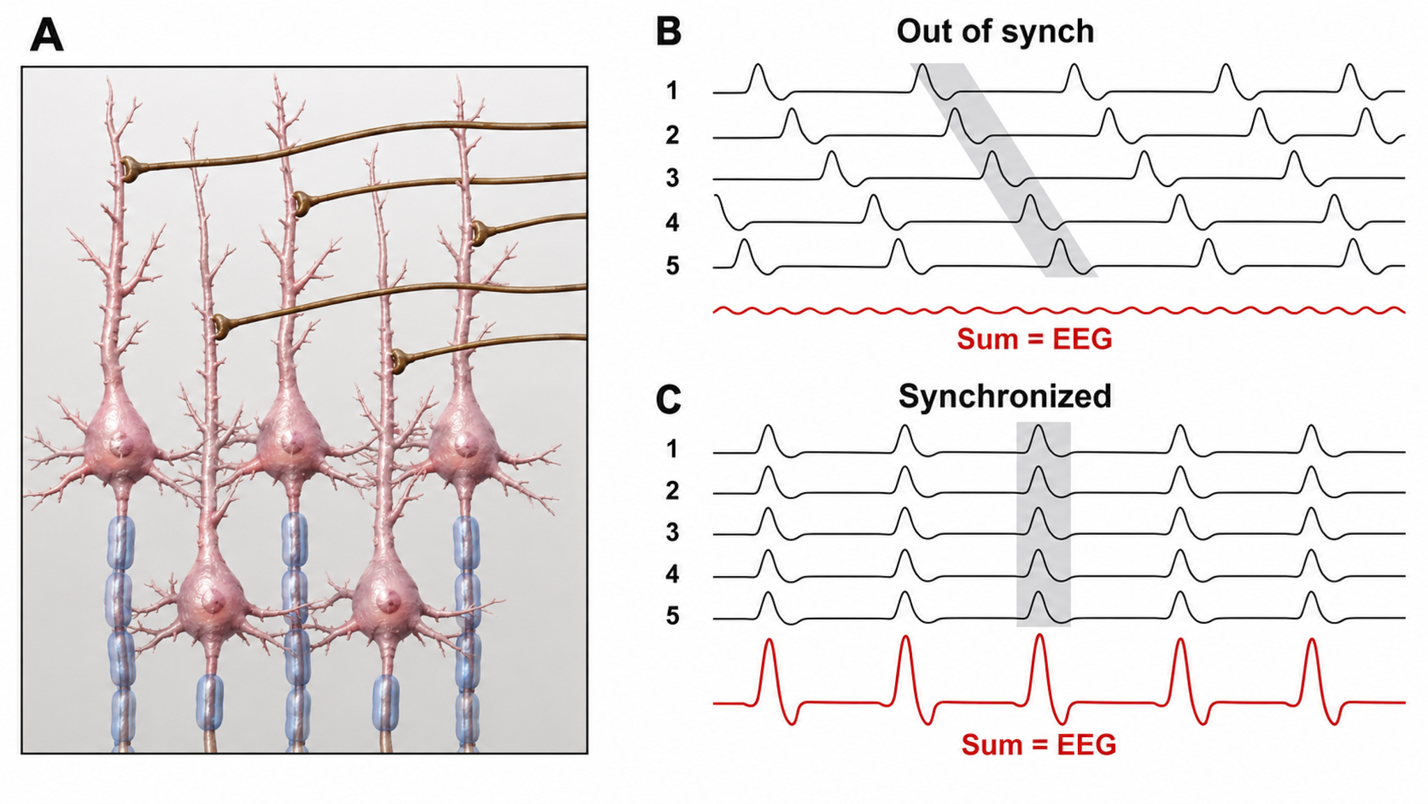
The "amount" of voltage, or amplitude reflects the number of neurons firing in synchrony at a given frequency. Lower neuron firing rates and fewer neurons firing in synchrony correspond to lower signal amplitude.
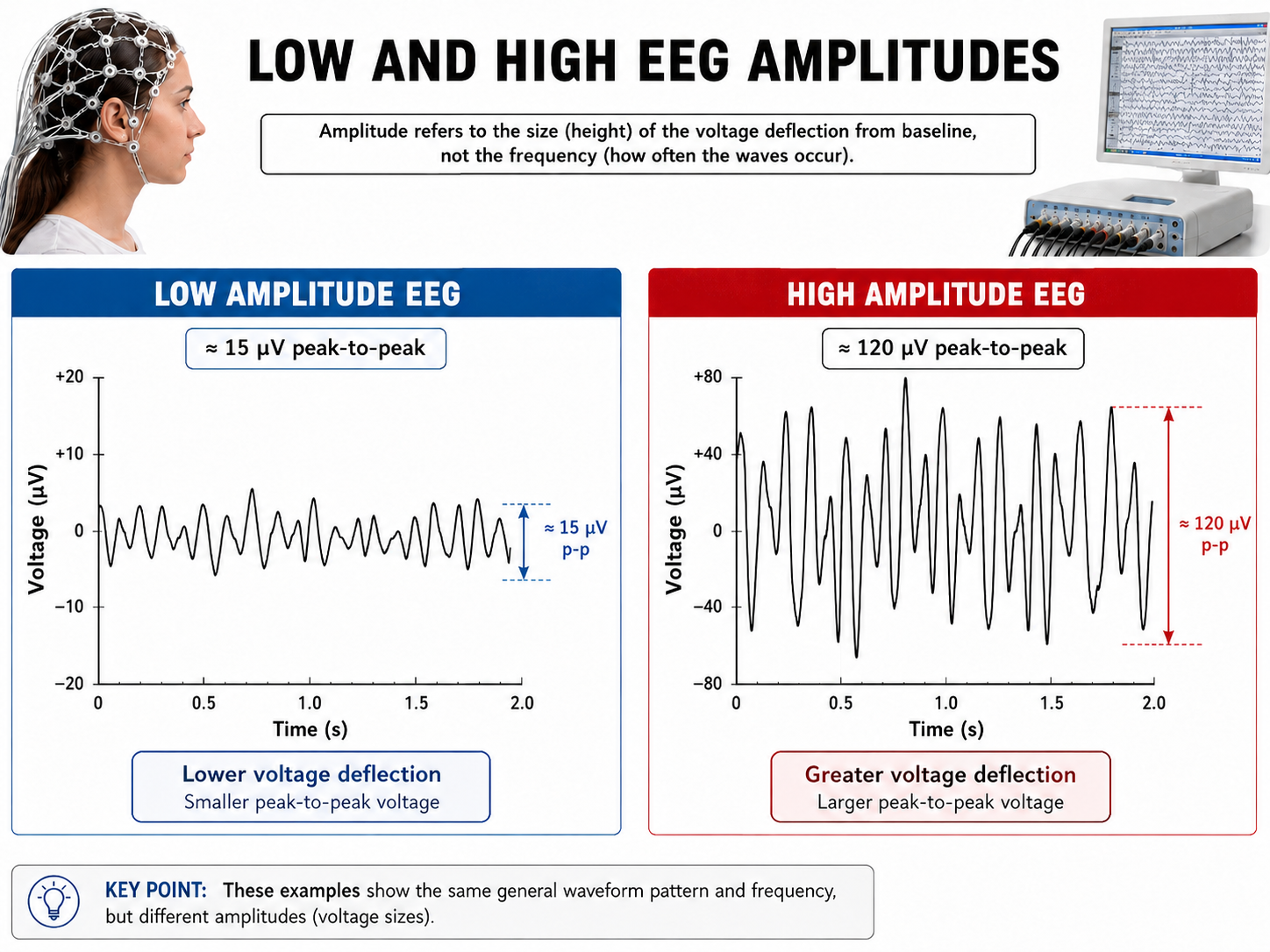
Amplitude measures the size of the voltage fluctuation and is expressed in microvolts. Amplitude is not the same quantity as power, which is proportional to the square of amplitude and is expressed in µV2.
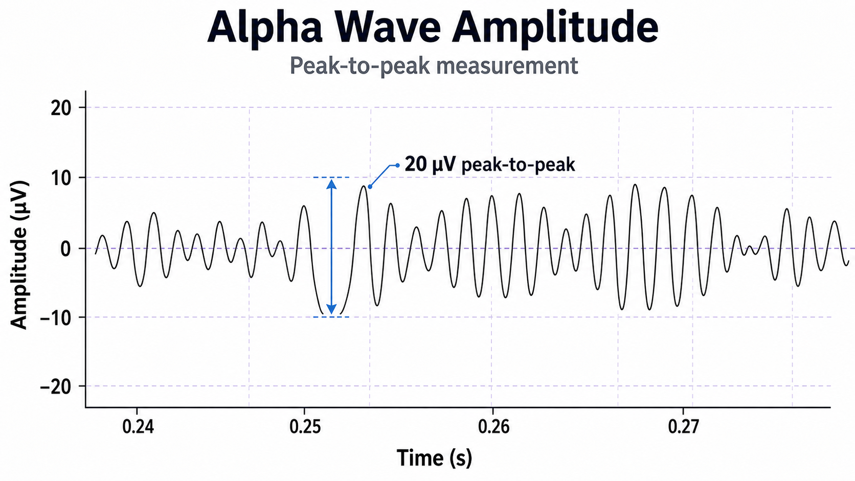
Greater synchrony in firing among neurons results in higher amplitudes detected at the scalp, as in the graphic below.
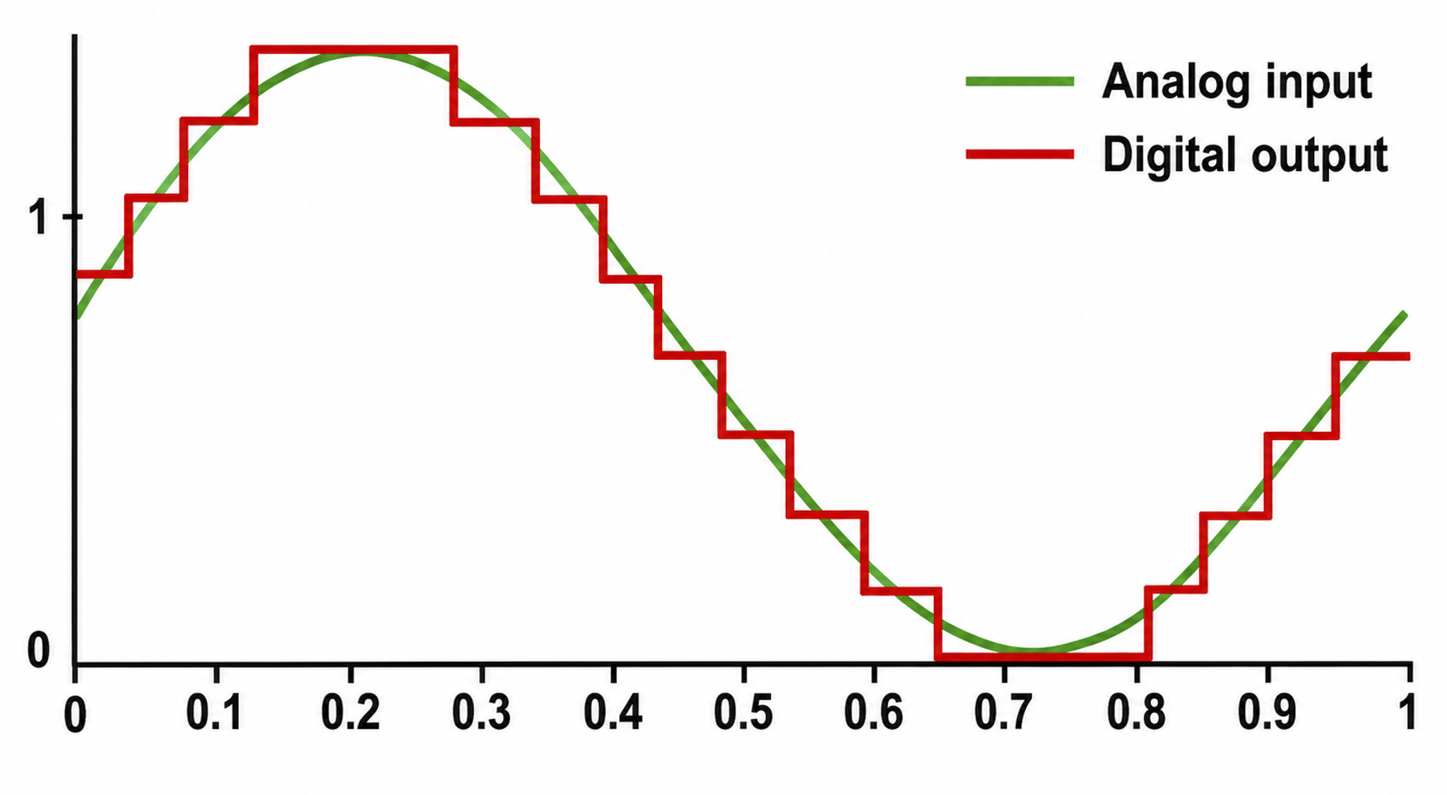
The EEG plots voltage changes over time, which can be displayed on a graph. The sampling rate is the number of measurements per second (Hz). Precision is the number of voltage gradations or steps.
According to the Nyquist theorem, the sampling rate must be at least twice the highest frequency of interest to represent that activity accurately. Sampling too slowly causes aliasing, in which fast activity masquerades as a slower, false rhythm. Recording 30-Hz beta therefore requires sampling well above 60 Hz, and modern systems sample at 256 Hz or higher (Collura, 2014).
The analog-to-digital (A/D) converters that transform voltages into numerical values vary in precision: more bits correspond to greater accuracy. Graphic adapted from © Fouad A. Saad/Shutterstock.com.
EEG Frequencies
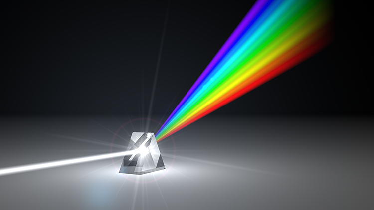
The raw EEG contains all EEG frequencies, just as white light contains all light frequencies. Digital filters separate the EEG frequencies just as a prism separates individual colors. Graphic © kmls/ Shutterstock.com.
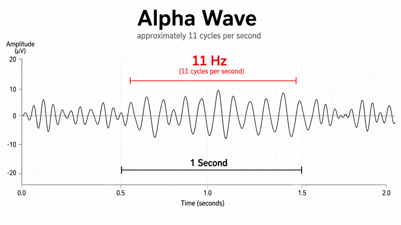
EEG frequency is measured in cycles per second or Hz. Count the number of peaks or count the number of zero (0.0) crossings divided by 2.
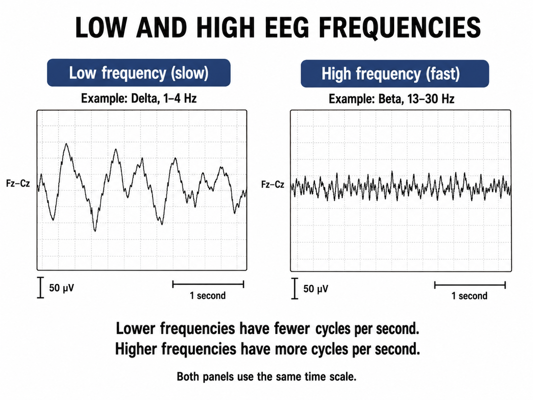
The slower the waves, the lower the EEG frequency.
The movie is a 19-channel BioTrace+ /NeXus-32 display of EEG activity from 1-64 Hz activity broken into its component delta, theta, alpha, and beta frequency bands by digital filters © John S. Anderson.
The movie is a 19-channel BioTrace+ /NeXus-32 display of alpha activity © John S. Anderson. Brighter colors represent higher alpha amplitudes. Frequency histograms are displayed for each channel. Notice the runs of high-amplitude alpha waves.
EEG Oscillations
The generation of oscillatory activity, sometimes called spindle behavior, is likely due to the interaction between thalamocortical relay neurons (TCR), reticular nucleus neurons (RE), and interneurons. These interactions are mediated by diverse neurotransmitters, including acetylcholine and GABA.
Circuits Contributing to the EEG
Feedforward, thalamocortical, and intra-cortical networks help generate the EEG.
Spindling or Bursting Activity
Spindling is a synaptically-generated oscillation in a circuit that necessarily includes reticular nucleus neurons (RE).
The movie below is a BioTrace+ /NeXus-32 display of EEG spindling activity © John S. Anderson.
The various spindle frequencies, which have often been interpreted as reflecting different types of oscillations, merely depend on various durations of the hyperpolarizations (negative shifts) in thalamic-cortical relay neurons. Long duration hyperpolarizations, as during ... deeply EEG-synchronized states, are associated with 7 Hz or even lower-frequency spindles, while relatively short hyperpolarizations result in ... higher frequencies (14 Hz) (Steriade, 2005).
The Purpose of Oscillatory Activity
A single neuron can influence multiple postsynaptic targets located between 0.5 and 5 mm away with conduction periods of between 1 and 10 ms. This time difference becomes progressively more pronounced when more complex events involve progressively larger assemblies of neurons. It may take hundreds of thousands of neurons, stimulating multiple postsynaptic targets, for the desired outcome to occur. When this many neurons are involved, it becomes increasingly clear that there is a need for organization and structure to manage this diverse activity.
Timing is everything since action potentials arrive from a large number of sources. The nervous system must correctly register arrival times to recognize a face, recall a name, or remember personal history and context. Oscillatory activity provides the temporal framework that coordinates this precisely timed communication.
Hierarchical Processing
Complex events require that the systems involved operate within a spatial and temporal hierarchy. Each oscillatory cycle is a window of time within which processing can occur, with a beginning and an end within which encoded or transferred messages must complete their tasks. Groups of neurons, close or distant, interact most effectively when firing windows are synchronous. The brain does not operate continuously but in discontinuous packets.
Multiple Oscillators
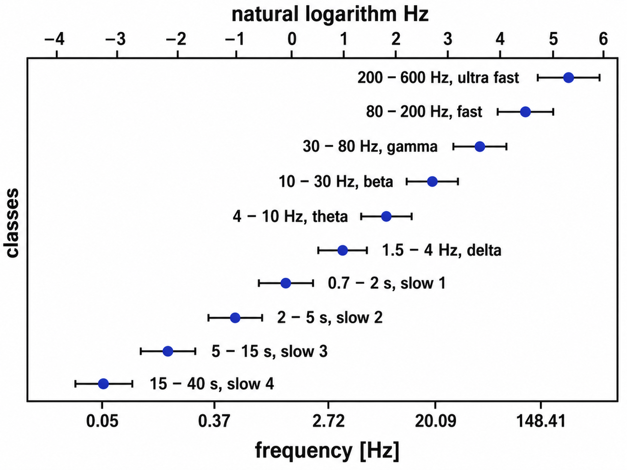
"Oscillatory classes in the cerebral cortex show a linear progression of the frequency classes on the log scale. In each class, the frequency ranges ('bandwidth') overlap with those of the neighboring classes, so that frequency coverage is more than four orders of magnitude" (Buzsáki, 2006). Graphic adapted from Buzsáki (2006).
Frequency Determines Complexity
The wavelength or frequency of the EEG band determines how long the processing window will remain open and, therefore, the size of the neuronal pool involved. Because of the distances involved, longer wavelengths (slower frequencies) allow larger groups of more distant neurons to be assembled and coordinated. Different frequencies organize different types of connections and different levels of computational complexity. This principle has direct clinical relevance: training slower frequencies influences broader, more distributed brain networks, while training faster frequencies targets more localized processing.
Local Versus Global Decision-Making
Short time windows of fast oscillators facilitate local integration, primarily because of the limitations of axon conduction delays. Fast oscillations favor local decisions. Slow oscillators can involve many neurons in large and/or distant brain areas. Slow oscillations favor complex, global decisions.
Complexity Versus Frequency
The organizing variable in this literature is the cortical distance over which activity must be integrated rather than task complexity as such. Long-range fronto-parietal integration during working-memory retention appears as 4-7 Hz synchronization (Sarnthein et al., 1998). Integration between neighboring temporal and parietal cortex during multimodal object processing, whether the object arrives as a spoken word, a written word, or a picture, appears in the 13-18 Hz beta1 range (von Stein et al., 1999). Integration local to a single area appears in the gamma range (von Stein & Sarnthein, 2000).
Traveling Waves Help Coordinate Widespread Brain Networks
Zhang et al. (2018) recorded oscillations between 2 and 15 Hz whose phase gradients showed them moving across the cortex at 0.25-0.75 meters per second, and suggested that such traveling waves help coordinate brain networks at a large scale and support connectivity.
Summary of EEG Oscillations
When the CNS processes incoming content, separate areas detect features of salient content, including visual, auditory, tactile, kinesthetic, and olfactory information. The CNS shares, integrates, and compares current with previous content, analyzes it, and makes decisions regarding memory and responses. Interacting networks linked by electrical and chemical signals perform this work, and we record the electrical potentials generated by this complex and dynamic network activity as the EEG.
The movie below of bursting alpha shows the sequential synchronization/desynchronization of groups of neurons. Higher voltage bursts are followed by voltage decreasing toward zero. These voltage fluctuations reflect rhythmic changes in the local field potential. This BioTrace+ /NeXus-32 video © John S. Anderson.
Key Takeaways
The scalp EEG is generated mainly by large pyramidal neurons in cortical columns whose apical dendrites, aligned perpendicular to the surface, sum their postsynaptic potentials into local field potentials. The EEG preferentially detects radial dipoles on gyral crowns and can miss tangential sources buried in sulci, and volume conduction further blurs each source across the scalp. Amplitude reflects how many neurons fire synchronously, while frequency reflects how fast they oscillate, with slower rhythms coordinating larger and more distant networks. Generators such as the thalamus pace much of this rhythmic activity, producing familiar patterns like the posterior dominant alpha rhythm. These principles explain both the power and the spatial limitations of the signal clinicians train.
Check Your Understanding
- Why are large pyramidal neurons in cortical columns the primary generators of the scalp EEG?
- How does the orientation of a dipole, radial versus tangential, affect whether the EEG can detect it?
- What does EEG amplitude reveal about the underlying neural population, and what does frequency reveal?
- According to the Nyquist theorem, why must the sampling rate be at least twice the highest frequency of interest?
- How does volume conduction limit the spatial resolution of the scalp EEG?
Definition of ERPs and SCPs
This section defines event-related potentials (ERPs) and slow cortical potentials (SCPs), two categories of brain electrical activity with important clinical applications. ERPs reveal how the brain processes specific stimuli, while SCPs reflect broader shifts in cortical excitability that neurofeedback clinicians can train.
Sensory evoked potentials are a subset of event-related potentials (ERPs)
Event-related potentials (ERPs) represent the brain's responses to external stimuli, events, or cognitive/motor tasks. ERPs can be detected throughout the cortex, and investigators monitor them by placing electrodes at midline sites (Fz, Cz, and Pz), sometimes using dense arrays of 64 to 256 electrodes. A computer analyzes a subject's EEG responses to the same stimulus or task over many trials to subtract random EEG activity. For a fixed stimulus, task, and electrode site, the averaged ERP is highly replicable, with negative and positive peaks at characteristic latencies following the stimulus. Morphology and scalp topography differ across modalities and tasks, so there is no single ERP waveform.
Sensory evoked potentials are a subset of ERPs elicited by external sensory stimuli (auditory, olfactory, somatosensory, and visual). In the auditory modality they show a negative peak near 80-100 ms and a positive peak near 170 ms following stimulus onset; the pattern-reversal visual evoked potential instead has its dominant positive peak near 100 ms. The orienting response ("What is it?") is a sensory ERP. The N1-P2 complex in the auditory cortex reveals whether an uncommunicative person can hear a stimulus, a clinically valuable application for patients who cannot report their own perceptual experiences. N refers to a negative potential and P to a positive potential.
Motor ERPs are detected over the primary motor cortex (precentral gyrus) during movement, and their amplitude is proportional to the force and rate of skeletal muscle contraction (Thompson & Thompson, 2015).
Slow cortical potentials modulate the excitability of associated neurons
Slow cortical potentials (SCPs) are gradual changes in the membrane potentials of cortical dendrites that last from 300 ms to several seconds. SCPs are characterized by low-frequency oscillations typically below 1 Hz, distinct from other brain rhythms such as delta (1-4 Hz) and spindling (7-14 Hz). SCPs have been observed at approximately 0.3 Hz, and their depolarizing-hyperpolarizing components have been extensively analyzed.

These potentials include the contingent negative variation (CNV), the readiness potential (Bereitschaftspotential), and movement-related potentials (MRPs). Slow cortical potentials are best understood as the low-frequency subclass of event-related potentials rather than as a category separate from them. The faster P300 and N400 components described below are event-related potentials but are not slow cortical potentials (Andreassi, 2007).
SCPs modulate the firing rate of cortical pyramidal neurons by exciting or inhibiting their apical dendrites and group the classical EEG rhythms using these synchronizing mechanisms (Steriade, 2005). For neurofeedback clinicians, SCPs represent a powerful training target because they regulate the very neural excitability that underlies faster EEG rhythms.
The movie is a 19-channel BioTrace+ /NeXus-32 display of SCPs © John S. Anderson. Brighter colors represent higher SCP amplitudes. As noted earlier, which direction a negative shift travels on screen depends on the software settings; the convention in electroencephalography is to plot negative up. Negative SCPs are produced by the depolarization of apical dendrites and increase the probability of neuron firing. Positive SCPs are produced by the hyperpolarization of these dendrites and decrease the likelihood of neuron firing.
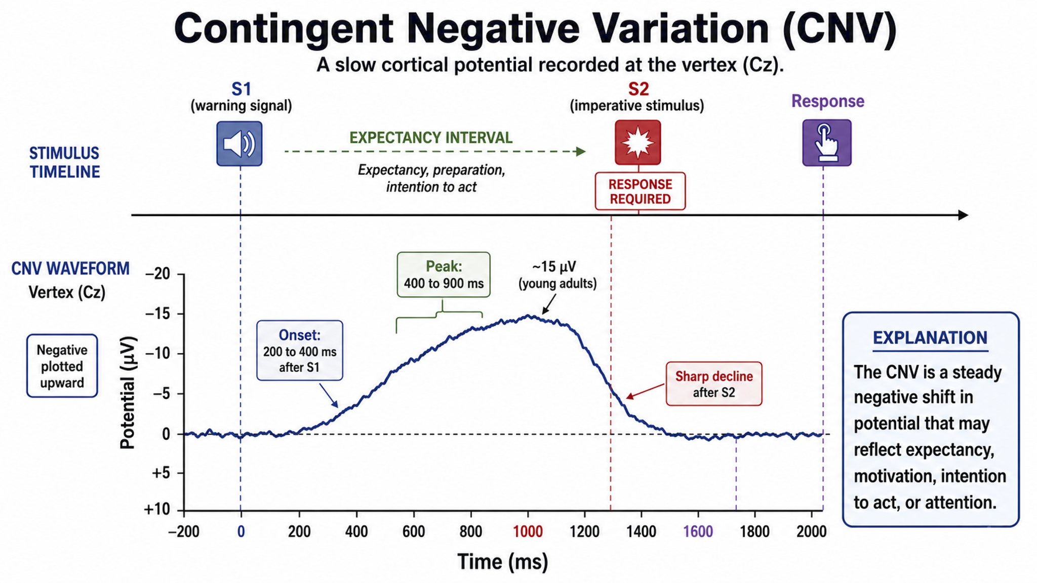
The contingent negative variation (CNV) is a steady, negative shift in potential (15 µV in young adults) detected at the vertex. This slow cortical potential may reflect expectancy, motivation, intention to act, or attention. The CNV appears 200-400 ms after a warning signal (S1), peaks within 400-900 ms in the short warning-interval paradigm shown below, and sharply declines after a second stimulus that requires a response (S2). With longer S1-S2 intervals the CNV becomes biphasic and its late component peaks just before S2.
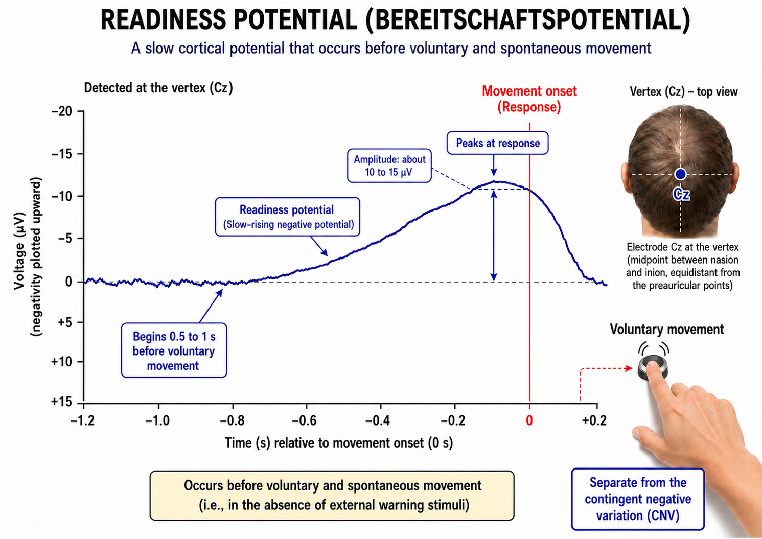
The readiness potential is a slow-rising, negative potential (10-15 µV) detected at the vertex before voluntary and spontaneous movement. This SCP precedes voluntary movement by 0.5 to 1 second in the late phase illustrated below and peaks at about the time the subject responds. Recorded over a longer epoch, an earlier component begins roughly 1.5 to 2 seconds before movement onset (Shibasaki & Hallett, 2006). It is separate from the CNV.
Movement-related potentials (MRPs) occur at 1 second as subjects prepare for unilateral voluntary movements. MRPs are distributed bilaterally with maximum amplitude at Cz. The supplementary motor area and primary motor and somatosensory cortices generate these potentials (Babiloni et al., 2002).
P300 and N400 ERPs are classified as long-latency potentials due to their extended latencies following stimulus onset.
The P300 potential is an ERP whose peak typically falls between 250 and 500 ms after stimulus onset, lengthening with task difficulty, age, and cognitive impairment. The largest amplitude positive peaks are located over the parietal lobe. Researchers elicit the P300 by exposing subjects to an odd-ball stimulus, a meaningful stimulus that differs from others in a series (a colored playing card presented in a series of monochrome cards). The P300 may reflect an event's subjective probability, meaning, and information transmission. Research shows this is separate from the CNV (Stern, Ray, & Quigley, 2001).
Shorter P300 latencies may reflect better allocation of attention, and researchers have measured longer P300 latencies in ADD than non-ADD samples. Experimental subjects show longer latencies when lying than when telling the truth (Farwell & Donchin, 1991; Thompson & Thompson, 2015).
The N400 potential is an ERP elicited when we encounter semantic violations like ending a sentence with an incongruent word ("The handsome prince married the beautiful fish"), or when the second word of a pair is unrelated to the first (BATTLE/GIRL). Warren and McIlvane (1998) speculate that the N400 is evoked whenever a conceptual system encounters a mismatch that violates equivalence relations. Halgren and colleagues (2002) consider it an index of the difficulty of semantic processing.
A Deep Dive Into SCPs
This section traces the history of SCP research from Richard Caton's 1875 observations through modern neurofeedback applications. It examines SCP generators, the paradox of scalp negativity during neural activation, and the clinical significance of SCPs across multiple disorders.
In 1875, Richard Caton identified what may have been the first evidence of SCPs in an article in the British Medical Journal titled "The Electric Currents of the Brain."
He reported that "feeble currents of varying direction pass through the multiplier when the electrodes are placed on two points of the external surface," and that "when any part of the grey matter is in a state of functional activity, its electric current usually exhibits negative variation." Working with a mirror galvanometer, Caton recorded deflections rather than calibrated voltages, so his one-page report contains no voltage figures. Some later researchers suggested that this signaled the discovery of the "steady potential" or the DC potential of the brain, though others have noted the possibility of equipment-based artifacts in his recordings (Niedermeyer, 1999).
From the late 1800s through the early 1900s, research into brain electrical activity turned toward observations of electrical stimulation and spontaneous electrical activity in animal studies. As technology improved, the ability of researchers to identify EEG rhythms also improved. Hans Berger is famous for his description of alpha-blocking with cognitive activity, made possible partly because of his use of more sensitive equipment (Niedermeyer, 1999).
Slow Cortical Potential Generators
Several neural mechanisms and structures within the brain generate SCPs. The generation of SCPs is primarily cortical, as evidenced by their persistence even after extensive thalamic destruction and corpus callosum transection (Steriade, Nuñez, & Amzica, 1993).
SCPs have been identified in cortical neurons, the thalamus, and glial cells. Cortical neurons in layers II to VI generate slow oscillations when the thalamus is removed or when cortical tissue is studied in vitro (in an artificial environment) or in vivo (within a living organism). Thalamic reticular neurons exhibit similar slow spontaneous oscillations in vitro, and synchronized intracortical oscillations may depend on a corticothalamic network.
The source and nature of SCPs remain in dispute. The prevailing theory holds that negative SCPs result from synchronous postsynaptic potentials in the apical dendrites of cortical pyramidal cells. Others hold that SCPs are produced by glial cells within the cortex. It appears that pyramidal neurons may be the source while the glial system is the "sink" (Strehl, 2005, personal communication).
Increased neuronal activity is associated with increased outflow of potassium ions leading to increased extracellular potassium concentrations. Glial cells depolarize when extracellular potassium concentrations increase, resulting in current flows similar to typical neuronal synaptic transmissions (Speckmann & Elger, 1999). Since glial cells are widely interconnected and have extensive processes, the glial system likely contributes to the potential changes producing SCP values recorded from the scalp.
Despite ongoing debate about SCP sources, scalp SCPs clearly represent cortical excitability. SCP negativity is associated with increased cortical excitability; high cortical negativity correlates with greater likelihood of seizures (Speckmann & Elger, 1984) and migraines (Siniatchkin et al., 2000) in susceptible individuals.
SCP positivity is associated with increased cortical inhibition. Higher-than-expected positive SCPs have been noted in children with elevated blood lead levels (Otto & Reiter, 1984). Children diagnosed with ADHD show deficient SCP self-regulation skills compared with controls (Heinrich et al., 2004). SCPs have been used to monitor anesthesia depth during surgical procedures (Sebel et al., 1997) because they are excellent indicators of arousal level.
Cortical Neurons
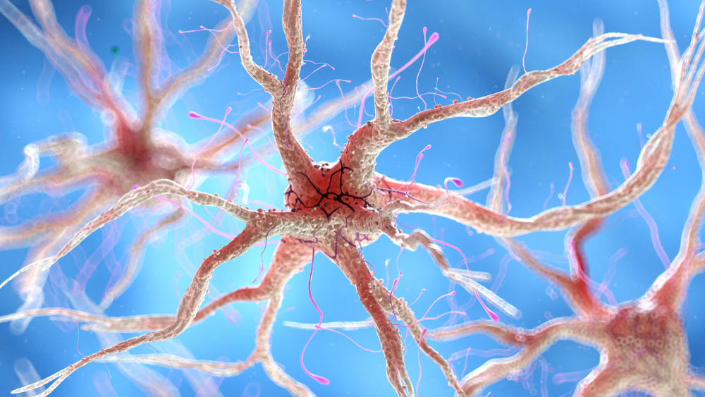
SCPs are primarily generated by the synchronized activity of large populations of cortical neurons. The slow shifts in membrane potential reflect changes in the overall excitability of cortical networks (Birbaumer et al., 1990). Neuron graphic © SciePro/Shutterstock.com.
Thalamocortical Interactions
Interactions between the thalamus and cortex also play a significant role in generating SCPs. Through its relay and integrative functions, the thalamus modulates cortical excitability and contributes to the slow potential changes observed (Lopes da Silva, 1991). Thalamocortical graphic © Netter.
Glial Cells
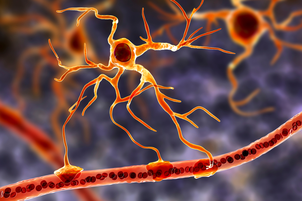
Emerging evidence suggests that glial cells, particularly astrocytes, influence SCPs by modulating the extracellular environment and supporting neuronal function (Amzica & Steriade, 2002).
Glia contribute to SCPs chiefly through the potassium mechanism described above: activity-driven rises in extracellular potassium depolarize astrocytes, and because glia are extensively interconnected, that depolarization spreads as a slow field change (Speckmann & Elger, 1999). The slow oscillations of glial cells may in turn influence the timing of neuronal firing through their control of potassium ion outflow (Steriade, 2005). Astrocyte graphic © Kateryna Kon/Shutterstock.com.
"The concept of a unified corticothalamic network that generates diverse types of brain rhythms grouped by the cortical slow oscillation is supported by EEG studies in humans" (Mölle et al., 2002).
The Meaning of SCP EEG Activity
SCPs indicate shifts in cortical excitability and are associated with various functional brain states. Surface-negative SCPs reflect synchronized depolarization of neuronal assemblies, indicating increased cortical activity. Surface-positive SCPs correspond to decreased cortical excitation, often involving inhibitory processes (Hinterberger et al., 2003).
The negative SCPs detected at the scalp during neuronal depolarization may seem counterintuitive at first.
When neurons are activated, their cell bodies become more positive internally due to the influx of positive ions. This leaves the immediate extracellular space more negative, and that negative charge is conducted through brain tissue, cerebrospinal fluid, skull, and scalp. EEG electrodes on the scalp detect this conducted negative potential, resulting in a negative deflection on the EEG trace.
This phenomenon is often referred to as paradoxical negativity in EEG literature (Birbaumer et al., 1990). What we record on the scalp is not a direct measure of neuronal membrane potential, but rather the result of complex electrical field propagation through various tissues (Elbert et al., 1980). Paradoxical positivity occurs when neurons are hyperpolarized.
This relationship is crucial for understanding the neurophysiological mechanisms underlying SCPs (Birbaumer et al., 1990). This paradoxical negativity graphic was adapted from Brienza and Mecarelli, 2019. Their original illustration is available under the license CC BY 3.0.
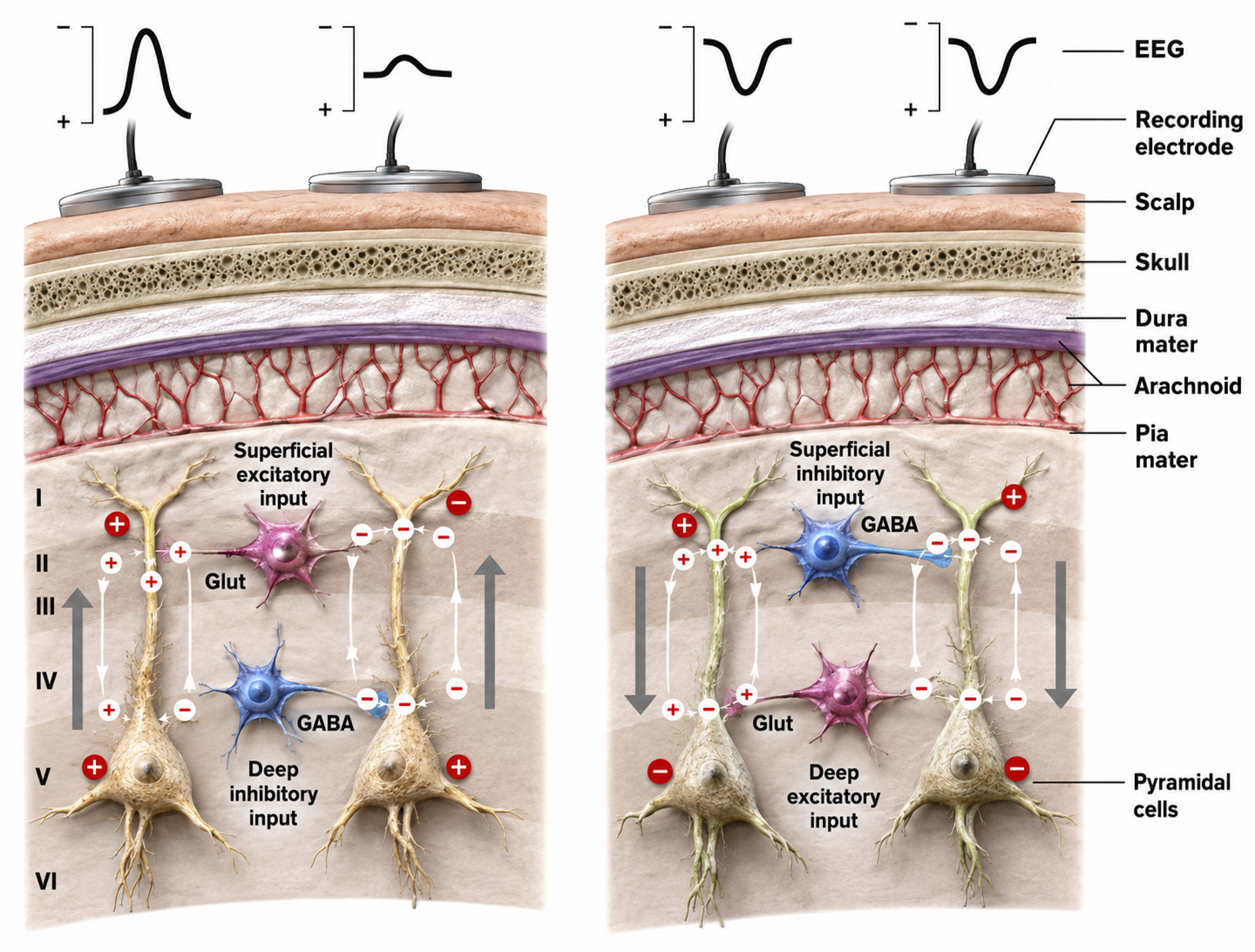
Schematic drawing of the scalp EEG registering negative (A) and positive (B) deflections elicited from summated EPSPs and IPSPs derived from pooled pyramidal cells. Cells releasing glutamate and GABA provide excitatory and inhibitory superficial and deep synaptic connections, resulting in an electrophysiological sink or source. EEG = electroencephalography; EPSPs = excitatory postsynaptic potentials; GABA = gamma-aminobutyric acid; IPSPs = inhibitory postsynaptic potentials. Figure courtesy of Anteneh Feyissa M.D. and Mayo Clinic.
Caton (1875) observed that cortical grey matter shows a negative variation in its electric current whenever it is functionally active. Underlying "tone" or valence factors determine the firing characteristics of neurons within a network. When SCPs are more positive, cortical neurons fire less due to hyperpolarization. When SCPs are more negative, firing increases due to depolarization.
SCPs participate in cognitive processes such as attention, preparation, and intention. Negative SCP shifts are linked to increased cortical excitability and readiness to respond, while positive shifts are associated with decreased excitability and relaxation (Birbaumer et al., 1990).
Some types of SCPs are event-related, including the Bereitschaftspotential (BP or readiness potential), contingent negative variation (CNV), and stimulus-preceding negativity (SPN). These represent slow negative waves related to anticipating a stimulus or preparing for a movement (Brunia et al., 2012). The BP occurs before executing a self-paced movement, CNV occurs when a preparatory stimulus foretells an imminent response demand, and SPN occurs after a movement when waiting for accuracy feedback.
The slow rhythm of SCPs is often combined with delta oscillations and these rhythms are phase-locked, suggesting close interaction between different frequency bands (Steriade, Nuñez, & Amzica, 1993).
SCPs play a crucial role in motor preparation and execution. The readiness potential (Bereitschaftspotential) precedes voluntary movements and reflects the planning and initiation of motor actions, particularly relevant for clinicians working with athletes and military personnel, where motor preparation timing can be a performance target.
SCPs are also associated with emotional and motivational states. Negative SCPs can indicate increased arousal and emotional engagement, whereas positive SCPs can reflect relaxation and disengagement (Hinterberger et al., 2003).
Psychological and Medical Disorders
SCPs have been extensively studied in various psychological and medical conditions. The following conditions illustrate the breadth of SCP-related assessment and intervention.
Attention-Deficit/Hyperactivity Disorder (ADHD)
Individuals with ADHD often exhibit abnormal SCP patterns, with a reduced ability to generate negative SCP shifts. Neurofeedback training targeting SCPs has shown promise in improving attention and reducing hyperactivity (Heinrich et al., 2004). This is one of the most well-studied applications of SCP neurofeedback.
Epilepsy
SCP neurofeedback has been explored as a treatment for epilepsy. Training individuals to increase positive SCP shifts can reduce cortical excitability and decrease seizure frequency (Rockstroh et al., 1993).
Parkinson's Disease
Studies have shown that patients with Parkinson's disease (PD) exhibit abnormal SCP patterns, particularly during motor tasks (Brittain & Brown, 2014). These abnormalities include altered amplitude and timing of SCPs, associated with impaired initiation and execution of voluntary movements.
During NREM sleep, cortico-basal slow wave delta activity increases while beta activity decreases. Deep brain stimulation (DBS) further modulates this altered activity, enhancing cortical delta and reducing alpha and low beta power. These findings suggest that SCPs and their interaction with other brain rhythms are significantly altered in PD, contributing to sleep dysfunction and spontaneous awakenings (Anjum et al., 2023).
SCPs are used to monitor the effects of therapeutic interventions such as DBS on cortical function in PD patients.
SCP neurofeedback has shown potential as a complementary treatment for PD, aiming to train self-regulation of brain activity associated with motor control.
Research suggests that SCP neurofeedback can improve motor function in PD patients (Kober & Wood, 2014). Some studies report better control over tremors and rigidity, along with improvements in non-motor symptoms including mood and cognitive function.
While promising, the current evidence is based on limited studies with small sample sizes. More extensive clinical trials are needed to establish long-term efficacy and generalizability.
Depression
SCP abnormalities are observed in depression, with patients often showing reduced amplitude of SCP shifts. Neurofeedback interventions aiming to normalize SCP patterns have shown potential in alleviating depressive symptoms (Strehl et al., 2017).
Sleep
SCPs play a role in sleep regulation and quality, making them relevant to clinicians who work with sleep-related complaints.
Sleep Onset and Maintenance
SCPs are involved in the transition from wakefulness to sleep. Positive SCP shifts are associated with sleep initiation and maintaining sleep stability (Sterman, 1996).
Sleep Disorders
SCPs are closely linked to sleep rhythms, particularly during NREM sleep (Anjum et al., 2023). The slow oscillations of SCPs facilitate the synchronization of neuronal activity essential for restorative sleep functions. In PD, the suppression of slow waves and the increase in subcortical beta activity before spontaneous awakenings highlight the critical role of SCPs in maintaining sleep quality.
Abnormal SCP patterns have been linked to insomnia. Neurofeedback training targeting SCPs can improve sleep onset latency and enhance overall sleep quality (Hoedlmoser et al., 2008).
Performance
Enhancing SCP activity through neurofeedback training has improved performance in various cognitive and motor tasks. This makes SCP training relevant not only for clinical populations but also for optimal performance programs serving athletes and military personnel.
Cognitive Performance
SCP training can enhance attention, memory, and executive function, likely due to improved cortical excitability regulation and better cognitive state management (Vernon et al., 2003).
Motor Performance
SCP neurofeedback improves motor performance, particularly in tasks requiring precise timing and coordination, attributed to the role of SCPs in motor preparation and execution (Gruzelier et al., 2014).
SCP Research
Research into human brain electrical characteristics became primarily focused on phasic phenomena from AC-coupled recordings, a trend continuing today with neurofeedback focusing primarily on training AC frequencies in the 1 to 60 Hz range.
The study of SCPs continued in physiology and animal research. Only recently has interest increased in observing SCP values in the human EEG and correlating them with cognitive activity, sensory processing, and motor activity. SCPs are distinguished from short-latency ERPs up to 500 ms and reflect cortical processes requiring more than one second to complete. Such changes occur in task-specific cortical areas and can be displayed using topographic maps, with areas of activation showing surface negative potential changes (Altenmuller & Gerloff, 1999).
Operant conditioning of SCP changes is an even more recent study area. One reason for increased interest is the excellent work by Birbaumer and colleagues (1999) at the University of Tübingen, demonstrating that SCPs can be operantly conditioned with positive outcomes for a variety of disorders. The recent availability of DC-coupled amplifiers has also contributed to this interest (Altenmuller & Gerloff, 1999).
According to Niedermeyer (1999), "DC" can mean several things. DC means direct current, a current without oscillations. From an electrophysiological perspective, "DC shifts" are ultra-slow potentials below the typical EEG oscillation frequency, generally around 0.1-0.2 cycles per second but potentially extending up to 1 cycle per second. So SCPs are not true direct current, though their oscillations are so slow that they are "DC-like" phenomena.
DC also refers to "direct coupling" (Niedermeyer, 1999) and describes an amplifier type that does not use capacitors between amplification stages and uses an infinite time constant for optimal DC recording. Until recently, this was difficult to achieve for EEG recording. Most conventional EEG amplifiers use capacitors in the input stage, which reject DC voltages and create a finite time constant that interferes with access to DC phenomena.
An approximation of DC information can be obtained from an AC amplifier by using a rectifier or extending the time constant to approximately 10 seconds (Kotchoubey et al., 1999). A thorough discussion of amplifier characteristics is beyond the scope of this article. Several excellent chapters on this subject can be found in Niedermeyer and Lopes da Silva (1999).
Recent studies have used SCPs to evaluate various task-oriented responses. Birbaumer and colleagues trained SCPs to reduce seizures (Daum et al., 1993; Kotchoubey et al., 1997, 1999, 2001, 2002), and other groups applied SCP feedback training to improve ADHD (Heinrich et al., 2004; Strehl, 2004, personal communication) and schizophrenia (Schneider et al., 1992).
SCP feedback training targets general arousal characteristics using a single measure, compared to other EEG training approaches that reward increases and/or decreases in certain frequency combinations. SCP feedback may provide a less complex approach to training neuronal activity, potentially offering greater accessibility through clinician-supervised home training devices.
Most research to date has used the Cz electrode site. However, at least one investigation trained left hemisphere language sites, demonstrating improved word processing following negativity training and diminished performance following positivity training (Pulvermüller, 2000). Studying the effects of SCP training at other electrode sites would be a productive research direction.
Some efforts have identified SCP values using multiple electrodes in a quantitative EEG paradigm. Basile and colleagues (2004) used four 32-channel DC-coupled amplifiers to compare SCP responses in schizophrenic patients with normal controls. They found significant variations, with controls showing simply-organized positivity and negativity patterns, while schizophrenic patients showed much more complex, fragmented patterns of activation and inhibition.
There are only a few clinically available DC-coupled amplifiers capable of accurately monitoring SCP activity. An Internet search yielded several research-grade devices with high prices and a couple of devices priced for clinical practice. A new 32-channel DC-coupled device for quantitative EEG assessments has also recently been released.
One attraction of DC amplifiers is the capability to monitor and/or train both SCPs and typical EEG frequencies. DC amplifiers are optimized for SCP and can also record faster frequencies, particularly those with better analog-to-digital conversion characteristics (bit size, not sampling rate).
Higher A/D conversion values (more bits per sample) allow newer DC amplifiers to resolve microvolt-level slow cortical potentials while simultaneously accommodating electrode offset potentials of tens of millivolts without saturating. The offset is what occupies the millivolt range; the slow cortical potentials themselves remain microvolt signals.
The training of SCP shifts is a fairly new endeavor. Much remains to be learned about the effects of training both positivity and negativity conditions at various electrode sites for individuals with diverse presenting concerns and neurophysiological characteristics.
This author's recent clinical experiences suggest that training SCP using newer, more accurate amplifiers may result in more pronounced changes occurring more quickly. Thus, it will be important to develop protocols with more specificity and flexibility to meet the needs of diverse client populations while considering changes in equipment and software characteristics that may affect skill acquisition rates and outcomes.
Training SCP
This section reviews approaches to training slow cortical potential shifts, from early event-related paradigms to modern infra-low frequency methods.
Various approaches to training slow cortical shifts have been applied. Early research (Birbaumer et al., 1990; Birbaumer, 1999) showed a correlation between cortical negativity and reaction time, signal detection, and short-term memory. This was identified through evoked and event-related potential research, leading to training paradigms involving sequences of 8-second trials that trained both positive and negative cortical gradient shifts using real-time visual and/or auditory feedback.
When greater cortical positivity was the goal, more positive shift trials were provided, and vice versa. Additional transfer trials without feedback tested skill acquisition. This approach was the primary paradigm during the early years of SCP research (Strehl, 2009).
Other clinicians and researchers, including Susan and Siegfried Othmer and Mark Smith, addressed slow gradient shifts with training called Infra-Low Frequency Neurofeedback (Othmer, 2020) and Infraslow Neurofeedback (Smith, 2013).
Post-traumatic stress, anxiety, and other conditions involving excessive cortical activation have been addressed by training to increase overall cortical positivity, using a 4-channel approach that rewards gradual shifts in the cortical gradient through proportional audio feedback. This has resulted in several client self-reports of an altered state characterized by decreased cognitive activity while retaining awareness; the positive shift in cortical gradient appears to correspond with reduced conscious thought while preserving self-awareness (multiple clinical observations shared with John Anderson).
Conclusion
Slow cortical potentials are characterized by low-frequency oscillations typically below 1 Hz, with significant depolarizing-hyperpolarizing components. Occurring at approximately 0.3 Hz, these potentials are crucial indicators of cortical excitability and are associated with various cognitive and motor processes. Generated by cortical neurons, thalamocortical interactions, and glial cells, SCPs reflect shifts in cortical excitability linked to attention, motor preparation, and emotional states. Negative SCP shifts indicate increased excitability and readiness to respond, while positive shifts are associated with relaxation and decreased excitability.
SCPs play roles in psychological and medical disorders like ADHD, epilepsy, and depression, and are vital in sleep regulation and performance enhancement. SCP neurofeedback shows promise in improving symptoms and cognitive functions across these conditions, making it an increasingly important tool in the neurofeedback clinician's repertoire.
The author would like to thank Ute Strehl of the University of Tübingen in Germany, Dave Siever of Mind Alive Inc. in Canada, and Erwin Hartsuiker of Mind Media BV in the Netherlands for technical assistance in preparing this article.
Consider a child referred for inattention and impulsivity. Children with ADHD often show a reduced ability to generate the negative slow cortical potential shifts that support cortical excitability and readiness to respond (Heinrich et al., 2004). Using SCP neurofeedback, the clinician rewards the child for producing and controlling these gradient shifts across repeated trials, including transfer trials without feedback that test whether the skill generalizes. Because SCP training targets a single, global measure of arousal regulation rather than a specific frequency band, it can offer an accessible entry point for young clients. Gains in attention and self-regulation reported in controlled trials illustrate how a slow, subthreshold cortical signal can become a practical training target.
Event-related potentials are averaged, time-locked responses to specific stimuli or tasks, whereas slow cortical potentials are gradual shifts in dendritic membrane potential lasting from hundreds of milliseconds to seconds. Negative SCP shifts reflect increased cortical excitability and readiness to respond, while positive shifts reflect reduced excitability and relaxation. Although the scalp records negativity during neuronal activation, this apparent paradox follows from how the extracellular field is conducted to the surface. SCP abnormalities appear across ADHD, epilepsy, depression, Parkinson's disease, and sleep disorders, and SCPs can be operantly conditioned. This makes SCP self-regulation a flexible target for both clinical treatment and performance enhancement.
Check Your Understanding
- How do event-related potentials differ from slow cortical potentials in their timing and in how they are measured?
- What do negative and positive SCP shifts indicate about cortical excitability?
- Why does the scalp often register negativity when cortical neurons are activated?
- Name three clinical conditions in which SCP self-regulation has been studied, and describe the general training goal in each.
- Why is the ability to operantly condition SCPs important for neurofeedback?
Neuroplasticity (LTD and LTP)
Neuroplasticity is the brain's ability to remodel its structure and function in response to experience, and it is the foundational mechanism that makes all neurofeedback training possible. Without it, the operant conditioning of brainwave activity would have no lasting effect. This section describes the levels at which neuroplastic change occurs and the two synaptic processes most central to learning, long-term depression (LTD) and long-term potentiation (LTP).
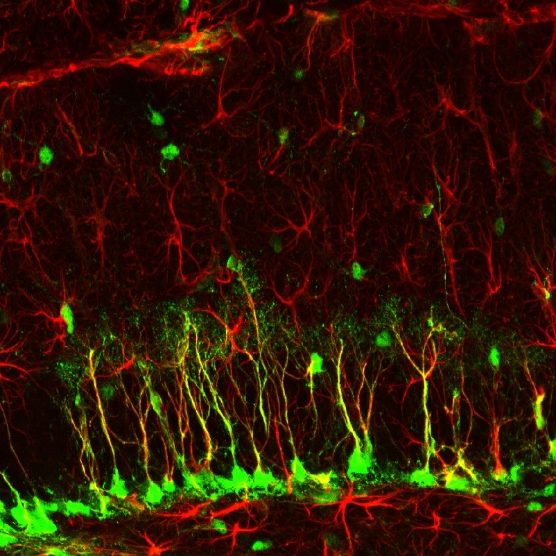
Neuroplasticity is responsible for learning and memory. Memory storage remodels neurons through changes in synaptic transmission, interneuron modulation, the formation of new synapses, and the rewiring of neural pathways (Bear et al., 2020). Because neurofeedback rewards moment-to-moment changes in central nervous system activity, it is itself a form of operant conditioning, and animal studies show that operant conditioning can induce astrogliogenesis (the creation of new astrocytes) and neurogenesis (the creation of new neurons) in structures such as the medial prefrontal cortex and hippocampus (Kerson et al., 2025; Rapanelli et al., 2011).
The graphic by Rebeca Cuesta is licensed under the Creative Commons Attribution-Share Alike 4.0 International license.
Levels of Neuroplastic Change
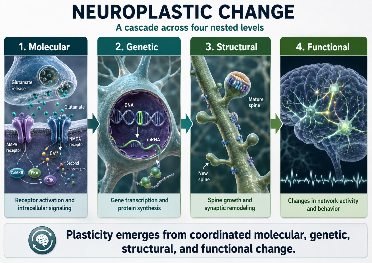
Neuroplastic change is not a single event but a cascade that unfolds across four nested levels: molecular, genetic, structural, and functional. The deeper levels make the more visible ones possible, so a durable training outcome reflects work at all four. Keeping these levels in mind helps clinicians explain why neurofeedback effects build gradually and why consistent practice matters.
At the molecular level, a rewarded shift in brain activity strengthens active synapses by trafficking receptors, activating calcium-dependent enzymes, and releasing growth factors such as brain-derived neurotrophic factor (BDNF). This strengthening is balanced locally, so that when some synapses are reinforced, neighboring synapses weaken to prevent runaway excitation, a process that depends on the regulatory protein Arc (El-Boustani et al., 2018). These molecular events are the first physical trace of what a client learns during a session.
At the genetic level, neural activity switches on immediate early genes within minutes, and their protein products convert short-lived synaptic changes into stable ones (Bear et al., 2020). This activity-dependent gene expression is why a single session produces only transient effects, while repeated sessions consolidate lasting change. It is the biological reason neurofeedback is delivered as a course of training rather than a one-time procedure.
At the structural level, neurons grow, prune, and reshape dendritic spines, add new connections through synaptogenesis, and extend dendritic branches (Breedlove & Watson, 2023). In some regions, the operant conditioning that drives training also supports neurogenesis and the formation of new glial cells (Rapanelli et al., 2011). These remodeled connections are the lasting hardware behind a stabilized training gain.
At the functional level, these changes alter how circuits behave, shifting synaptic efficacy, the balance of excitation and inhibition, and the connectivity between regions (Collura, 2014; Thompson & Thompson, 2015). Self-regulation of cortical rhythms can produce measurable, durable changes in cortical excitability; in one study, 30 minutes of alpha-suppression neurofeedback raised corticospinal excitability and reduced intracortical inhibition for at least 20 minutes afterward (Ros et al., 2010). Reviews of the field describe neurofeedback as a closed-loop method that reshapes the targeted networks through this kind of self-directed plasticity (Sitaram et al., 2017).
Whether training a veteran with PTSD to reduce excessive high-beta activity or an athlete to optimize alpha and theta activity, the underlying mechanism is neuroplastic change working from the molecular level upward. To learn more about neuroplasticity, view the Khan Academy video Neuroplasticity.
Long-Term Depression and Long-Term Potentiation
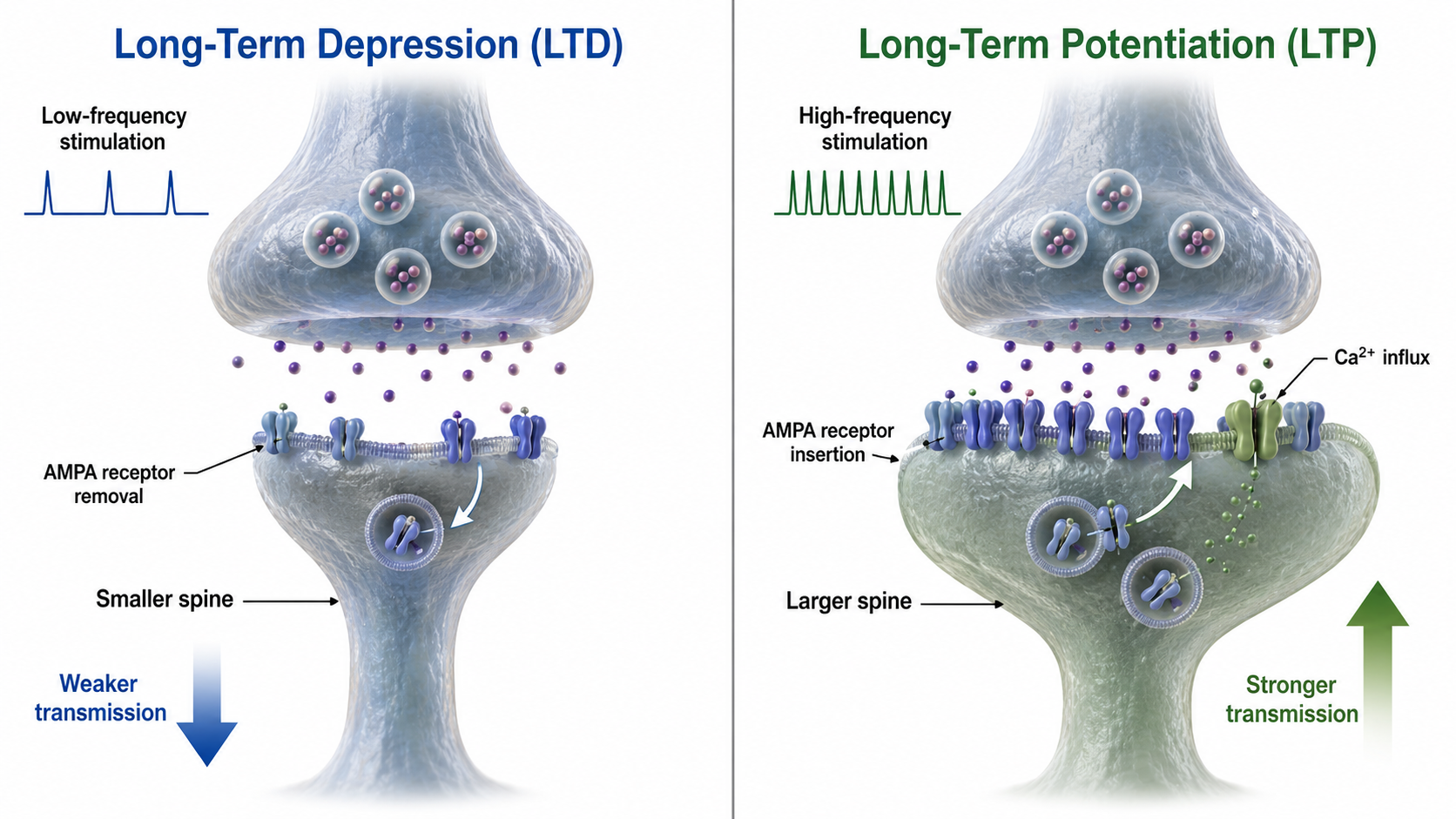
Two synaptic processes carry out functional plasticity: long-term depression and long-term potentiation. Together they provide the bidirectional control that learning requires, allowing the brain to both weaken and strengthen specific connections.
In long-term depression (LTD), synaptic transmission that coincides with slight depolarization of the postsynaptic neuron weakens the synapse, and low-frequency stimulation of afferent neurons reduces their response to future input. LTD is not simply forgetting. It is an active process that prunes less-used connections to keep neural networks efficient.
In long-term potentiation (LTP), synaptic transmission that coincides with strong depolarization strengthens the synapse, producing a stable increase in effectiveness that can persist for weeks or longer. LTP creates new synapses, enhances existing ones, and builds new dendritic branches and spines (Breedlove & Watson, 2023). Together, LTD and LTP are the cellular mechanisms through which neurofeedback training produces lasting changes in brain function.
To learn more, watch the Khan Academy video Long Term Potentiation and Synaptic Plasticity.
Integration: Why Neuroplasticity Matters for Neurofeedback
Neuroplasticity is the thread that connects every topic in this chapter, from the postsynaptic potentials that generate the EEG to the slow cortical shifts that clients learn to control. The four levels of neuroplastic change describe one continuous process in which a rewarded moment of brain activity leaves a molecular trace, recruits the gene expression that stabilizes it, reshapes synapses and dendrites, and finally alters how whole circuits behave. Long-term depression and long-term potentiation are the synaptic engines of that process, giving the brain the bidirectional control it needs to prune unhelpful connections and reinforce useful ones. Together these mechanisms explain how a stream of feedback tones and visual rewards becomes a lasting change in cortical function.
For the clinician, this account reframes neurofeedback as the deliberate engineering of neuroplastic change through operant conditioning (Kerson et al., 2025; Sitaram et al., 2017). Because the deeper molecular and genetic levels make the visible functional gains possible, durable outcomes depend on consistent practice across many sessions rather than a single dramatic result. The same principle explains why training effects build gradually, why consolidation continues between sessions, and why generalization to daily life reflects genuine structural remodeling rather than a temporary state (Ros et al., 2010). Whether the goal is reducing excessive high-beta activity in a veteran with PTSD or optimizing alpha and theta activity in a performing athlete, the clinician is guiding the brain through plasticity from the molecular level upward.
Neuroplasticity is the brain's capacity to remodel its structure and function with experience, and it is the mechanism that makes the effects of neurofeedback last. Change unfolds across four nested levels, molecular, genetic, structural, and functional, so that a rewarded moment of activity leaves a trace that gene expression stabilizes, synapses and dendrites embody, and circuits express as altered behavior. Long-term depression and long-term potentiation supply the bidirectional control that lets the brain weaken unhelpful connections and strengthen useful ones. Because neurofeedback is a form of operant conditioning that drives this cascade, durable outcomes depend on consistent practice that allows molecular and genetic changes to consolidate into stable structural and functional gains.
Check Your Understanding
- How do the four levels of neuroplastic change, molecular, genetic, structural, and functional, build on one another to produce a durable neurofeedback outcome?
- Why does activity-dependent gene expression help explain why neurofeedback is delivered as a course of training rather than a single session?
- How do long-term depression and long-term potentiation together provide the bidirectional control that learning requires?
- In what sense is neurofeedback a form of operant conditioning, and how does that connect to the neurogenesis and astrogliogenesis reported in animal studies?
- How would you explain to a client why neurofeedback effects build gradually and why consistent practice between and across sessions matters?
Cutting-Edge Topics in Neurophysiology
Silent Synapses in the Adult Brain
One of the most striking recent findings is the discovery of silent synapses on filopodia in the adult mouse brain (Vardalaki et al., 2022). Because these dormant connections can be recruited without disturbing existing synapses, they may reconcile the brain's competing needs for stability and flexibility. Establishing whether the adult human brain contains silent synapses could reshape how we understand learning-based interventions in mature clients.
Traveling Waves Coordinate Distant Networks
Zhang and colleagues (2018) reported that oscillations between roughly 2 and 15 Hz travel across the human neocortex as waves moving at about a quarter to three-quarters of a meter per second. Rather than treating rhythms as standing oscillations confined to one region, this work frames them as propagating signals that coordinate activity across distant areas. Traveling waves offer a fresh way to think about how large-scale connectivity is organized in time, with implications for how network-level training effects spread.
Neurofeedback as Closed-Loop, Self-Directed Plasticity
Contemporary reviews describe neurofeedback as a closed-loop method in which a person uses real-time feedback to reshape targeted brain networks through self-directed plasticity (Sitaram et al., 2017). Supporting this view, a single session of alpha-suppression training raised corticospinal excitability and reduced intracortical inhibition for at least twenty minutes afterward (Ros et al., 2010). Framing neurofeedback as deliberate, closed-loop engineering of neuroplastic change helps explain why consistent practice consolidates lasting gains.
Assignment
Now that you have completed this unit, how would you explain the relationship between local field potentials and the EEG? How does anatomy explain why the EEG is comprised of EPSPs and IPSPs instead of action potentials?
Glossary
amplitude: the magnitude of a signal's excursion from a specified reference level, reported using a stated convention such as peak, peak-to-peak, or RMS.
astrocytes: central nervous system glial cells that support metabolic homeostasis, ion and neurotransmitter regulation, blood-brain barrier function, and synaptic modulation.
cognitive performance: the ability to use mental processes to perform tasks, including memory, attention, and executive function.
contingent negative variation (CNV): a slow negative event-related potential developing between a warning stimulus and an anticipated imperative stimulus, associated with expectancy, attention, and motor preparation.
cortical negativity: a state in which the cortical surface carries a negative electrical potential, associated with depolarization and increased excitability.
cortical neurons: nerve cells in the cortex responsible for generating and transmitting electrical impulses.
cortical positivity: a state in which the cortical surface carries a positive electrical potential, associated with hyperpolarization and decreased excitability.
current sink: a region of inward positive transmembrane current that produces relative negativity in the surrounding extracellular field.
current source: a region of outward positive transmembrane current that produces relative positivity in the surrounding extracellular field.
deep brain stimulation (DBS): a neurosurgical procedure involving the implantation of electrodes in specific brain areas to modulate neuronal activity.
depolarization: a shift in membrane potential toward a less negative or more positive value.
dipole: an electrical source with separated regions of positive and negative charge; for example, the eye is electropositive at the front and electronegative at the back.
EEG power: the squared amplitude of EEG activity quantified over time or frequency, expressed as µV² for band power or µV²/Hz for power spectral density.
electrode polarization: charge accumulation and electrochemical potential at the electrode-electrolyte interface that can produce drift, offset, or reduced signal fidelity.
electroencephalograph: an instrument that acquires, amplifies, filters, digitizes, displays, or records electroencephalographic signals.
event-related potential (ERP): a voltage change in the averaged EEG that is time-locked to a sensory, cognitive, or motor event.
evoked potential: an event-related potential elicited by a controlled sensory stimulus and extracted from EEG by time-locked averaging across repeated trials.
excitability: the ability of neurons to respond to stimuli and generate action potentials.
excitatory postsynaptic potential (EPSP): a graded postsynaptic depolarization that increases the probability of an action potential, commonly through cation influx.
glial cell: a non-neuronal nervous-system cell that supports homeostasis, myelination, immune defense, synaptic function, and neural signaling.
hyperpolarization: a change in membrane potential that makes the cell interior more negative relative to the exterior.
inhibitory postsynaptic potential (IPSP): a graded postsynaptic potential that reduces the probability of action-potential generation, often by increasing chloride or potassium conductance.
local field potential (LFP): a low-frequency extracellular voltage reflecting summed transmembrane currents from a local neural population, especially synaptic and dendritic activity.
local synchrony: synchrony that occurs when the coordinated firing of cortical neurons produces high-amplitude EEG signals.
long-latency potential: an event-related potential component occurring relatively late after an event and commonly reflecting higher-order cognitive processing.
long-term depression (LTD): a persistent activity-dependent reduction in synaptic efficacy.
long-term potentiation (LTP): a persistent activity-dependent increase in synaptic efficacy.
motor control: the process by which humans and animals use their brains and muscles to perform movements.
motor ERP: an event-related potential recorded over primary motor cortex during movement, whose amplitude scales with the force and rate of skeletal muscle contraction.
movement-related cortical potential (MRCP): a slow cortical potential associated with preparation, initiation, and execution of voluntary movement, including the readiness potential.
N1-P2: a sensory event-related potential in the auditory cortex of the temporal cortex that reveals whether an uncommunicative person can hear a stimulus.
N400: a negative event-related potential component peaking near 400 ms and sensitive to semantic access, expectancy, and integration.
neuroplasticity: experience- or injury-related change in neural structure, function, connectivity, or representation.
Nyquist theorem: the principle that a signal must be sampled at more than twice its highest frequency component to be reconstructed without distortion.
oddball stimulus: an infrequent or task-relevant event embedded among frequent standard events, commonly used to elicit a P300 response.
orienting response: a coordinated attentional and physiological response to a novel, unexpected, or significant stimulus that facilitates information intake.
P300: a positive event-related potential component, commonly maximal centroparietally, whose latency and amplitude vary with attention, task relevance, probability, and context.
paradoxical negativity: the surface-negative EEG shift recorded when neurons depolarize, produced by volume conduction of the negative extracellular potential from the cortex to the scalp.
paradoxical positivity: the surface-positive EEG shift recorded when neurons hyperpolarize, produced by volume conduction of the positive extracellular potential from the cortex to the scalp.
Parkinson's disease (PD): a progressive neurodegenerative disorder characterized primarily by motor symptoms such as tremor, rigidity, bradykinesia (slowness of movement), and postural instability.
precision: the closeness of repeated measurements or estimates to one another, reflected statistically by sampling variability and confidence-interval width.
radial dipole: a current dipole oriented perpendicular to the scalp, as in the crown of a gyrus. Because its field projects directly toward the electrodes, the scalp EEG detects it well.
readiness potential: a slow negative cortical potential beginning up to several seconds before self-initiated voluntary movement and maximal over central regions.
sampling rate: the number of discrete signal samples acquired per unit time, usually expressed in samples per second or hertz.
scalp EEG: the noninvasive recording of the brain's electrical activity from electrodes placed on the scalp, reflecting the summed synchronous activity of large neuronal populations.
sensory event-related potentials (ERPs): event-related potentials evoked by external auditory, olfactory, somatosensory, or visual stimuli, with exogenous peaks at characteristic latencies such as an auditory negativity near 100 ms and a positivity near 170 ms.
slow cortical potential (SCP): a very slow EEG voltage shift lasting hundreds of milliseconds to seconds and reflecting changes in cortical excitability and preparation.
source: the place at the end of the neuron opposite the sink where current leaves, making the surrounding extracellular area electrically positive.
spindle behavior: the waxing-and-waning, fusiform amplitude envelope of a transient oscillatory burst in which amplitude grows to a peak and then tapers, rather than holding a constant level.
stimulus-preceding negativity (SPN): a slow negative potential shift observed before a stimulus that carries important information, such as feedback about the accuracy of a movement, reflecting anticipatory attention and affective processing.
surface-negative: a negative slow cortical potential shift, associated with synchronized depolarization, increased cortical excitability, and readiness to respond.
surface-positive: a positive slow cortical potential shift, associated with hyperpolarization, decreased cortical excitability, and relaxation.
synchronization: the coordinated, in-phase activity of neuronal populations within or across brain regions, which increases EEG amplitude at the shared frequency.
tangential dipole: a current dipole oriented parallel to the scalp, as in the wall of a sulcus. Much of its field runs sideways beneath the electrodes, and opposing sulcal walls can cancel, so the scalp EEG detects it poorly.
thalamocortical interactions: the reciprocal connections and communications between the thalamus and the cerebral cortex.
traveling waves: EEG oscillations that move across the cortex that may mediate large-scale coordination of brain networks and support connectivity.
voltage: electric potential difference between two points, expressed in volts.
volume conduction: passive spread of electrical fields through conductive biological tissues, allowing a source to influence electrodes located at a distance.
Test Yourself on ClassMarker
Click the button below to take a 10-question exam over this entire unit. There is no password.
Review Flash Cards on Quizlet
Click the button below to review our chapter flash cards.
References
Altenmuller, E. O., & Gerloff, C. (1999). Psychophysiology and the EEG. In E. Niedermeyer & F. Lopes da Silva (Eds.), Electroencephalography: Basic principles, clinical applications, and related fields (4th ed.). Williams and Wilkins.
Amzica, F., & Steriade, M. (2002). The functional significance of K-complexes. Sleep Medicine Reviews, 6(2), 139-149. https://doi.org/10.1053/smrv.2001.0181
Andreassi, J. L. (2007). Psychophysiology: Human behavior and physiological response (5th ed.). Lawrence Erlbaum and Associates, Inc.
Anjum, M., Smyth, C., Dijk, D., Starr, P., Denison, T., & Little, S. (2023). Multi-night cortico-basal recordings reveal mechanisms of NREM slow wave suppression and spontaneous awakenings at high-temporal resolution in Parkinson's disease. Research Square. https://doi.org/10.21203/rs.3.rs-3484527/v1
Babiloni, C., Babiloni, F., Carducci, F., Cincotti, F., Del Percio, C., Hallett, M., Moretti, D. V., Romani, G. L., & Rossini, P. M. (2002). High resolution EEG of sensorimotor brain functions: Mapping ERPs or mu ERD? In R. C. Reisin, M. R. Nuwer, M. Hallett, & C. Medina (Eds.), Advances in clinical neurophysiology (Supplements to Clinical Neurophysiology Vol. 54). Elsevier Science B. V.
Basile, L. F. H., Yacubian, J., Ferreira, B. L. C., Valim, A. C., & Gattaz, W. F. (2004). Topographic abnormality of slow cortical potentials in schizophrenia. Brazilian Journal of Medical and Biological Research, 37(1), 97-109. https://doi.org/10.1590/s0100-879x2004000100014
Bear, M. F., Connors, B. W., & Paradiso, M. A. (2020). Neuroscience: Exploring the brain (4th ed.). Jones & Bartlett Learning.
Bear, M. F., Connors, B. W., & Paradiso, M. A. (2026). Neuroscience: Exploring the brain (5th ed.). Jones & Bartlett Learning.
Birbaumer, N. (1999). Slow cortical potentials: Plasticity, operant control, and behavioral effects. The Neuroscientist, 5, 74-78. https://doi.org/10.1177/1073858499005002
Birbaumer, N., Elbert, T., Canavan, A. G., & Rockstroh, B. (1990). Slow potentials of the cerebral cortex and behavior. Physiological Reviews, 70(1), 1-41. https://doi.org/10.1152/physrev.1990.70.1.1
Breedlove, S. M., & Watson, N. V. (2023). Behavioral neuroscience (10th ed.). Sinauer Associates, Inc.
Brienza, M., & Mecarelli, O. (2019). Neurophysiological basis of EEG. In O. Mecarelli (Ed.), Clinical electroencephalography (pp. 25-45). Springer. https://doi.org/10.1007/978-3-030-04573-9_2
Brittain, J.-S., & Brown, P. (2014). Oscillations and the basal ganglia: Motor control and beyond. NeuroImage, 85(2), 637-647. https://doi.org/10.1016/j.neuroimage.2013.05.084
Brunia, C. H. M., van Boxtel, G. J. M., & Böcker, K. B. E. (2012). Negative slow waves as indices of anticipation: The Bereitschaftspotential, the contingent negative variation, and the stimulus-preceding negativity. In E. S. Kappenman & S. J. Luck (Eds.), The Oxford handbook of event-related potential components (pp. 190-208). Oxford Academic. https://doi.org/10.1093/oxfordhb/9780195374148.013.0108
Buzsáki, G. (2006). Rhythms of the brain. Oxford University Press.
Caton, R. (1875). The electric currents of the brain. British Medical Journal, 2, 278.
Collura, T. F. (2014). Technical foundations of neurofeedback. Taylor & Francis.
Daum, I., Rockstroh, B., Birbaumer, N., Elbert, T., Canavan, A., & Lutzenberger, W. (1993). Behavioural treatment of slow cortical potentials in intractable epilepsy: Neuropsychological predictors of outcome. Journal of Neurology, Neurosurgery, and Psychiatry, 56(1), 94-97. https://doi.org/10.1136/jnnp.56.1.94
Elbert, T., Rockstroh, B., Lutzenberger, W., & Birbaumer, N. (1980). Biofeedback of slow cortical potentials. I. Electroencephalography and Clinical Neurophysiology, 48(3), 293-301. https://doi.org/10.1016/0013-4694(80)90265-5
El-Boustani, S., Ip, J., Breton-Provencher, V., Knott, G., Okuno, H., Bito, H., & Sur, M. (2018). Locally coordinated synaptic plasticity of visual cortex neurons in vivo. Science, 360(6395), 1349-1354. https://doi.org/10.1126/science.aao0862
Farwell, L. A., & Donchin, E. (1991). The truth will out: Interrogative polygraphy ("lie detection") with event-related brain potentials. Psychophysiology, 28, 531-547. https://doi.org/10.1111/j.1469-8986.1991.tb01990.x
Fisch, B. J. (1999). Fisch and Spehlmann's EEG primer: Basic principles of digital and analog EEG (3rd ed.). Elsevier.
Gruzelier, J. H. (2014). EEG-neurofeedback for optimising performance. I: A review of cognitive and affective outcome in healthy participants. Neuroscience and Biobehavioral Reviews, 44, 124-141. https://doi.org/10.1016/j.neubiorev.2013.09.015
Halgren, E., Dhond, R. P., Christensen, N., Van Petten, C., Marinkovic, K., Lewine, J. D., & Dale, A. M. (2002). N400-like magnetoencephalography responses modulated by semantic context, word frequency, and lexical class in sentences. NeuroImage, 17(3), 1101-1116. https://doi.org/10.1006/nimg.2002.1268
Heinrich, H., Gevensleben, H., Freisleder, F. J., Moll, G. H., & Rothenberger, A. (2004). Training of slow cortical potentials in attention-deficit/hyperactivity disorder: Evidence for positive behavioral and neurophysiologic effects. Biological Psychiatry, 55(7), 772-775. https://doi.org/10.1016/j.biopsych.2003.11.013
Hinterberger, T., Veit, R., Wilhelm, B., Weiskopf, N., Vatine, J. J., & Birbaumer, N. (2005). Neuronal mechanisms underlying control of a brain-computer interface. The European Journal of Neuroscience, 21(11), 3169-3181. https://doi.org/10.1111/j.1460-9568.2005.04092.x
Hoedlmoser, K., Pecherstorfer, T., Gruber, G., Anderer, P., Doppelmayr, M., Klimesch, W., & Schabus, M. (2008). Instrumental conditioning of human sensorimotor rhythm (12-15 Hz) and its impact on sleep as well as declarative learning. Sleep, 31(10), 1401-1408. https://doi.org/10.5665/sleep/31.10.1401
Jackson, A., & Stoney, S. D. (2006). Thalamocortical and corticothalamic organization. In Advances in clinical neurophysiology. Elsevier.
Kober, S. E., & Wood, G. (2014). Changes in hemodynamic signals accompanying motor imagery and motor execution of swallowing: A near-infrared spectroscopy study. NeuroImage, 93(Pt 1), 1-10. https://doi.org/10.1016/j.neuroimage.2014.02.011
Kotchoubey, B., Blankenhorn, V., Fröscher, W., Strehl, U., & Birbaumer, N. (1997). Stability of cortical self-regulation in epilepsy patients. NeuroReport, 8(8), 1867-1870. https://doi.org/10.1097/00001756-199705260-00015
Kotchoubey, B., Busch, S., Strehl, U., & Birbaumer, N. (1999). Changes in EEG power spectra during biofeedback of slow cortical potentials in epilepsy. Applied Psychophysiology and Biofeedback, 24(4), 213-233. https://doi.org/10.1023/a:1022226412991
Kotchoubey, B., Kubler, A., Strehl, U., Flor, H., & Birbaumer, N. (2002). Can humans perceive their brain states? Consciousness and Cognition, 11(1), 98-113. https://doi.org/10.1006/ccog.2001.0535
Kotchoubey, B., Schneider, D., Schleichert, H., Strehl, U., Uhlmann, C., Blankenhorn, V., Fröscher, W., & Birbaumer, N. (1996). Self-regulation of slow cortical potentials in epilepsy: A retrial with analysis of influencing factors. Epilepsy Research, 25(3), 269-276. https://doi.org/10.1016/s0920-1211(96)00082-4
Kotchoubey, B., Strehl, U., Holzapfel, S., Blankenhorn, V., Fröscher, W., & Birbaumer, N. (1999). Negative potential shifts and the prediction of the outcome of neurofeedback therapy in epilepsy. Clinical Neurophysiology, 110(4), 683-686. https://doi.org/10.1016/s1388-2457(99)00005-x
Kotchoubey, B., Strehl, U., Uhlmann, C., Holzapfel, S., Konig, M., Fröscher, W., Blankenhorn, V., & Birbaumer, N. (2001). Modification of slow cortical potentials in patients with refractory epilepsy: A controlled outcome study. Epilepsia, 42(3), 406-416. https://doi.org/10.1046/j.1528-1157.2001.22200.x
Lopes da Silva, F. (1991). Neural mechanisms underlying brain waves: From neural membranes to networks. Electroencephalography and Clinical Neurophysiology, 79(2), 81-93. https://doi.org/10.1016/0013-4694(91)90044-5
Mölle, M., Marshall, L., Gais, S., & Born, J. (2002). Grouping of spindle activity during slow oscillations in human non-rapid eye movement sleep. The Journal of Neuroscience, 22(24), 10941-10947. https://doi.org/10.1523/JNEUROSCI.22-24-10941.2002
Niedermeyer, E. (1999). Historical aspects. In E. Niedermeyer & F. Lopes da Silva (Eds.), Electroencephalography: Basic principles, clinical applications, and related fields (4th ed.). Williams and Wilkins.
Niedermeyer, E., & Lopes da Silva, F. (Eds.). (1999). Electroencephalography: Basic principles, clinical applications, and related fields (4th ed.). Williams and Wilkins.
Othmer, S., & Othmer, S. (2020). Toward a theory of infra-low frequency neurofeedback. In H. W. Kirk (Ed.), Restoring the brain. Routledge. eBook ISBN 9780429275760
Otto, D., & Reiter, L. (1984). Developmental changes in slow cortical potentials of young children with elevated body lead burden: Neurophysiological considerations. Annals of the New York Academy of Sciences, 425(1), 377-383. https://doi.org/10.1111/j.1749-6632.1984.tb23559.x
Pulvermüller, F., Mohr, B., Schleichert, H., & Veit, R. (2000). Operant conditioning of left-hemispheric slow cortical potentials and its effect on word processing. Biological Psychology, 53(2-3), 177-215. https://doi.org/10.1016/S0301-0511(00)00046-6
Rapanelli, M., Frick, L. R., & Zanutto, B. S. (2011). Learning an operant conditioning task differentially induces gliogenesis in the medial prefrontal cortex and neurogenesis in the hippocampus. PLoS ONE, 6(2), e14713. https://doi.org/10.1371/journal.pone.0014713
Rockstroh, B., Elbert, T., Birbaumer, N., Wolf, P., Düchting-Röth, A., Reker, M., Daum, I., Lutzenberger, W., & Dichgans, J. (1993). Cortical self-regulation in patients with epilepsies. Epilepsy Research, 14(1), 63-72. https://doi.org/10.1016/0920-1211(93)90075-i
Sarnthein, J., Petsche, H., Rappelsberger, P., Shaw, G. L., & von Stein, A. (1998). Synchronization between prefrontal and posterior association cortex during human working memory. Proceedings of the National Academy of Sciences, 95(12), 7092-7096. https://doi.org/10.1073/pnas.95.12.7092
Schneider, F., Rockstroh, B., Heimann, H., Lutzenberger, W., Mattes, R., Elbert, T., Birbaumer, N., & Bartels, M. (1992). Self-regulation of slow cortical potentials in psychiatric patients: Schizophrenia. Biofeedback and Self-Regulation, 17(4), 277-292. https://doi.org/10.1007/bf01000051
Sebel, P. S., Lang, E., Rampil, I. J., White, P. F., Cork, R., Jopling, M., Smith, N. T., Glass, P. S., & Manberg, P. (1997). A multicenter study of bispectral electroencephalogram analysis monitoring anesthetic effect. Anesthesia and Analgesia, 84(4), 891-899. https://doi.org/10.1097/00000539-199704000-00035
Shibasaki, H., & Hallett, M. (2006). What is the Bereitschaftspotential? Clinical Neurophysiology, 117(11), 2341-2356. https://doi.org/10.1016/j.clinph.2006.04.025
Siniatchkin, M., Hierundar, A., Kropp, P., Kuhnert, R., Gerber, W.-D., & Stephani, U. (2000). Self-regulation of slow cortical potentials in children with migraine: An exploratory study. Applied Psychophysiology and Biofeedback, 25(1), 13-32. https://doi.org/10.1023/a:1009581321624
Smith, M. L. (2013). Infra-slow fluctuation training: On the down-low in neuromodulation. NeuroConnections.
Speckmann, E.-J., & Elger, C. E. (1984). The neurophysiological basis of epileptic activity: A condensed overview. In R. Degen & E. Niedermeyer (Eds.), Epilepsy, sleep, and sleep deprivation (pp. 23-34). Elsevier. PMID: 1760082
Speckmann, E.-J., & Elger, C. E. (1999). Introduction to the neurophysiological basis of the EEG and DC potentials. In E. Niedermeyer & F. Lopes da Silva (Eds.), Electroencephalography: Basic principles, clinical applications, and related fields (4th ed.). Williams and Wilkins.
Steriade, M. (2005). Cellular substrates of brain rhythms. In E. Niedermeyer & F. Lopes da Silva (Eds.), Electroencephalography: Basic principles, clinical applications, and related fields (5th ed.). Lippincott Williams & Wilkins.
Steriade, M., Nuñez, A., & Amzica, F. (1993). Intracellular analysis of relations between the slow (< 1 Hz) neocortical oscillation and other sleep rhythms of the electroencephalogram. The Journal of Neuroscience, 13(8), 3266-3283. https://doi.org/10.1523/JNEUROSCI.13-08-03266.1993
Sterman, M. B. (1996). Physiological origins and functional correlates of EEG rhythmic activities: Implications for self-regulation. Biofeedback and Self-Regulation, 21(1), 3-33. https://doi.org/10.1007/BF02214147
Stern, R. M., Ray, W. J., & Quigley, K. S. (2001). Psychophysiological recording (2nd ed.). Oxford University Press.
Strehl, U. (2009). Slow cortical potentials neurofeedback. Journal of Neurotherapy, 13(2), 117-126. https://doi.org/10.1080/10874200902885936
Strehl, U., Aggensteiner, P., Wachtlin, D., Brandeis, D., Albrecht, B., Arana, M., Bach, C., Banaschewski, T., Bogen, T., Flaig-Röhr, A., Freitag, C. M., Fuchsenberger, Y., Gest, S., Gevensleben, H., Herde, L., Hohmann, S., Legenbauer, T., Marx, A. M., Millenet, S., Pniewski, B., … Holtmann, M. (2017). Neurofeedback of slow cortical potentials in children with attention-deficit/hyperactivity disorder: A multicenter randomized trial controlling for unspecific effects. Frontiers in Human Neuroscience, 11, 135. https://doi.org/10.3389/fnhum.2017.00135
Thompson, M., & Thompson, L. (2015). The neurofeedback book: An introduction to basic concepts in applied psychophysiology (2nd ed.). Association for Applied Psychophysiology and Biofeedback.
Vernon, D., Egner, T., Cooper, N., Compton, T., Neilands, C., Sheri, A., & Gruzelier, J. (2003). The effect of training distinct neurofeedback protocols on aspects of cognitive performance. International Journal of Psychophysiology, 47(1), 75-85. https://doi.org/10.1016/s0167-8760(02)00091-0
von Stein, A., Rappelsberger, P., Sarnthein, J., & Petsche, H. (1999). Synchronization between temporal and parietal cortex during multimodal object processing in man. Cerebral Cortex, 9(2), 137-150. https://doi.org/10.1093/cercor/9.2.137
von Stein, A., & Sarnthein, J. (2000). Different frequencies for different scales of cortical integration: From local gamma to long range alpha/theta synchronization. International Journal of Psychophysiology, 38(3), 301-313. https://doi.org/10.1016/s0167-8760(00)00172-0
Warren, A. M., & McIlvane, W. J. (1998). Stimulus equivalence and the N400 effect [Poster presentation]. Annual Meeting of the Cognitive Neuroscience Society, San Francisco, CA, United States.
Zhang, H., Watrous, A. J., Patel, A., & Jacobs, J. (2018). Theta and alpha oscillations are traveling waves in the human neocortex. Neuron, 98(6), 1269-1281. https://doi.org/10.1016/j.neuron.2018.05.019