
Ascending Sensory Pathways to the Cortex
What You Will Learn in This Chapter
Every qEEG you record is a picture of a brain that is busy listening, looking, and feeling. Before you can interpret that picture, you need to know how sensory information reaches the cortex in the first place, and why the simple act of opening the eyes can reshape an entire posterior recording.
This unit follows sensory signals from the retina, the cochlea, and the skin up through the thalamus to their cortical destinations. You will meet the specialized thalamic relay nuclei, the dorsal and ventral processing streams that each modality shares, and the massive descending projections that let the cortex edit its own inputs. You will also see why sensory input and arousal desynchronize the EEG, producing the alpha blocking you will observe in nearly every client.
IQCB Blueprint Coverage: This unit addresses II. Neuroscience: Basic Anatomy of Ascending Sensory Pathways to the Cortex.
Learning Objectives
After completing this section, you will be able to:
Describe the general organization of the classical routes for EEG activation, including the role of specialized thalamic relay nuclei and the single sensory modality that bypasses the thalamus.
Trace the visual pathway from the retinal ganglion cells through the optic chiasm and the lateral geniculate nucleus to the primary visual cortex.
Trace the auditory pathway from the inner hair cells through the superior olivary nuclei, the inferior colliculi, and the medial geniculate nucleus to the auditory cortex.
Trace the somatosensory touch pathway from the dorsal horn through decussation in the medulla and the ventral posterior nucleus to the primary somatosensory cortex.
Compare and contrast the dorsal and ventral processing streams in the visual and auditory systems.
Describe the somatic nervous system, including where somatic motoneuron and sensory cell bodies are located.
Explain how top-down corticothalamic control shapes perception, and why the roughly ten-to-one ratio of descending to ascending fibers undermines a purely feedforward model.
Predict how sensory input and arousal change the EEG, and identify alpha blocking when you see it in a recording.
Full-Length Lecture: Ascending Sensory Pathways
Basic Anatomy of Ascending Sensory Pathways to the Cortex
This section examines how sensory information travels from the body's receptors to the cortex, focusing on the visual, auditory, and somatosensory systems and on the somatic nervous system that feeds them. Understanding these pathways is essential because sensory input directly shapes the EEG patterns you will assess and train. Sensory input produced by activities like reading a novel or listening to music can desynchronize cortical activity, resulting in lower-amplitude, higher-frequency EEG waveforms (Neumann et al., 2003). Arousal and specific forms of cognitive activity may reduce alpha amplitude or eliminate it entirely, a phenomenon called alpha blocking, while increasing EEG power in the beta range (Andreassi, 2007).
The classical routes for EEG activation consist of ascending sensory pathways that distribute information to specialized thalamic nuclei, which then project the results of thalamic processing to the appropriate cortical regions. A hallmark of these pathways is their hierarchical structure, which preserves the spatial location from which signals arise, with specialized thalamic nuclei serving as critical relay points. The one exception is olfaction (smell), which bypasses the thalamus and projects directly to the primary olfactory cortex.
The old-school view held that these ascending pathways exercise purely bottom-up control of perception as feedforward circuits. This overlooks the fact that ten times more cortical efferent neurons target the sensory thalamus than thalamic afferent neurons project to the cortex. The new-school view recognizes extensive interconnections between the thalamus and cortex that permit a substantial degree of top-down cortical control over perception (Kandel et al., 2021). Subcortical areas, including the midbrain, thalamus, and cerebellum, also participate in ascending and descending transmission and processing of neural messages.
In summary, sensory pathways are not one-way streets but dynamic, bidirectional systems in which the cortex actively shapes the information it receives. We will now examine the visual, auditory, and somatosensory systems to illustrate these principles, and then look at the somatic nervous system that carries the signals in from the body.
Visual System
Retinal ganglion cells, whose axons form the optic nerves, ascend to the midline optic chiasm where the two optic nerves meet. At the chiasm, temporal axons (those toward the side of the head) continue on their own side's optic tract, while nasal axons cross over to join the opposite side's optic tract. Most optic tract axons project to the lateral geniculate nucleus (LGN) of the thalamus.
While the LGN relays visual information to the cortex, brainstem and cortical neurons actively modulate its activity. Brainstem neurons that mediate alertness and attention can adjust the LGN's response to visual input. The cortex can also exert top-down selection to increase attention to a salient region of the visual field at the expense of others (Bear et al., 2020). This is clinically relevant because it demonstrates that even at the earliest relay station, perception is shaped by attention and arousal, the very processes you aim to assess and train.
LGN neurons form the optic radiations and project to cortical layer IV of the primary visual cortex (V1) in the occipital lobe. A minority of retinal ganglion cell axons target the dorsal midbrain superior colliculus, which directs visual gaze and selective attention to visual objects (Breedlove & Watson, 2023). The cortex contains many specialized regions for processing visual properties like color, shape, location, motion, and orientation, organized into two major streams originating in V1.
The dorsal stream projects from V1 to the parietal lobe, helping us localize objects and guide movements. The adjacent motor association cortex contains neurons with both visual and motor properties, called mirror neurons, whose networks may play a role in learning actions by observing others, understanding intentions, and empathy (Buccino et al., 2006). The lower ventral stream projects to inferior temporal and frontal areas and allows us to identify objects and faces.
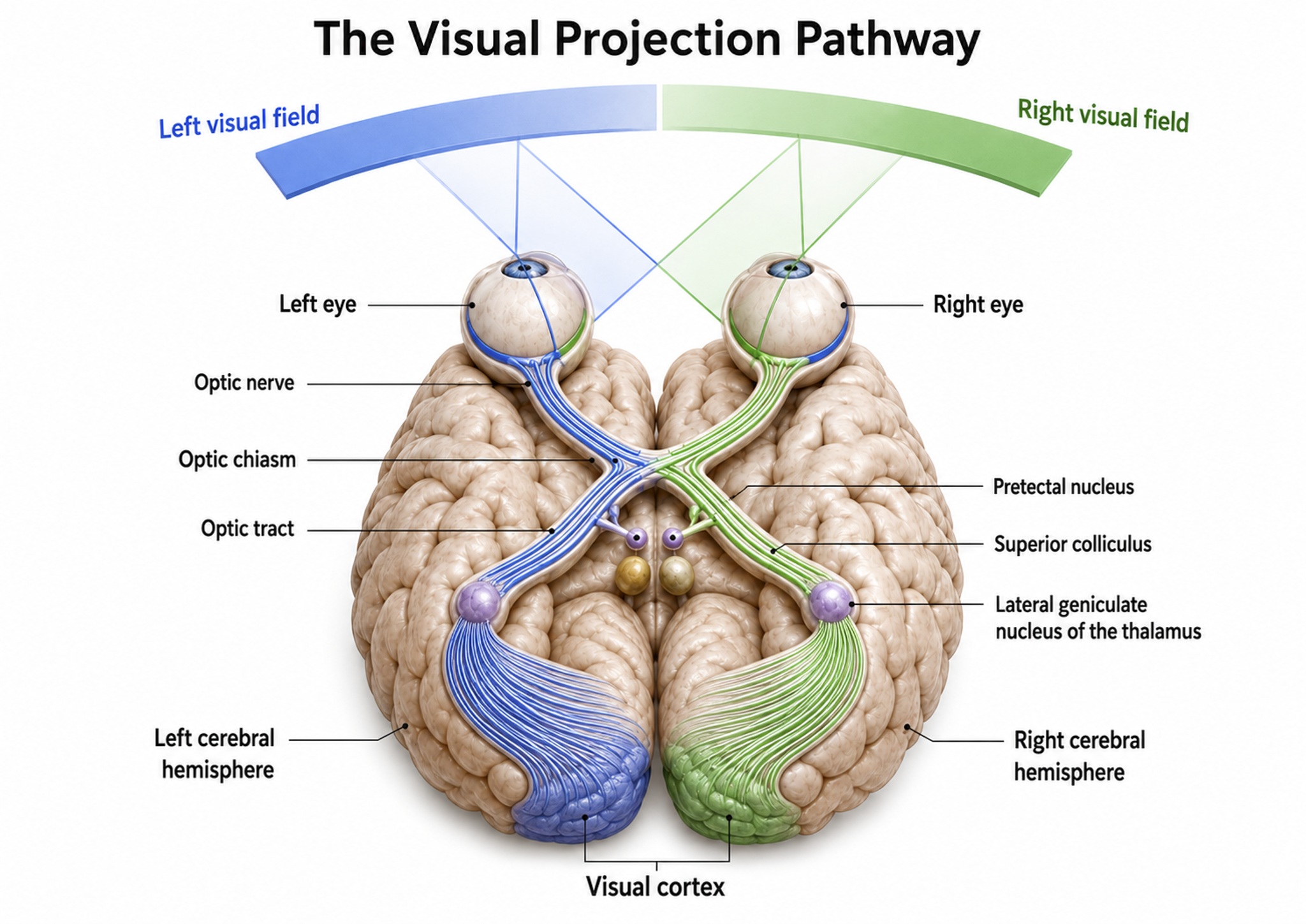
Visual pathway graphic adapted from © Alila Medical Media/Shutterstock.com.
Auditory System
The cochlea's inner hair cells within the organ of Corti send approximately 30,000 auditory fibers to several destinations: the superior olivary nuclei of the pons, the inferior colliculi of the midbrain, and the medial geniculate nucleus of the thalamus. The superior olivary nuclei process binaural (two-ear) information to localize sound, while all ascending auditory neurons ultimately innervate the inferior colliculi, some via intermediate relays.
The inferior colliculi integrate information about spatial localization and multiple sensory modalities, including somatosensory information. They project to the thalamus' medial geniculate nucleus (MGN), which in turn projects to several cortical auditory areas using two separate pathways. The MGN mainly relays frequency, amplitude, and binaural information to the auditory cortex in the temporal lobe.
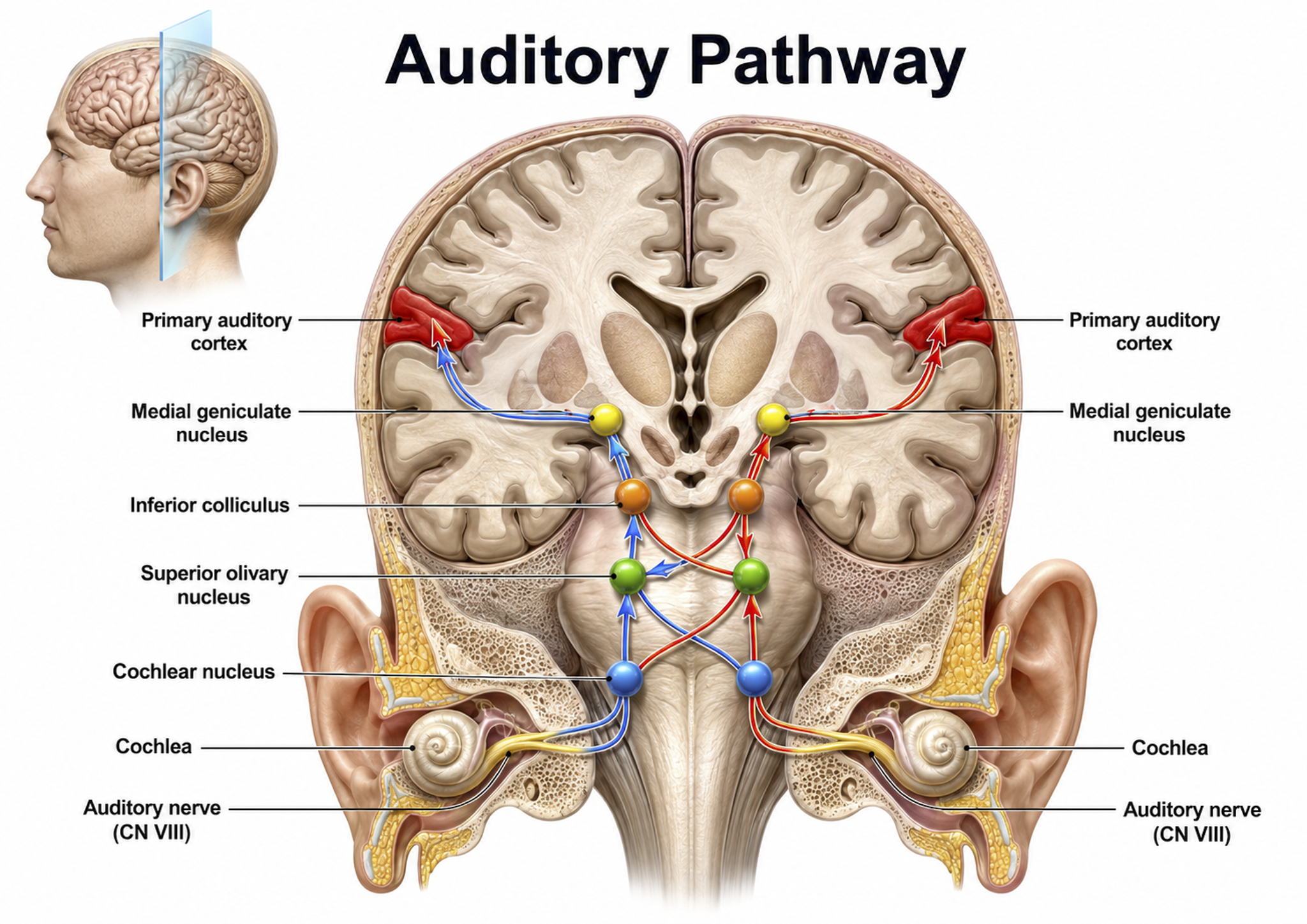
Auditory pathway graphic © medicalstocks/Shutterstock.com.
The auditory cortex processes auditory information within dorsal and ventral streams, paralleling the visual system's organization. The dorsal stream, extending to the parietal lobe, helps us spatially localize sounds. The lower ventral stream, projecting to the temporal lobe, analyzes sound components, possibly including speech sounds (Breedlove & Watson, 2023). As with the visual pathways, the auditory system involves extensive feedback: brainstem neurons innervate outer hair cells that adjust basilar membrane sensitivity to specific frequencies, and auditory cortex axons innervate both the inferior colliculi and MGN to exercise top-down control (Bear et al., 2020).
Somatosensory System
The somatosensory system employs specialized receptors to perceive itch, pain, temperature, and touch. For touch, the axon of a unipolar neuron enters the dorsal horn of the spinal cord and synapses with a dorsal column neuron in the medulla. Axons from this neuron decussate (cross the midline) and innervate the thalamus' ventral posterior nucleus (VPN), which in turn distributes this information to the primary somatosensory cortex (S1).
While each hemisphere's S1 maps touch information from the opposite side of the body, the secondary somatosensory cortex (S2) maps both sides. The maps are overlaid so that the left and right arms are represented in the same region of the body surface map (Breedlove & Watson, 2023). This bilateral representation in S2 may help explain why somatosensory processing can produce widely distributed EEG changes that clinicians observe during assessment.
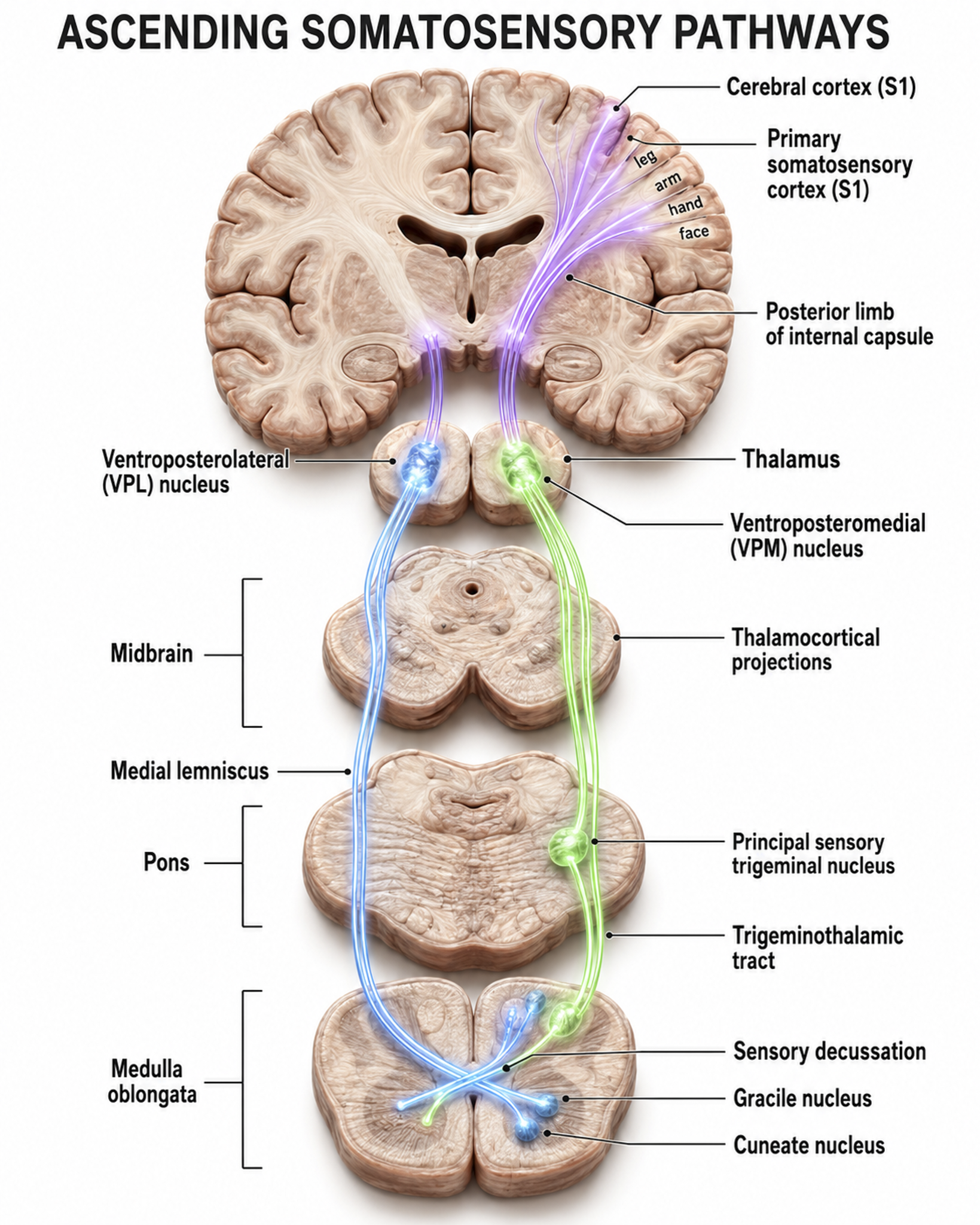
Ascending somatosensory pathway from the dorsal horn to the primary somatosensory cortex.
As with the visual and auditory systems, the ascending somatosensory pathways do far more than relay information. These networks process and alter sensory information at each successive synapse, and the cortex exercises top-down control over neurons in the dorsal column and VPN to dynamically adjust cortical inputs (Bear et al., 2020). The recurring theme across all three sensory systems is bidirectional communication: the cortex is not a passive receiver but an active participant that shapes the sensory data it processes.
Somatic Nervous System
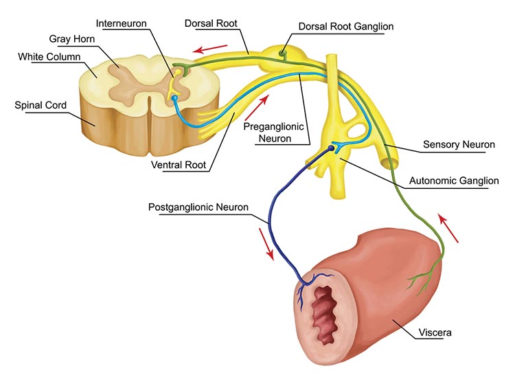
Before somatosensory signals can ascend to the thalamus, something has to carry them in from the body. That job belongs to the somatic nervous system, which comprises the spinal nerves that innervate somatosensory receptors in the skin, joints, and skeletal muscles.
Notice how this division straddles the two halves of the nervous system. Somatic motoneuron cell bodies lie in the central nervous system while most of their axons run in the peripheral nervous system, and the cell bodies of somatic sensory neurons sit in the dorsal root ganglia of the peripheral nervous system. The somatosensory association area, which lies posterior to S1 in the parietal lobe, is where this incoming information is integrated with other cortical input.
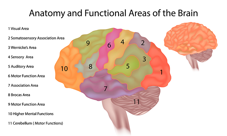
Somatosensory association area graphic © Sakurra/Shutterstock.com.
Dorsal root graphic © stihii/Shutterstock.com.
Imagine Maria, a graduate student who closes her eyes during an assessment and produces a strong posterior alpha rhythm. The moment you ask her to solve a mental arithmetic problem, her alpha amplitude drops and beta activity rises. This is alpha blocking, the desynchronization that sensory input and focused cognition produce (Andreassi, 2007). Recognizing that ordinary attention reshapes the EEG helps you set realistic baselines and interpret changes during an assessment rather than mistaking them for pathology.
Ascending sensory pathways relay information through specialized thalamic nuclei to the appropriate cortical regions, preserving the spatial location from which signals arise. Olfaction is the exception, bypassing the thalamus to reach the primary olfactory cortex directly. These pathways are not one-way streets, because roughly ten times more cortical neurons project back to the sensory thalamus than the other way, giving the cortex substantial top-down control. The visual, auditory, and somatosensory systems each split into a dorsal stream for locating stimuli and a ventral stream for identifying them. Sensory input and arousal desynchronize the EEG, lowering alpha and raising beta, a change clinicians see as alpha blocking.
Check Your Understanding
- Which sensory modality bypasses the thalamus, and where does it project instead?
- What does the roughly ten-to-one ratio of corticothalamic to thalamocortical projections tell us about how perception is controlled?
- Trace the visual pathway from the retinal ganglion cells to the primary visual cortex, naming the thalamic relay.
- How does the auditory dorsal stream differ in function from the auditory ventral stream?
- How does sensory input change the EEG, and what is the phenomenon called when alpha is reduced or eliminated?
Cutting-Edge Topics in qEEG Research
Layer 6 Feedback Can Retune a Single Sensory Circuit
If the cortex really does edit its own inputs, how local is that editing? Borbély and colleagues (2025) recorded from thalamocortical circuits while selectively driving layer 6 corticothalamic neurons, the descending population that outnumbers the ascending relay by roughly ten to one.
Different patterns of layer 6 activity pushed the circuit toward light sleep-like states, deep sleep-like states, or a desynchronized waking-like state. The striking result was how contained the effect was, because the state change stayed within the activated circuit rather than sweeping the whole brain.
Consider what that means for your surface recordings. A regional shift in slowing or desynchronization does not have to reflect a global change in arousal, since a single sensory loop can change state on its own. When you see a focal finding, treat local corticothalamic dynamics as a live candidate alongside the usual global explanations.
Where the Alpha Rhythm Actually Comes From
The posterior alpha you watch disappear when a client opens the eyes has long been credited to thalamocortical loops, but the details have been hard to pin down in humans. Nestvogel and McCormick (2022) showed that state-dependent visual cortical activity depends on ongoing thalamocortical communication, because inactivating either the thalamus or the cortex abolished the low-frequency oscillation entirely.
Human evidence has now sharpened the picture. Using simultaneous scalp EEG and intracranial recordings, Wang and colleagues (2026) found that low alpha (8 to 10 Hz) arises from occipital regions during eyes-closed wakefulness, while high alpha (10 to 13 Hz) emerges globally during anesthesia-induced loss of consciousness. They traced the shift to periodic rather than aperiodic activity, and modeled it as a change in inhibitory neurotransmission.
That distinction matters at the report-writing stage. This tutorial divides the band into alpha 1 (8 to 10 Hz) and alpha 2 (10 to 12 Hz), which is the same split these researchers call low and high alpha. If the two halves have different generators and different state dependencies, then collapsing them into a single alpha measure can hide the very difference you are trying to describe.
Top-Down Inference as the Default, Not the Exception
The new-school view of sensory pathways says the cortex predicts as much as it receives, and computational work is now explaining why that architecture exists. Csikor and colleagues (2025) trained a deep generative model on natural images and found that top-down influence falls out of hierarchical inference automatically, without being built in.
In their model, higher visual areas supply contextual priors that shape both the average responses and the noise correlations of primary visual cortex. Feedback connections, in other words, are not a refinement bolted onto a feedforward system. They are what make perception work at all.
For you, this reframes what a resting baseline is. A client lying still with eyes closed is not producing a stimulus-free recording, because the cortex is still running predictions, and those predictions are part of what your electrodes pick up.
Assignment
Now that you have completed this unit, trace the visual pathway from the retinal ganglion cells to the primary visual cortex, naming what happens at the optic chiasm and identifying the thalamic relay. Then do the same for the auditory pathway from the inner hair cells to the auditory cortex, and for the somatosensory touch pathway from the dorsal horn to the primary somatosensory cortex.
Next, compare the dorsal and ventral streams. Explain what each stream contributes in the visual system and in the auditory system, and describe how the somatic nervous system delivers the signals that the somatosensory pathway carries upward.
Finally, explain why the ratio of corticothalamic to thalamocortical projections argues against a purely bottom-up account of perception, and describe what you would expect to see in a posterior recording when a client opens the eyes, and why.
Glossary
afferent: a nerve fiber or pathway that carries information toward the central nervous system.
alpha blocking: the attenuation or disappearance of posterior alpha activity with eye opening, sensory stimulation, movement, or active cognitive processing.
alpha rhythm: an 8 to <13 Hz EEG rhythm dominant posteriorly during relaxed wakefulness with eyes closed and attenuated by eye opening.
arousal: a multidimensional state of wakefulness, alertness, and physiological activation supported by distributed brainstem, hypothalamic, and forebrain systems.
auditory cortex: the region of the temporal lobe that processes auditory information within the dorsal and ventral streams, supporting sound localization, pitch discrimination, and speech comprehension.
beta rhythm: a 13 to <30 Hz EEG rhythm, usually low amplitude, associated with alert wakefulness and active cognitive or motor processing.
contralateral: located on or relating to the side opposite a specified structure or event.
corticothalamic network: reciprocal circuits between cerebral cortex and thalamus that regulate sensory processing, excitability, sleep-wake states, and oscillatory activity.
decussate: to cross the midline, as when axons carrying touch information from one side of the body cross over in the medulla before ascending to the opposite thalamus.
desynchronization: the replacement of large, synchronized EEG rhythms by faster, lower-amplitude activity when the brain engages with a stimulus or task.
dorsal stream (auditory): the auditory "where" pathway, projecting from the auditory cortex of the temporal lobe to the parietal lobe, that helps spatially localize sounds.
dorsal stream (visual): the visual "where" pathway, extending from the primary visual cortex (V1) to the parietal lobe, that supports spatial perception, motion analysis, and visuomotor guidance, helping us localize objects and guide movements towards them.
efferent: a nerve fiber or pathway carrying signals away from the central nervous system or another reference center toward an effector.
inferior colliculi: the paired midbrain nuclei of the auditory pathway that integrate ascending auditory input for sound localization and auditory reflexes.
ipsilateral: located on, originating from, or affecting the same side of the body as another referenced structure or event.
lateral geniculate nucleus (LGN): the thalamic relay nucleus for vision, which receives input from the retina and projects visual information to the primary visual cortex.
medial geniculate nucleus (MGN): the thalamic relay nucleus for hearing, which receives input from the inferior colliculi and projects to several cortical auditory areas using two separate pathways.
midbrain: the middle division called the mesencephalon, which includes the inferior colliculi, superior colliculi, and substantia nigra.
mirror neuron: a neuron that fires both when an individual performs a movement and when the individual observes another performing the same action.
nucleus reticularis paragigantocellularis: a medullary reticular nucleus involved in autonomic and descending pain-modulatory circuits through noradrenergic and other pathways.
occipital lobe: the posterior cerebral lobe containing primary and association visual cortices.
parietal lobe: a cerebral lobe posterior to the central sulcus involved in somatosensation, spatial representation, attention, body schema, and sensorimotor integration.
postcentral gyrus: the gyrus immediately posterior to the central sulcus, containing primary somatosensory cortex (Brodmann areas 3, 1, and 2).
primary somatosensory cortex (S1): cortex in the postcentral gyrus that receives organized thalamic somatosensory input and represents touch, proprioception, and nociceptive features.
primary visual cortex (V1): the occipital lobe region located in the calcarine sulcus (BA 17, also called striate cortex) that receives most visual information from the lateral geniculate nucleus of the thalamus and performs the initial processing of visual information received from the retinas.
secondary somatosensory cortex (S2): parietal-opercular cortex receiving bilateral somatosensory input and contributing to integration, learning, and higher-order perception of bodily stimuli.
sensory EEG activation: attenuation or reorganization of ongoing EEG rhythms during sensory input or task engagement through distributed thalamocortical and cortical processing.
sensory nerves: peripheral nerves whose afferent fibers carry impulses from sensory receptors toward the central nervous system.
somatic nervous system: the peripheral sensory and motor system mediating conscious somatic sensation, spinal reflexes, and skeletal-muscle control.
superior colliculus: the dorsal midbrain structure that receives visual information and directs visual gaze and attention to selected stimuli.
superior olivary nuclei: pontine structures within the brainstem that process binaural information to localize sound and contribute to acoustic reflex modulation.
temporal lobe: a cerebral lobe inferior to the lateral sulcus involved in auditory processing, language, memory, and recognition of objects and faces.
thalamus: a paired diencephalic structure that relays and modulates most sensory and motor information reaching cortex and contributes to arousal and cortical rhythms.
ventral posterior nucleus (VPN): the thalamic relay nucleus for body sensation, which receives somatosensory information following crossover at the medulla and projects to the primary somatosensory cortex (S1).
ventral stream (auditory): the auditory "what" pathway, projecting from the auditory cortex to the temporal lobe, that appears to analyze sound components, including speech sounds, and supports sound identification.
ventral stream (visual): the visual "what" pathway, extending from the primary visual cortex (V1) to the inferior temporal and frontal areas, that allows us to identify objects, recognize faces, and process meaning.
Test Yourself on ClassMarker
Click the button below to take a 10-question exam over this entire unit. There is no password.
Review Flash Cards on Quizlet
Click the button below to review our chapter flash cards.
References
Andreassi, J. L. (2007). Psychophysiology: Human behavior and physiological response (5th ed.). Lawrence Erlbaum and Associates, Inc.
Bear, M. F., Connors, B. W., & Paradiso, M. A. (2020). Neuroscience: Exploring the brain (Enhanced 4th ed.). Jones & Bartlett Learning.
Borbély, S., Zalatnai, A., Gulyás, É., Balogh, V., Csernai, M., & Barthó, P. (2025). Corticothalamic feedback locally modulates network state. Scientific Reports, 15, Article 20606. https://doi.org/10.1038/s41598-025-05592-y
Breedlove, S. M., & Watson, N. V. (2023). Behavioral neuroscience (10th ed.). Sinauer Associates, Inc.
Buccino, G., Solodkin, A., & Small, S. L. (2006). Functions of the mirror neuron system: Implications for neurorehabilitation. Cognitive and Behavioral Neurology, 19(1), 55-63. https://doi.org/10.1097/00146965-200603000-00007
Csikor, F., Meszéna, B., Ócsai, K., & Orbán, G. (2025). Top-down perceptual inference shaping the activity of early visual cortex. Nature Communications, 16, Article 9998. https://doi.org/10.1038/s41467-025-64967-x
Kandel, E., Koester, J. D., Mack, S. H., & Siegelbaum, S. (2021). Principles of neural science (6th ed.). McGraw-Hill Education.
Nestvogel, D. B., & McCormick, D. A. (2022). Visual thalamocortical mechanisms of waking state-dependent activity and alpha oscillations. Neuron, 110(1), 120-138.e4. https://doi.org/10.1016/j.neuron.2021.10.005
Neumann, N., Strehl, U., & Birbaumer, N. (2003). A primer of electroencephalographic instrumentation. In M. Schwartz & F. Andrasik (Eds.), Biofeedback: A practitioner's guide (3rd ed.). Guilford Press.
Wang, R., Jiang, S., Cai, Q., Lang, L., Che, X., He, J., Wang, Y., Hu, J., & Han, C. (2026). Distinct origins of human low and high alpha rhythms revealed by simultaneous EEG-SEEG. Communications Biology, 9, Article 503. https://doi.org/10.1038/s42003-026-09769-7
Return to Top