
Brodmann Areas and Their Functional Attributes
What You Will Learn in This Chapter
Imagine slicing a piece of cortex thinner than a sheet of paper, staining it so that the cell bodies leap out in dark violet, and then studying it under a microscope for a decade. That is roughly how Korbinian Brodmann spent the opening years of the twentieth century. What he found is that the cortex is not a uniform sheet: the size, density, and layering of its neurons shift from one patch to the next, and those shifts occur at borders you can actually see. The map he drew from those borders is still the vocabulary you use when you report where an abnormality lives.
This unit walks you through that map. You will learn how Brodmann defined his 52 areas from cellular architecture alone, why a purely anatomical scheme turned out to predict function so well, and what the most studied areas actually do. You will examine areas 1, 2, and 3 in the postcentral gyrus, area 4 in the precentral gyrus, area 17 in the occipital lobe, area 22 in the superior temporal gyrus, area 40 in the parietal lobe, and area 46 in the prefrontal cortex.
You will also see why this century-old map still appears in modern qEEG reports. When source localization software estimates where scalp-recorded activity originated, it names the answer in Brodmann coordinates, so reading a current-source report means reading Brodmann. By the end of the chapter you should be able to move comfortably between an electrode label, a gyrus, and an area number.
IQCB Blueprint Coverage: This unit addresses Basic Knowledge of Brodmann Areas (V.E) within qEEG (V).
Learning Objectives
After completing this section, you will be able to:
Define Brodmann areas and explain the cytoarchitectonic criteria Brodmann used to draw their borders.
Describe the role of Nissl staining and cortical layering in the construction of the 1909 map.
Explain why an anatomical parcellation corresponds so closely to functional organization.
Identify the functions of Brodmann areas 1, 2, 3, 4, 17, 22, 40, and 46.
Distinguish somatotopic organization in the primary motor cortex from retinotopic organization in the primary visual cortex.
Relate Wernicke's area to language comprehension and predict the consequences of damage to it.
Explain how Brodmann areas are used to report source localization findings in qEEG.
Listen to the Full-Length Lecture
What Brodmann Areas Are
Brodmann areas are a system of classification for the cerebral cortex based on the organization of neurons in different regions. They were defined by the German anatomist Korbinian Brodmann in the early twentieth century. These areas have become foundational in understanding brain function and neuroanatomy.
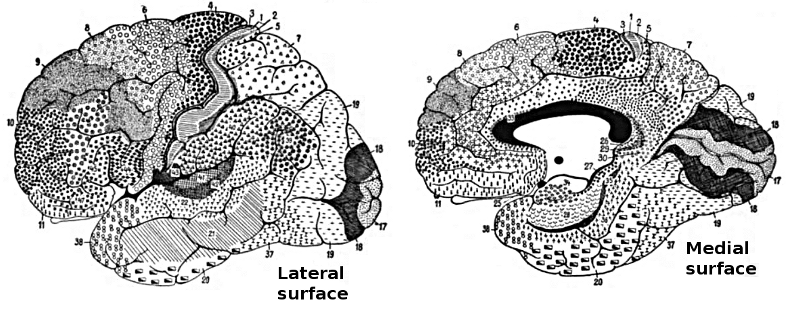
Definition of Brodmann Areas
Korbinian Brodmann defined the areas of the cerebral cortex based on their cytoarchitecture, which involves the microscopic appearance and organization of cells. Using Nissl stain to highlight cell bodies, Brodmann examined the cortical layers and identified variations in cellular structure across different regions. In 1909, he published a map dividing the human cortex into 52 distinct areas, each with unique structural features (Brodmann, 1909).
Brodmann's classification was primarily anatomical, focusing on the differences in cell types, densities, and arrangements within the cortical layers. He was not looking for function at all; he was looking for borders. Despite its anatomical basis, the Brodmann map has shown significant correlations with functional areas of the brain, making it a valuable tool in both neuroanatomy and neurophysiology (Amunts & Zilles, 2015).
That correspondence is not a coincidence, and it is worth pausing on. Cortical regions that perform different jobs receive different inputs, send different outputs, and therefore need different cellular hardware. A region that receives dense thalamic input develops a thick layer IV to house it, while a region that sends long motor projections develops large layer V pyramidal cells. Brodmann was reading the consequences of function in the tissue, even though he could not measure the function itself.
Brodmann areas are a historically important division of the cerebral cortex into distinct regions based on their cytoarchitecture or neuronal arrangement and connections. The concept of Brodmann areas was introduced by the German neurologist Korbinian Brodmann in the early 20th century (Brodmann, 1909). Brodmann's work has significantly impacted the understanding of the functional organization of the cortex and remains influential in contemporary neuroscience research.Brodmann's classification was based on his observations of differences in the cellular organization of the cortex across various mammalian species, including humans. He identified 43 numbered areas in the human brain by examining the cellular organization, cell types, and layer thickness (Zilles & Amunts, 2010). Although some refinements have been made since Brodmann's initial classification, many identified areas retain their original numbering.
The Importance of Brodmann Areas
The identification of Brodmann areas has facilitated the investigation of functional specialization within the cortex. Most cortical functions involve the networked activity of multiple Brodmann areas. Several Brodmann areas are now associated with specific functions, such as primary sensory and motor areas, as well as higher cognitive functions like language processing and decision-making (Glasser et al., 2016). For instance, Brodmann area 4 corresponds to the primary motor cortex, area 17 to the primary visual cortex, and areas 44 and 45 to Broca's area, which is crucial for speech production. While Brodmann areas provide a valuable framework for understanding cortical organization, they do not capture the full complexity of the brain's functional architecture.Relevance and Limitations
Brodmann areas continue to serve as a foundational framework for understanding cortical organization and function in both clinical and research contexts, from neurosurgical planning to the study of cognitive processes and neuropsychiatric disorders. Advances in neuroimaging techniques have identified additional areas and functional networks, highlighting the intricate organization of the cortex beyond Brodmann's classification (Glasser et al., 2016). Advances in neuroimaging and connectomics are gradually supplementing this early anatomical schema with more detailed and individualized models of brain organization. For neurofeedback practitioners, Brodmann areas remain the standard language for communicating about cortical regions, but clinicians should recognize that individual brains vary and that a given Brodmann area may not occupy the exact same cortical territory in every client.
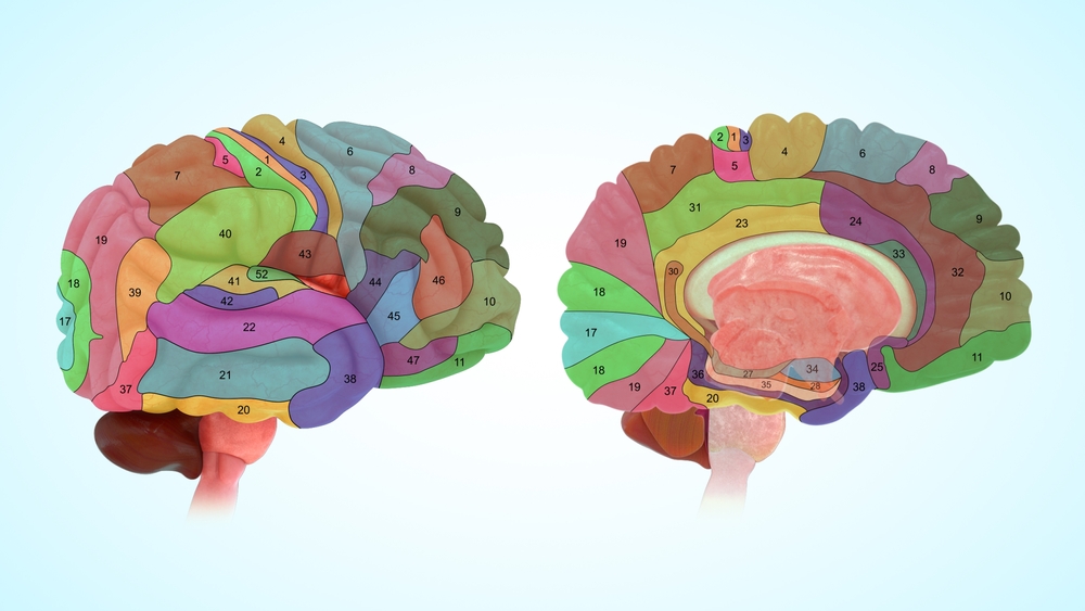
Researchers have revised the Brodmann maps and correlated areas with their functions. The Brodmann maps below were contributed by Mark Dow, Research Assistant at the Brain Development Lab, the University of Oregon to Wikimedia Commons.
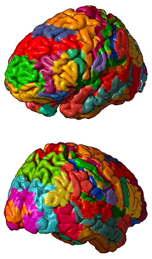
Brodmann areas are cortical regions defined by cytoarchitecture, meaning the types, densities, and layered arrangement of neurons revealed by Nissl staining. Brodmann published his map of 52 areas in 1909 using purely anatomical criteria. The map nonetheless predicts functional organization closely, because cortical regions with different jobs require different cellular hardware. That anatomical-to-functional correspondence is why a map drawn before EEG existed still organizes modern electrophysiology.
Areas 3, 1, and 2: Primary Somatosensory Cortex (S1)
The primary somatosensory cortex (S1) is a critical region for processing somatosensory information in the brain. It is involved in processing touch, proprioception, and temperature. These four cortical areas contain separate somatotopic maps (Purves, 2018). Graphic © Big8/Shutterstock.com..jpg)
Brodmann areas
The S1 is located in the postcentral gyrus, mainly in Brodmann areas 3, 1, and 2. These areas have distinct functions; Brodmann area 3 receives and processes cutaneous and proprioceptive inputs, area 1 processes tactile stimuli, and area 2 integrates proprioceptive and tactile inputs (Kaas, 2008).Location
The S1 is located in the parietal lobe, immediately posterior to the central sulcus. It is bordered by the primary motor cortex (M1) anteriorly and the secondary somatosensory cortex (S2) posteriorly. The closest sites are C3 and C4, which overlie the central sulcus (Jasper, 1958).Connections
The S1 strongly connects with other cortical and subcortical areas, including the M1, premotor cortex, supplementary motor area, posterior parietal cortex, and thalamus (Lemon, 2008). These connections are essential for sensorimotor integration and control.Participation in brain networks
The S1 is a critical node in the somatosensory network, which includes other areas like the S2, insular cortex, and parietal operculum. It also participates in the sensorimotor network, interacting with the motor and premotor cortices (Sepulcre, 2012).Functions
The S1 is crucial for processing somatosensory information like touch, proprioception, and temperature. It plays a significant role in perceiving object features, body awareness, and sensorimotor integration.Role in clinical disorders
The altered functioning of the S1 has been implicated in various clinical conditions, including neuropathic pain (Baliki et al., 2011), phantom limb pain (Makin et al., 2013), and stroke-related sensory deficits (Carey et al., 2002).Area 4: Primary Motor Cortex (M1)
The primary motor cortex (M1) is a key region in the brain responsible for the execution of voluntary movements. Graphic © Big8/Shutterstock.com.Brodmann areas
The M1 is located in the precentral gyrus, mainly in Brodmann area 4. It contains large pyramidal neurons, known as Betz cells, essential for motor control (Geyer et al., 1996).Location
The M1 is situated in the frontal lobe, immediately anterior to the central sulcus. It is bordered by the posteriorly primary somatosensory cortex (S1) and anteriorly premotor cortex. The closest sites are C3 and C4, which overlie the central sulcus (Jasper, 1958).Connections
The M1 strongly connects with various cortical and subcortical areas, including the S1, premotor cortex, supplementary motor area, posterior parietal cortex, and thalamus (Lemon, 2008). These connections are critical for sensorimotor integration and control.Participation in brain networks
The M1 is a central node in the sensorimotor network, interacting with the somatosensory cortex, premotor cortex, and other motor-related areas (Sepulcre, 2012).Functions
The primary function of the M1 is the execution of voluntary movements. M1 neurons primarily control movements rather than discrete muscles (Breedlove & Watson, 2023). It is critical in planning, controlling, and coordinating complex motor tasks.Role in clinical disorders
Alterations in M1 function have been implicated in various clinical conditions, including motor deficits following stroke (Ward, 2004), Parkinson's disease (Wu & Hallett, 2013), and motor neuron diseases like amyotrophic lateral sclerosis (ALS; Kew & Leigh, 1997).Areas 5 and 7: Somatosensory Association Cortex (SAC)
The somatosensory association cortex (SAC) is involved in the integration and interpretation of somatosensory information coming from the primary somatosensory cortex (S1). Graphic © Big8/Shutterstock.com.Brodmann areas
The SAC is mainly located in Brodmann areas 5 and 7 within the posterior parietal cortex (Culham & Kanwisher, 2001).Location
The SAC is situated in the parietal lobe, superior to the primary somatosensory cortex (S1), and posterior to the postcentral gyrus. The closest sites are likely P3 and P4, which overlie the parietal cortex.Connections
The SAC strong connects with various cortical and subcortical regions, including the S1, primary motor cortex (M1), premotor cortex, supplementary motor area, posterior parietal cortex, and thalamus (Cavada & Goldman-Rakic, 1989). These connections are critical for sensorimotor integration, spatial awareness, and higher-order sensory processing.Participation in brain networks
The SAC is a key node in the somatosensory network, including areas like the S1, S2, and insular cortex. Additionally, it is part of the dorsal attention network, which is involved in attentional control and spatial processing (Corbetta & Shulman, 2002).Functions
The SAC is essential for integrating and interpreting somatosensory information, including tactile and proprioceptive stimuli. It plays a significant role in sensorimotor integration, spatial awareness, and attention.Role in clinical disorders
Alterations in SAC function have been implicated in various clinical conditions, including somatosensory neglect (Vallar et al., 2003), spatial processing deficits (Whitlock et al., 2012), and somatosensory deficits in autism spectrum disorder (Cascio et al., 2012).Area 6: Supplementary Motor Cortex and Premotor Cortex
The supplementary motor cortex (SMA) and premotor cortex (PMC) are critical regions for planning and executing voluntary movements. Graphic © Big8/Shutterstock.com.Brodmann areas
The SMA is primarily located in Brodmann area 6, on the medial aspect of the frontal lobe (Picard & Strick, 2001). The PMC is also predominantly found in Brodmann area 6 but is located on the lateral aspect of the frontal lobe (Wise et al., 1997).Location
The SMA is located in the medial part of the frontal lobe, superior to the cingulate sulcus and anterior to the paracentral lobule. The PMC is situated in the lateral part of the frontal lobe, anterior to the primary motor cortex (M1). The closest sites are likely FC3 and FC4, which overlie the dorsolateral prefrontal cortex.Connections
Both the SMA and PMC have strong connections with various cortical and subcortical areas, including the M1, primary somatosensory cortex (S1), posterior parietal cortex, and basal ganglia (Lemon, 2008; Nachev et al., 2008). These connections are critical for sensorimotor integration, movement planning, and execution.Participation in brain networks
The SMA and PMC are central nodes in the sensorimotor network, interacting with the M1, S1, and other motor-related areas (Sepulcre, 2012).Functions
The SMA and PMC are essential for motor planning, execution, and coordination of complex movements. The SMA is particularly involved in initiating and controlling internally generated movements, while the PMC is more concerned with the planning and executing visually-guided movements (Wise et al., 1997; Picard & Strick, 2001).Role in clinical disorders
Alterations in SMA and PMC function have been implicated in various clinical conditions, including movement disorders like Parkinson's disease (Wu & Hallett, 2013), apraxia (Haaland et al., 2000), and motor deficits following stroke (Ward, 2004).Area 8: Frontal Eye Field (FEF)
The frontal eye field (FEF) is essential for the control of eye movements and visual attention. Graphic © Big8/Shutterstock.com.Brodmann areas
The FEF is predominantly in Brodmann area 8, located in the dorsolateral prefrontal cortex (Paus, 1996).Location
The FEF is located in the anterior bank of the precentral sulcus within the dorsolateral prefrontal cortex, close to the border with the primary motor cortex (M1) (Paus, 1996). The closest sites are likely F3 and F4, which overlie the dorsolateral prefrontal cortex.Connections
The FEF has extensive connections with other cortical and subcortical regions, including the parietal cortex, superior colliculus, thalamus, and extrastriate visual areas (Schall, 2002; Stanton et al., 2005). These connections are crucial for visual attention, saccadic eye movements, and smooth pursuit.Participation in brain networks
The FEF is a key node in the dorsal attention network responsible for goal-directed attention and eye movement control. This network also includes the intraparietal sulcus and superior parietal lobule (Corbetta & Shulman, 2002).Functions
The FEF is crucial in controlling saccadic eye movements, smooth pursuit, and visual attention. It is involved in the initiation, planning, and execution of eye movements, as well as the allocation of attention to relevant visual stimuli (Schall, 2002). The FEF is vital in cognitive functions, including attention orientation, visual consciousness, access to our conscious experience, perceptual performance, and decision-making (Vernet et al., 2014).Role in clinical disorders
Alterations in FEF function have been implicated in various clinical conditions, including attention deficit hyperactivity disorder (ADHD; Mahone et al., 2011), oculomotor apraxia (Rizzo et al., 1996), and progressive supranuclear palsy (Burrell et al., 2012).Areas 9 and 46: Dorsolateral Prefrontal Cortex (DLPFC)
The dorsolateral prefrontal cortex (DLPFC), which consists of distinct regions, is essential for higher-order cognitive functions, including working memory, executive control, and decision-making (Ahuja & Rodriguez, 2022). Graphic © Big8/Shutterstock.com.Brodmann areas
The DLPFC is primarily located in Brodmann areas 9 and 46 within the lateral aspect of the frontal lobe (Petrides, 2005).Location
The DLPFC is situated in the lateral portion of the frontal lobe, superior and anterior to the premotor and primary motor cortex (M1). The closest sites are likely F3 and F4.Connections
The DLPFC has extensive connections with other cortical and subcortical regions, including the parietal cortex, medial prefrontal cortex, anterior cingulate cortex, orbitofrontal cortex, thalamus, and basal ganglia (Petrides & Pandya, 2002). These connections are critical for cognitive control, working memory, and decision-making.Participation in brain networks
The DLPFC is a key node in the frontoparietal control network, which is responsible for executive control, and the working memory network, which maintains and manipulates information (Cabeza & Nyberg, 2000; Vincent et al., 2008).Functions
The DLPFC plays a crucial role in higher-order cognitive functions such as working memory, executive control, and decision-making. It allocates cognitive resources, goal-directed behavior, task switching, and the flexible adaptation of behavior in response to changing demands (Breedlove & Watson, 2023; Petrides, 2005).Role in clinical disorders
Alterations in DLPFC function have been implicated in various clinical conditions, including schizophrenia (Barch, 2005), attention deficit hyperactivity disorder (ADHD; Cortese et al., 2012), and major depressive disorder (MDD; Drevets et al., 2008).Area 10: Anterior Prefrontal Cortex (aPFC)
The anterior prefrontal cortex (aPFC), also referred to as the frontopolar cortex, is involved in higher-order cognitive processes such as decision-making, planning, and reasoning. Graphic © Big8/Shutterstock.com.Brodmann areas
The aPFC is primarily located in Brodmann area 10, at the most anterior part of the frontal lobe (Ramnani & Owen, 2004).Location
The aPFC is located at the most rostral part of the frontal lobe, anterior to the dorsolateral prefrontal cortex (DLPFC) and orbitofrontal cortex. The closest sites are likely Fp1 and Fp2, which overlie the frontal pole.Connections
The aPFC has extensive connections with other cortical and subcortical regions, including the DLPFC, orbitofrontal cortex, medial prefrontal cortex, posterior parietal cortex, temporal cortex, and thalamus (Burgess et al., 2007). These connections are essential for complex cognitive tasks, multitasking, and mentalizing.Participation in brain networks
The aPFC is a key node in the frontoparietal control network, which is responsible for executive control, as well as the default mode network (DMN), which is involved in self-referential processing and mentalizing (Vincent et al., 2008; Spreng et al., 2009).Functions
The aPFC involves higher-order cognitive processes such as decision-making, planning, reasoning, multitasking, and mentalizing. It is crucial in coordinating and integrating information from various cognitive domains and is responsible for goal-directed behavior and social cognition (Ramnani & Owen, 2004). The aPFC is engaged in various tasks, such as problem-solving, memory recall, future-oriented memory, source and context memory, task-switching, and attention reallocation (Ramnani & Owen, 2004). The aPFC contributes to high-level nociception and pain processing (Peng et al., 2018).Role in clinical disorders
Alterations in aPFC function have been implicated in various clinical conditions, including autism spectrum disorder (ASD; Gilbert et al., 2008), schizophrenia (Perlstein et al., 2001), and major depressive disorder (MDD; Drevets et al., 2008).Areas 11, 12, 13, and 47: Orbitofrontal Cortex (OFC)
The orbitofrontal cortex (OFC) processes reward, emotion, and decision-making and integrates sensory information with emotional valence. Graphics © Big8/Shutterstock.com..jpg)
Brodmann areas
The OFC primarily encompasses Brodmann areas 11, 12, 13, and 47, located in the ventral portion of the frontal lobe (Kringelbach, 2005).Location
The OFC is situated in the ventral part of the frontal lobe, just above the orbits (eye sockets). The anterior prefrontal cortex and the medial prefrontal cortex border it. The closest sites are likely Fp1 and Fp2, which overlie the ventral and rostral portions of the frontal lobe.Connections
The OFC has extensive connections with other cortical and subcortical regions, including the amygdala, insula, cingulate cortex, hippocampus, thalamus, striatum, and sensory cortices (Kringelbach, 2005; Price, 2007). These connections are essential for emotion processing, reward-based decision-making, and social cognition.Participation in brain networks
The OFC is a key node in the salience network, which is responsible for detecting and integrating emotionally and motivationally salient stimuli, and the default mode network (DMN), which is involved in self-referential processing and social cognition (Seeley et al., 2007; Spreng et al., 2009).Functions
The OFC is crucial in processing reward, emotion, and decision-making. It integrates sensory information with emotional valence, evaluates outcomes and actions, and represents social and emotional information (Kringelbach, 2005).Role in clinical disorders
Alterations in OFC function have been implicated in various clinical conditions, including obsessive-compulsive disorder (OCD) (Menzies et al., 2008), major depressive disorder (MDD) (Drevets, 2007), bipolar disorder (BD) (Blumberg et al., 2003), and addiction (Volkow & Fowler, 2000). Depression may be associated with heightened responsiveness and increased connectivity in the lateral orbitofrontal cortex (not linked to rewards), while it is connected to reduced responsiveness and connectivity in the medial orbitofrontal cortex (related to rewards; Rolls, Cheng, & Feng, 2020).Areas 13-16 and 52: Insular Cortex (Insula)
The insular cortex, or insula, is involved in diverse functions, including interoception, emotion processing, pain perception, and cognitive control. Graphics © Big8/Shutterstock.com. Brodmann areas
The insular cortex comprises Brodmann areas 13, 14, 15, 16, and parts of area 52. These include sensorimotor, central-olfactogustatory, socio-emotional, and cognitive anterior-dorsal regions (Kurth et al., 2010).Location
The insular cortex is situated deep within the lateral sulcus, which separates the frontal and parietal lobes from the temporal lobe. The opercula of the frontal, parietal, and temporal lobes cover it.Connections
The insular cortex has extensive connections with various cortical and subcortical regions, including the amygdala, anterior cingulate cortex (ACC), prefrontal cortex, primary and secondary somatosensory cortices, orbitofrontal cortex (OFC), and thalamus (Nieuwenhuys, 2012). These connections contribute to the diverse functions of the insula.Participation in brain networks
The insular cortex is a key node in the salience network, which is responsible for detecting and integrating emotionally and motivationally salient stimuli, and the central autonomic network (CAN), which is involved in autonomic regulation (Seeley et al., 2007; Thayer et al., 2012).Functions
The insular cortex is crucial in interoception, emotion processing, pain perception, and cognitive control. It represents internal bodily states, integrates sensory and emotional information, and modulates cognitive and affective processes (Craig, 2009).Role in clinical disorders
Alterations in insular cortex function have been implicated in various clinical conditions, including anxiety disorders (Paulus & Stein, 2006), major depressive disorder (MDD; Sliz & Hayley, 2012), addiction (Naqvi & Bechara, 2010), and autism spectrum disorder (ASD; Di Martino et al., 2009).Area 17: Primary Visual Cortex (V1)
The primary visual cortex (V1), or the striate cortex, is responsible for processing basic visual information, such as orientation, spatial frequency, and color. Graphic © Big8/Shutterstock.com.Brodmann areas
The primary visual cortex is primarily located in Brodmann area 17, in the occipital lobe (Horton & Adams, 2005).Location
The primary visual cortex is located in the occipital lobe, along the calcarine sulcus, which runs horizontally through the medial part of the lobe. The closest sites are likely O1 and O2, which overlie the occipital lobe.Connections
The primary visual cortex receives input from the lateral geniculate nucleus (LGN) of the thalamus and sends output to the secondary visual cortex (V2) and other extrastriate areas (V3, V4, V5/MT). These connections are essential for the hierarchical processing of visual information (Felleman & Van Essen, 1991).Participation in brain networks
The primary visual cortex is a key node in the visual processing network responsible for processing and interpreting visual information from the retina. This network includes other areas of the occipital lobe and extends to the parietal and temporal cortices (Nassi & Callaway, 2009).Functions
The primary visual cortex processes basic visual information, such as orientation, spatial frequency, and color. It forms the initial stage of the hierarchical processing of visual information and is critical for visual perception (Horton & Adams, 2005).Role in clinical disorders
Alterations in primary visual cortex function have been implicated in various clinical conditions, including amblyopia (lazy eye; Hess et al., 2010), cortical blindness (Celesia, 2005), and visual hallucinations in conditions like Charles Bonnet syndrome (Griffiths, 2000).Areas 18 and 19: Secondary Visual Cortex (V2)
The secondary visual cortex (V2), also known as the prestriate cortex, is involved in the further processing and integration of visual information received from the primary visual cortex (V1). Graphic © Big8/Shutterstock.com.Brodmann areas
The secondary visual cortex is primarily located in Brodmann areas 18 and 19, in the occipital lobe (Tootell et al., 1998).Location
The secondary visual cortex is located in the occipital lobe, surrounding the primary visual cortex along the calcarine sulcus, extending to the lateral parts of the occipital lobe. The closest sites are likely O1 and O2.Connections
The V2 receives input from the primary visual cortex (V1). It sends output to higher-order extrastriate areas (V3, V4, V5/MT) and other cortical regions involved in visual processing, including the parietal and temporal cortices (Felleman & Van Essen, 1991).Participation in brain networks
The secondary visual cortex is a key node in the visual processing network responsible for processing and interpreting visual information from the retina. This network includes other areas of the occipital lobe and parietal and temporal cortices (Nassi & Callaway, 2009).Functions
The secondary visual cortex is involved in further processing and integrating visual information from the primary visual cortex. It is crucial in processing complex visual attributes, such as form, color, and motion (Tootell et al., 1998).Role in clinical disorders
Alterations in secondary visual cortex function have been implicated in various clinical conditions, including visual agnosia, characterized by the inability to recognize objects despite normal visual acuity and intact primary visual cortex function (Milner & Goodale, 2008).Areas 18, 19, 37, 21, and 22: Visual Association Cortex (V3, V4, V5)
The visual association cortex, also known as the higher-order extrastriate cortex, is responsible for the advanced processing of visual information, such as object recognition, face perception, and processing of complex visual scenes. Graphic © Big8/Shutterstock.com.Brodmann areas
The visual association cortex comprises several Brodmann areas, including areas 18, 19, 37, 21, and 22, mainly in the occipital and temporal lobes (Tootell et al., 1998; Kanwisher & Yovel, 2006).Location
The visual association cortex is located primarily in the occipital and temporal lobes, surrounding the primary (V1) and secondary (V2) visual cortices. It includes regions such as the fusiform face area (FFA), the parahippocampal place area (PPA), and the lateral occipital complex (LOC; Epstein & Kanwisher, 1998; Kanwisher & Yovel, 2006; Malach et al., 1995). The closest sites are likely O1, O2, T5, and T6, which overlie the occipital and temporal lobes.Connections
The visual association cortex receives input from the primary (V1) and secondary (V2) visual cortices and has extensive connections with other cortical and subcortical regions, including the parietal lobe, prefrontal cortex, hippocampus, and amygdala (Felleman & Van Essen, 1991; Kravitz et al., 2013).Participation in brain networks
The visual association cortex is a key component of the ventral visual processing stream, also known as the "what" pathway, responsible for object recognition and processing of complex visual scenes (Kravitz et al., 2011).Functions
The visual association cortex is involved in advanced visual processing, including object recognition, face perception, processing of complex visual scenes, and integration of visual information with other sensory modalities (Kanwisher & Yovel, 2006; Tootell et al., 1998).Role in clinical disorders
Alterations in visual association cortex function have been implicated in various clinical conditions, including prosopagnosia (face blindness; Duchaine & Nakayama, 2006), visual agnosia (Milner & Goodale, 2008), and higher-order visual processing deficits in conditions such as autism spectrum disorder (ASD; Simmons et al., 2009).Areas 20 and 37: Inferior Temporal Gyrus (ITG)
The inferior temporal gyrus (ITG) is a part of the temporal lobe involved in high-level visual processing and object recognition. Graphic © Big8/Shutterstock.com.Brodmann areas
The inferior temporal gyrus primarily includes Brodmann areas 20 and 37 (Amunts et al., 2000).Location
The inferior temporal gyrus is located in the ventral part of the temporal lobe, below the middle temporal gyrus and superior temporal sulcus, and above the fusiform gyrus. The closest sites are likely T5 (or TP7) and T6 (or TP8), which overlie the temporal lobes.Connections
The ITG has extensive connections with other cortical and subcortical regions, including the primary and secondary visual cortices, fusiform gyrus, parahippocampal gyrus, hippocampus, amygdala, and prefrontal cortex (Kravitz et al., 2013; Felleman & Van Essen, 1991).Participation in brain networks
The ITG is a key component of the ventral visual processing stream, also known as the "what" pathway, responsible for object recognition and processing of complex visual scenes (Kravitz et al., 2011).Functions
The ITG is involved in high-level visual processing, object recognition, semantic processing, and the integration of visual information with other sensory modalities (Kanwisher & Yovel, 2006).Role in clinical disorders
Alterations in ITG function have been implicated in various clinical conditions, including visual agnosia, prosopagnosia (face blindness), and higher-order visual processing deficits in conditions such as autism spectrum disorder (ASD; Duchaine & Nakayama, 2006; Simmons et al., 2009).Areas 21 and 39: Middle Temporal Gyrus (MTG)
The middle temporal gyrus (MTG) is a part of the temporal lobe involved in various functions, such as semantic processing, language, and high-level visual processing. Graphics © Big8/Shutterstock.com. Brodmann areas
The MTG primarily includes Brodmann areas 21 and 39 (Amunts et al., 2000).Location
The MTG is located in the lateral part of the temporal lobe, between the superior temporal gyrus (above) and the inferior temporal gyrus (below), and adjacent to the superior temporal sulcus.Connections
The MTG has extensive connections with other cortical and subcortical regions, including the primary and secondary visual cortices, the angular gyrus, the fusiform gyrus, the parahippocampal gyrus, the hippocampus, the amygdala, and the prefrontal cortex (Kravitz et al., 2013; Felleman & Van Essen, 1991).Participation in brain networks
The MTG participates in various brain networks, including the ventral visual processing stream ("what" pathway) for object recognition and processing of complex visual scenes (Kravitz et al., 2011), and the language network for semantic processing and word retrieval (Binder et al., 2009).Functions
The MTG is involved in various functions, such as semantic processing, language comprehension, word retrieval, and high-level visual processing, including object and face recognition (Binder et al., 2009; Kanwisher & Yovel, 2006).Role in clinical disorders
Alterations in MTG function have been implicated in various clinical conditions, including semantic dementia (Hodges et al., 1992), language impairments in aphasia (Dronkers et al., 2004), and higher-order visual processing deficits in conditions such as autism spectrum disorder (ASD; Simmons et al., 2009).Areas 22, 39, and 40: Superior Temporal Gyrus (STG)
The superior temporal gyrus (STG), including Wernicke's area, is a part of the temporal lobe involved in various functions such as language comprehension, auditory processing, and social cognition. Graphic © Big8/Shutterstock.com.Brodmann areas
Wernicke's area primarily includes Brodmann area 22 and, to some extent, areas 39 and 40 (Amunts et al., 2000).Location
Wernicke's area is located in the posterior part of the superior temporal gyrus, usually in the left hemisphere, near the lateral sulcus. The STG runs laterally along the temporal lobe, above the middle temporal gyrus. The closest site is likely T5 (or TP7) for the left hemisphere, where Wernicke's area is typically located.Connections
Wernicke's area has extensive connections with other language-related regions, including Broca's area (via the arcuate fasciculus), the angular gyrus, and other parts of the superior temporal gyrus (Friederici, 2009). The STG also connects with the primary and secondary auditory cortices, social cognition, and memory regions.Participation in brain networks
Wernicke's area participates in the language network, playing a crucial role in language comprehension and semantic processing (Binder et al., 2009). The STG is also involved in the auditory processing network and the social cognition network.Functions
Wernicke's area involves language comprehension, semantic processing, and integrating auditory information into meaningful speech (Price, 2012). The STG also plays a role in auditory processing, social cognition, and memory.Role in clinical disorders
Alterations in the function of Wernicke's area and the STG have been implicated in various clinical conditions, such as Wernicke's aphasia, characterized by impaired language comprehension and fluent but nonsensical speech (Dronkers et al., 2004). The STG has also been implicated in auditory processing deficits and social cognition impairments in conditions such as autism spectrum disorder (ASD; Boddaert et al., 2004).Area 23: Ventral Posterior Cingulate Cortex (vPCC)
The ventral posterior cingulate cortex (vPCC) is a region within the posterior cingulate cortex (PCC), a part of the limbic system involved in various functions such as memory, emotion, and self-referential processing. Graphic © Big8/Shutterstock.com. Brodmann areas
The vPCC primarily includes Brodmann area 23 (Vogt et al., 2006).Location
The vPCC is located in the medial part of the brain, in the posterior cingulate cortex, and ventral to the dorsal posterior cingulate cortex (dPCC). It is positioned between the precuneus and the corpus callosum. The closest sites are likely Pz and CPz, located over the midline parietal and central regions, respectively.Connections
The vPCC connects with various brain regions, including the medial prefrontal cortex (mPFC), hippocampus, parahippocampal gyrus, and lateral parietal regions (Leech & Sharp, 2014; Utevsky et al., 2014).Participation in brain networks
The vPCC is a key component of the default mode network (DMN), which is active during rest and involved in self-referential thinking, autobiographical memory, and social cognition (Raichle et al., 2001; Buckner et al., 2008).Functions
The vPCC is involved in various functions, such as self-referential thinking, autobiographical memory, social cognition, and emotional processing (Leech & Sharp, 2014; Utevsky et al., 2014).Role in clinical disorders
Alterations in vPCC function have been implicated in various clinical conditions, including Alzheimer's disease (Buckner et al., 2005), major depressive disorder (Sheline et al., 2010), and autism spectrum disorder (ASD; Padmanabhan et al., 2017).Areas 24 and 25: Ventral Anterior Cingulate Cortex (vACC)
The ventral anterior cingulate cortex (vACC) is a region within the anterior cingulate cortex (ACC), which is part of the limbic system and involved in various functions, such as emotion processing, reward-based learning, and decision-making. Graphic © Big8/Shutterstock.com. Brodmann areas
The vACC primarily includes Brodmann areas 24 and 25 (Vogt, 2005).Location
The vACC is located in the medial part of the brain, in the anterior cingulate cortex, ventral to the dorsal anterior cingulate cortex (dACC). It is positioned anterior to the genu of the corpus callosum. The closest sites are likely FCz and Cz, located over the midline frontal and central regions, respectively.Connections
The vACC has connections with various brain regions, including the amygdala, hippocampus, medial prefrontal cortex (mPFC), orbitofrontal cortex (OFC), and nucleus accumbens (Bush et al., 2000; Etkin et al., 2011).Participation in brain networks
The vACC is a key component of the salience network, which detects and integrates salient emotional and sensory stimuli and modulates attention and cognitive control (Menon, 2011; Seeley et al., 2007).Functions
The vACC is implicated in various cognitive and emotional functions, including error detection, conflict monitoring, emotion regulation, empathy, and social cognition (Bush et al., 2000; Etkin et al., 2011).Role in clinical disorders
Abnormalities in the vACC have been implicated in several psychiatric and neurological disorders, such as depression, anxiety, schizophrenia, bipolar disorder, attention deficit hyperactivity disorder (ADHD), and autism spectrum disorders (Drevets et al., 2008; Etkin et al., 2010).Areas 25 and 24b: Subgenual Ventromedial Prefrontal Cortex (vmPFC)
The subgenual region of the ventromedial prefrontal cortex (vmPFC) is an important brain region involved in various cognitive and emotional processes. Graphic © Big8/Shutterstock.com. Brodmann areas
The subgenual region of the vmPFC primarily consists of Brodmann areas 25 and 24b (Ongür et al., 2003).Location
The subgenual region of the vmPFC is located in the medial prefrontal cortex, ventral to the genu of the corpus callosum, and adjacent to the anterior cingulate cortex (Mayberg, 2003). Its nearby EEG electrode positions include Fp1, Fp2, Fz, and AFz, located along the scalp's midline (Jasper, 1958).Connections
The subgenual region of the vmPFC has extensive connections with other brain regions, including the amygdala, hippocampus, hypothalamus, nucleus accumbens, thalamus, and other prefrontal areas (Ongür et al., 2003; Price & Drevets, 2010).Participation in brain networks
The subgenual vmPFC is a key component of the default mode network (DMN) and the affective network, involved in self-referential processing, emotion regulation, and decision-making (Buckner et al., 2008; Rudebeck et al., 2014).Functions
The subgenual vmPFC is implicated in various cognitive and emotional functions, including value-based decision-making, emotion regulation, self-referential processing, and social cognition (Rudebeck et al., 2014; Roy et al., 2012).Role in clinical disorders
Abnormalities in the subgenual vmPFC have been implicated in several psychiatric disorders, such as major depressive disorder, bipolar disorder, anxiety disorders, and post-traumatic stress disorder (Mayberg, 2003; Price & Drevets, 2010).Areas 29 and 30: Ectosplenial Retrosplenial Cerebral Cortex
The ectosplenial region is not a widely recognized or well-established region within the human retrosplenial cortex. However, the retrosplenial cortex is a crucial brain area involved in various cognitive processes, particularly spatial memory, and navigation. Graphic © Big8/Shutterstock.com. Brodmann areas
The retrosplenial cortex mainly comprises Brodmann areas 29 and 30, in the posterior cingulate cortex (Vogt et al., 2006).Location
The retrosplenial cortex is situated in the medial parietal lobe, posterior to the splenium of the corpus callosum, and adjacent to the posterior cingulate cortex (Vann et al., 2009). Its nearby EEG electrode positions include Pz, CPz, and Oz, located along the midline of the scalp (Jasper, 1958).Connections
The retrosplenial cortex has extensive connections with other brain regions, including the hippocampus, parahippocampal cortex, thalamus, anterior cingulate cortex, and other parietal and frontal areas (Vann et al., 2009).Participation in brain networks
The retrosplenial cortex is a key component of the default mode network (DMN) and is involved in spatial memory, episodic memory, and self-referential processing (Buckner et al., 2008).Functions
The retrosplenial cortex is implicated in various cognitive functions, including spatial memory, navigation, episodic memory, and scene construction (Epstein, 2008; Vann et al., 2009).Role in clinical disorders
Abnormalities in the retrosplenial cortex have been implicated in several neurological and psychiatric disorders, such as Alzheimer's disease, amnesia, and schizophrenia, which often involve impairments in spatial memory and navigation (Maguire, 2001; Mendez & Cherrier, 2003).Areas 29 and 30: Retrosplenial Cingulate Cortex
The retrosplenial cingulate cortex is an important brain region involved in various cognitive processes, particularly related to spatial memory and navigation. Graphic © Big8/Shutterstock.com. Brodmann areas
The retrosplenial cortex mainly comprises Brodmann areas 29 and 30, which are located in the posterior cingulate cortex (Vogt et al., 2006).Location
The retrosplenial cortex is situated in the medial parietal lobe, posterior to the splenium of the corpus callosum, and adjacent to the posterior cingulate cortex (Vann et al., 2009). Its nearby EEG electrode positions include Pz, CPz, and Oz, located along the midline of the scalp (Jasper, 1958).Connections
The retrosplenial cortex has extensive connections with other brain regions, including the hippocampus, parahippocampal cortex, thalamus, anterior cingulate cortex, and other parietal and frontal areas (Vann et al., 2009).Participation in brain networks
The retrosplenial cortex is a key component of the default mode network (DMN) and is involved in spatial memory, episodic memory, and self-referential processing (Buckner et al., 2008).Functions
The retrosplenial cortex is implicated in various cognitive functions, including spatial memory, navigation, episodic memory, and scene construction (Vann et al., 2009; Epstein, 2008).Role in clinical disorders
Abnormalities in the retrosplenial cortex have been implicated in several neurological and psychiatric disorders, such as Alzheimer's disease, amnesia, and schizophrenia, which often involve impairments in spatial memory and navigation (Maguire, 2001; Mendez & Cherrier, 2003).Areas 23, 24, and 31: Dorsal Posterior Cingulate Cortex (dPCC)
The dorsal posterior cingulate cortex (dPCC) is an important brain region involved in various cognitive processes, particularly related to attention and memory. Graphic © Big8/Shutterstock.com. Brodmann areas
The dPCC is primarily composed of Brodmann areas 23 and 31. These areas are associated with the cingulate cortex and form part of the limbic system, which plays a crucial role in emotion formation, processing, learning, and memory (Vogt, Finch, & Olson, 1992).Location
The dPCC is located in the medial aspect of the brain, towards the back. It's located directly above the corpus callosum, a nerve fiber bundle connecting the left and right cerebral hemispheres.Connections
The dPCC has numerous connections with other areas of the brain. It connects with other regions of the cingulate cortex, the medial prefrontal cortex, the parahippocampal gyrus, and the precuneus. It also connects with the thalamus and various parts of the temporal and parietal lobes. These connections make the dPCC a central hub for information processing and transfer (Margulies et al., 2009).Participation in brain networks
The dPCC is part of several crucial brain networks. It is an integral part of the default mode network (DMN), which is most active when the brain is at rest and not focused on the outside world. The dPCC also interacts with the salience network, which is crucial for determining the sensory or emotional inputs most relevant to our goals and current situation (Leech & Sharp, 2014).Functions
The functions of the dPCC are diverse and complex due to its involvement in various brain networks and its wide-ranging connections. These functions include self-referential thought, episodic memory retrieval, and consciousness. It also plays a role in internally directed thought, such as daydreaming, future planning, and moral reasoning (Andrews-Hanna et al., 2010).Role in clinical disorders
Abnormalities or dysfunction in the dPCC have been linked to several clinical disorders. These include Alzheimer's disease, where decreased activity in the dPCC has been associated with the early stages of the disease (Buckner, R. L., et al., 2005). The dPCC has also been implicated in various psychiatric disorders, such as depression, anxiety, and schizophrenia, where altered connectivity within and between networks involving the dPCC is often seen (Greicius et al., 2007).Areas 24, 25, 32, and 33: Anterior Cingulate Cortex (ACC)
The anterior cingulate cortex (ACC) is a crucial brain region involved in various cognitive, emotional, and regulatory processes. Graphic © Big8/Shutterstock.com.Brodmann areas
The ACC is divided into several subregions, including the dorsal ACC (dACC; Brodmann areas 24 and 32) and the ventral ACC (vACC; Brodmann areas 25 and 33; Vogt, 2009).Location
The ACC is located in the medial aspect of the cerebral cortex, surrounding the corpus callosum, with the dACC situated dorsally and the vACC situated ventrally (Vogt, 2009).Connections
The ACC has extensive connections with other brain regions, including the prefrontal cortex, parietal cortex, amygdala, hippocampus, thalamus, and other limbic areas (Devinsky et al., 1995).Participation in brain networks
The ACC is a key component of several brain networks, including the default mode network (DMN), the salience network, and the executive control network, which are involved in cognitive, emotional, and behavioral processing (Bressler & Menon, 2010).Functions
The ACC is implicated in various cognitive functions, including attention, error detection, conflict monitoring, emotion regulation, and decision-making (Bush et al., 2000).Role in clinical disorders
Abnormalities in the ACC have been implicated in several neurological and psychiatric disorders, such as Alzheimer's disease, depression, anxiety, and schizophrenia, which often involve impairments in cognitive, emotional, and behavioral processing (Vogt, 2005).Areas 24, 32, and 33: Cingulate Cortex
The cingulate cortex is an important brain region involved in various cognitive, emotional, and behavioral processes. Graphic © Big8/Shutterstock.com.
Brodmann areas
The cingulate cortex is divided into several subregions, including the anterior cingulate cortex (ACC; Brodmann areas 24, 32, and 33) and the posterior cingulate cortex (PCC; Brodmann areas 23, 29, and 30; Vogt, 2009).Location
The cingulate cortex is located in the medial aspect of the cerebral cortex, surrounding the corpus callosum, with the ACC situated anteriorly and the PCC situated posteriorly (Vogt, 2009). EEG electrode positions near the cingulate cortex include Fz, FCz, and Cz, located along the scalp's midline (Jasper, 1958).Connections
The cingulate cortex has extensive connections with other brain regions, including the prefrontal cortex, parietal cortex, amygdala, hippocampus, thalamus, and other limbic areas (Devinsky et al., 1995).Participation in brain networks
The cingulate cortex is a key component of several brain networks, including the default mode network (DMN), the salience network, and the executive control network, which are involved in cognitive, emotional, and behavioral processing (Bressler & Menon, 2010).Functions
The cingulate cortex is implicated in various cognitive functions, including attention, error detection, conflict monitoring, emotion regulation, and decision-making (Bush et al., 2000).Role in clinical disorders
Abnormalities in the cingulate cortex have been implicated in several neurological and psychiatric disorders, such as Alzheimer's disease, depression, anxiety, and schizophrenia, which often involve impairments in cognitive, emotional, and behavioral processing (Vogt, 2005).Area 27: Pyriform (Piriform) Cortex
The pyriform cortex, also known as the primary olfactory cortex, is a crucial brain region that processes olfactory information. Graphic © Big8/Shutterstock.com. Brodmann areas
The pyriform cortex is not typically associated with specific Brodmann areas. It is part of the allocortex, which has a simpler organization than the isocortex, where Brodmann areas are usually defined (Shepherd, 2007).Location
The pyriform cortex is in the medial temporal lobe, anterior to the perirhinal cortex and lateral to the amygdala (Neville & Haberly, 2004).Connections
The pyriform cortex has extensive connections with other brain regions, including the olfactory bulb, amygdala, thalamus, orbitofrontal cortex, and hippocampus, which are involved in processing and integrating olfactory information (Gottfried, 2010).Participation in brain networks
The pyriform cortex is a key component of the olfactory network, which processes and integrates olfactory information from the environment and plays a role in memory, emotion, and decision-making (Gottfried, 2010).Functions
The pyriform cortex primarily processes olfactory information, including odor discrimination, odor memory, and odor-guided behavior (Neville & Haberly, 2004).Role in clinical disorders
Abnormalities in the piriform cortex have been implicated in several neurological and psychiatric disorders, such as Alzheimer's disease, Parkinson's disease, and schizophrenia, which often involve impairments in olfactory function (Doty, 2008).Area 28: Ventral Entorhinal Cortex (vEC)
The ventral entorhinal cortex (vEC) is an important brain region for various cognitive processes, particularly memory and spatial navigation. Graphic © Big8/Shutterstock.com. Brodmann areas
The entorhinal cortex is not typically associated with specific Brodmann areas, as it is part of the allocortex, which has a simpler organization than the isocortex, where Brodmann areas are usually defined (Witter et al., 2000).Location
The ventral entorhinal cortex is located in the medial temporal lobe, anterior to the hippocampus and posterior to the perirhinal cortex (Witter et al., 2000).Connections
The ventral entorhinal cortex has extensive connections with other brain regions, including the hippocampus, perirhinal cortex, parahippocampal cortex, and prefrontal cortex, which are involved in memory and spatial navigation (van Strien et al., 2009; Witter et al., 2000).Participation in brain networks
The ventral entorhinal cortex is a key component of the medial temporal lobe memory system, crucial for episodic memory and spatial navigation (Eichenbaum et al., 2007).Functions
The ventral entorhinal cortex is implicated in various cognitive functions, including episodic memory and spatial navigation (Eichenbaum et al., 2007; Hafting et al., 2005).Role in clinical disorders
Abnormalities in the ventral entorhinal cortex have been implicated in several neurological and psychiatric disorders, such as Alzheimer's disease, temporal lobe epilepsy, and schizophrenia, which often involve impairments in memory and spatial navigation (Braak & Braak, 1991; Du et al., 2017).Areas 28 and 34: Dorsal Entorhinal Cortex (dEC)
The dorsal entorhinal cortex (dEC) is an important brain region in spatial memory and navigation. Graphic © Big8/Shutterstock.com. Brodmann areas
Brodmann areas do not easily define the entorhinal cortex (EC), as it is an evolutionarily conserved structure that does not map neatly onto the cytoarchitectonic divisions. However, it is often associated with Brodmann areas 28 and 34 (Van Strien et al., 2009).Location
The dEC is located in the medial temporal lobe, situated dorsal to the ventral entorhinal cortex (vEC) (Van Strien et al., 2009).Connections
The dEC has extensive connections with other brain regions, including the hippocampus, perirhinal cortex, parahippocampal cortex, and other medial temporal lobe structures (Witter et al., 2000).Participation in brain networks
The dEC is involved in the medial temporal lobe memory system, which plays a crucial role in spatial memory and navigation (Eichenbaum, 2000).Functions
The dEC is implicated in various cognitive functions, including spatial memory, navigation, and contextual processing (Hafting et al., 2005).Role in clinical disorders
Abnormalities in the dEC have been implicated in several neurological disorders, such as Alzheimer's, which involves memory and navigation impairments (Khan et al., 2014).Areas 29 and 30: Ectosplenial Retrosplenial Cerebral Cortex
The ectosplenial region is not a widely recognized or well-established region within the human retrosplenial cortex. However, the retrosplenial cortex is a crucial brain area involved in various cognitive processes, particularly spatial memory and navigation. Graphic © Big8/Shutterstock.com. Brodmann areas
The retrosplenial cortex mainly comprises Brodmann areas 29 and 30, located in the posterior cingulate cortex (Vogt et al., 2006).Location
The retrosplenial cortex is situated in the medial parietal lobe, posterior to the splenium of the corpus callosum, and adjacent to the posterior cingulate cortex (Vann et al., 2009). Its nearby EEG electrode positions include Pz, CPz, and Oz, located along the midline of the scalp (Jasper, 1958).Connections
The retrosplenial cortex has extensive connections with other brain regions, including the hippocampus, parahippocampal cortex, thalamus, anterior cingulate cortex, and other parietal and frontal areas (Vann et al., 2009).Participation in brain networks
The retrosplenial cortex is a key component of the default mode network (DMN) and is involved in spatial memory, episodic memory, and self-referential processing (Buckner et al., 2008).Functions
The retrosplenial cortex is implicated in various cognitive functions, including spatial memory, navigation, episodic memory, and scene construction (Epstein, 2008; Vann et al., 2009).Role in clinical disorders
Abnormalities in the retrosplenial cortex have been implicated in several neurological and psychiatric disorders, such as Alzheimer's disease, amnesia, and schizophrenia, which often involve impairments in spatial memory and navigation (Maguire, 2001; Mendez & Cherrier, 2003).Areas 35 and 36: Perirhinal Cortex (PRC)
The perirhinal cortex (PRC) is a significant brain region involved in various cognitive processes, such as object recognition and memory. Graphic © Big8/Shutterstock.com.Brodmann areas
The PRC is associated with Brodmann areas 35 and 36, located within the medial temporal lobe (Van Hoesen & Pandya, 1975).Location
The PRC is located in the medial temporal lobe, adjacent to the entorhinal and parahippocampal cortex (Van Hoesen & Pandya, 1975).Connections
The PRC has extensive connections with other brain regions, including the entorhinal cortex, hippocampus, parahippocampal cortex, amygdala, and other medial temporal lobe structures (Suzuki & Amaral, 1994).Participation in brain networks
The PRC is a crucial component of the medial temporal lobe memory system, playing an essential role in object recognition, associative memory, and familiarity-based recognition (Eichenbaum et al., 2007).Functions
The PRC is implicated in various cognitive functions, including object recognition, associative memory, and familiarity-based recognition (Eichenbaum et al., 2007).Role in clinical disorders
Abnormalities in the PRC have been implicated in several neurological disorders, such as Alzheimer's disease and other memory-related disorders (Khan et al., 2014).Areas 37 and 19: Fusiform Gyrus
The fusiform gyrus is a key brain region involved in various cognitive processes, such as face and object recognition. Graphic © Big8/Shutterstock.com.Brodmann areas
The fusiform gyrus is associated with Brodmann areas 37 and 19, located on the ventral surface of the temporal and occipital lobes (Grill-Spector et al., 2001).Location
The fusiform gyrus is located on the ventral surface of the temporal and occipital lobes, medial to the inferior temporal gyrus, and lateral to the parahippocampal gyrus (Grill-Spector et al., 2001).Connections
The fusiform gyrus has extensive connections with other brain regions, including the inferior temporal cortex, occipital cortex, parietal cortex, amygdala, and other medial temporal lobe structures (Catani et al., 2003).Participation in brain networks
The fusiform gyrus is involved in the ventral visual processing stream, playing a crucial role in object and face recognition and other high-level visual processes (Grill-Spector et al., 2001).Functions
The fusiform gyrus is implicated in various cognitive functions, including object recognition, face recognition, and high-level visual processing (Grill-Spector et al., 2001).Role in clinical disorders
Abnormalities in the fusiform gyrus have been implicated in several neurological disorders, such as prosopagnosia, autism spectrum disorders, and Alzheimer's disease (Avidan & Behrmann, 2009).Area 38: Temporopolar Area (Temporal Pole)
The temporopolar area, also known as the temporal pole, involves various cognitive and emotional processes. Graphic © Big8/Shutterstock.com.Brodmann areas
The temporopolar area is associated with Brodmann area 38, located at the most anterior part of the temporal lobe (Öngür et al., 2003).Location
The temporopolar area is located at the most anterior part of the temporal lobe, anterior to the superior, middle, and inferior temporal gyri (Öngür et al., 2003).Connections
The temporopolar area has extensive connections with other brain regions, including the amygdala, hippocampus, orbitofrontal cortex, insula, and other temporal lobe structures (Olson et al., 2007).Participation in brain networks
The temporopolar area is involved in various brain networks, including the default mode and salience networks, playing crucial roles in social cognition, emotional processing, and semantic memory (Roy et al., 2009).Functions
The temporopolar area is implicated in various cognitive functions, including social cognition, emotional processing, and semantic memory (Roy et al., 2009).Role in clinical disorders
Abnormalities in the temporopolar area have been implicated in several neurological disorders, such as frontotemporal dementia, Alzheimer's disease, and other memory-related disorders (Seeley et al., 2009).Area 39: Angular Gyrus
The angular gyrus is involved in various cognitive processes, such as language, attention, and spatial cognition. Graphic © Big8/Shutterstock.com.Brodmann areas
The angular gyrus is associated with Brodmann area 39 in the parietal lobe (Caspers et al., 2006).Location
The angular gyrus is located in the parietal lobe, at the junction of the superior temporal and occipital lobes, and is bordered by the supramarginal gyrus and the occipital cortex (Caspers et al., 2006). The angular gyrus is near the P3 and P4 electrode sites of the International 10-20 system (Jasper, 1958).Connections
The angular gyrus has extensive connections with other brain regions, including the prefrontal cortex, posterior cingulate cortex, superior temporal sulcus, and other regions within the parietal lobe (Seghier, 2013).Participation in brain networks
The angular gyrus plays crucial roles in attention, memory, and language processing in several brain networks, such as the default mode and frontoparietal control networks (Seghier, 2013).Functions
The angular gyrus is implicated in various cognitive functions, including language processing, attention, spatial cognition, and mathematical processing (Seghier, 2013).Role in clinical disorders
Abnormalities in the angular gyrus have been implicated in several neurological disorders, such as dyslexia, aphasia, and Gerstmann syndrome, which involve impairments in language, calculation, and other cognitive processes (Hoeft et al., 2007).Area 40: Supramarginal Gyrus
The supramarginal gyrus is a brain region involved in various cognitive processes, such as language, attention, and sensorimotor integration. Graphic © Big8/Shutterstock.com.Brodmann areas
The supramarginal gyrus is associated with Brodmann area 40, located in the parietal lobe (Caspers et al., 2006).Location
The supramarginal gyrus is located in the parietal lobe at the posterior end of the Sylvian fissure and is bordered by the angular gyrus and the postcentral gyrus (Caspers et al., 2006). It is situated near the P3 and P4 electrode sites of the International 10-20 system (Jasper, 1958).Connections
The supramarginal gyrus is involved in several brain networks, such as the frontoparietal control and dorsal attention networks, playing crucial roles in attention, language processing, and sensorimotor integration (Caspers et al., 2011).Participation in brain networks
The supramarginal gyrus is involved in several brain networks, such as the frontoparietal control network and the dorsal attention network, playing crucial roles in attention, language processing, and sensorimotor integration (Caspers et al., 2011).Functions
The supramarginal gyrus is implicated in various cognitive functions, including language processing, attention, sensorimotor integration, and working memory (Caspers et al., 2011).Role in clinical disorders
Abnormalities in the supramarginal gyrus have been implicated in several neurological disorders, such as dyslexia, apraxia, and other cognitive impairments involving language and sensorimotor processing (Hoeft et al., 2007).Areas 41 and 42: Auditory Cortex
The auditory cortex is a brain region involved in processing auditory information. Graphic © Big8/Shutterstock.com.Brodmann areas
The auditory cortex comprises several Brodmann areas, including the primary auditory cortex (Brodmann areas 41 and 42) and the surrounding secondary auditory cortex (Brodmann area 22; Morosan et al., 2001).Location
The auditory cortex is located in the superior temporal gyrus within the Sylvian fissure, extending into the lateral sulcus in the temporal lobe (Morosan et al., 2001).Connections
The auditory cortex has extensive connections with other brain regions, including the thalamus (specifically, the medial geniculate nucleus), inferior colliculus, and other cortical regions involved in language, attention, and multisensory integration (Bizley & Cohen, 2013).Participation in brain networks
The auditory cortex participates in several brain networks, such as the auditory processing, language, and attention networks, playing crucial roles in sound processing, speech perception, and auditory attention (Griffiths & Warren, 2002).Functions
The auditory cortex involves various functions, including sound processing, speech perception, scene analysis, and attention (Griffiths & Warren, 2002).Role in clinical disorders
Abnormalities in the auditory cortex have been implicated in several neurological disorders, such as tinnitus, auditory processing disorders, and language-related impairments like dyslexia (Sedley et al., 2015).Area 43: Primary Gustatory Cortex (PGC)
The primary gustatory cortex (PGC) is a brain region that processes taste information. Graphic © Big8/Shutterstock.com.Brodmann areas
The PGC is associated with Brodmann area 43, also known as the opercular part of the inferior frontal gyrus and part of the insular cortex (Brodmann area 13; Ogawa, 2012; Small et al., 1999).Location
The PGC is situated in the insular cortex, specifically in the anterior insula, and extends into the adjacent opercular part of the inferior frontal gyrus (Small et al., 1999).Connections
The PGC connects with various brain regions, including the thalamus (specifically, the ventroposteromedial nucleus), orbitofrontal cortex, amygdala, and other cortical regions involved in multisensory integration, emotion, and memory (Rolls, 2006).Participation in brain networks
The PGC is part of the gustatory processing network, which involves taste perception and associated emotional and cognitive processes (Rolls, 2006).Functions
The PGC processes taste information, including taste perception, taste discrimination, and integration with other sensory modalities (Small et al., 1999).Role in clinical disorders
Abnormalities in the PGC have been implicated in several neurological disorders, such as taste-related disorders (ageusia) and eating disorders (anorexia nervosa; Frank et al., 2016).Area 44: Pars Opercularis (inferior temporal gyrus and part of Broca's area)
The pars opercularis is a brain region involved in language processing and motor control. Graphic © Big8/Shutterstock.com.Brodmann areas
The pars opercularis is part of Brodmann area 44, also known as the opercular part of the inferior frontal gyrus (Amunts et al., 1999).Location
The pars opercularis is situated in the inferior frontal gyrus, posterior to the pars triangularis, and anterior to the precentral gyrus in the frontal lobe (Amunts et al., 1999). It is located near the F7 and F8 electrode sites of the International 10-20 system (Jasper, 1958).Connections
The pars opercularis connects with various brain regions, including the posterior superior temporal gyrus (Wernicke's area), precentral gyrus, supplementary motor area, and other cortical regions involved in language processing and motor control (Friederici, 2011).Participation in brain networks
The pars opercularis is part of the language and motor networks, playing crucial roles in speech production, syntactic processing, and motor control (Friederici, 2011).Functions
The pars opercularis is involved in various functions, including speech production, syntactic processing, and motor control (Friederici, 2011).Role in clinical disorders
Abnormalities in the pars opercularis have been implicated in several neurological disorders, such as developmental language disorders, stuttering, and apraxia of speech (Neef et al., 2018; Watkins et al., 2002).Area 45: Pars Triangularis (inferior temporal gyrus and part of Broca's area)
The pars triangularis is a brain region involved in language processing and executive functions. Graphic © Big8/Shutterstock.com.
Brodmann areas
The pars triangularis is part of Brodmann area 45, also known as the triangular part of the inferior frontal gyrus (Amunts et al., 1999).Location
The pars triangularis is situated in the inferior frontal gyrus, anterior to the pars opercularis, and posterior to the pars orbitalis in the frontal lobe (Amunts et al., 1999). It is located near the F7 and F8 electrode sites of the International 10-20 system (Jasper, 1958).Connections
The pars triangularis connects with various brain regions, including the posterior superior temporal gyrus (Wernicke's area), dorsolateral prefrontal cortex, anterior cingulate cortex, and other cortical regions involved in language processing and executive functions (Friederici, 2011).Participation in brain networks
The pars triangularis is part of the language and executive control networks, playing crucial roles in semantic processing, working memory, and cognitive control (Friederici, 2011).Functions
The pars triangularis is involved in various functions, including semantic processing, working memory, and cognitive control (Friederici, 2011).Role in clinical disorders
Abnormalities in the pars triangularis have been implicated in several neurological disorders, such as developmental language disorders, aphasia, and ADHD (Booth et al., 2005; Watkins et al., 2002).Areas 9, 46, 8, and 10: Dorsolateral Prefrontal Cortex (DLPFC)
The dorsolateral prefrontal cortex (DLPFC) is a critical brain region involved in various cognitive and executive functions. Graphic © Big8/Shutterstock.com.
Brodmann areas
The DLPFC mainly includes Brodmann areas 9 and 46 and parts of areas 8 and 10 (Rajkowska & Goldman-Rakic, 1995).Location
The DLPFC is situated in the lateral and superior part of the frontal lobe, encompassing the middle and superior frontal gyri (Rajkowska & Goldman-Rakic, 1995). The DLPFC is located near the F3 and F4 electrode sites of the International 10-20 system (Jasper, 1958).Connections
The DLPFC connects with various brain regions, including the parietal cortex, anterior cingulate cortex, thalamus, and striatum, forming key nodes within the fronto-parietal and cingulo-opercular networks (Fuster, 2001; Dosenbach et al., 2007).Participation in brain networks
The DLPFC is part of the central executive network, playing crucial roles in cognitive control, working memory, decision-making, and goal-directed behavior (Fuster, 2001; Niendam et al., 2012).Functions
The DLPFC is involved in various functions, including cognitive control, working memory, decision-making, and goal-directed behavior (Fuster, 2001; Niendam et al., 2012).Role in clinical disorders
Abnormalities in the DLPFC have been implicated in several neurological disorders, such as schizophrenia, depression, and ADHD (Broyd et al., 2009; Cao et al., 2021; Liston et al., 2011).Area 47: Pars Orbitalis (part of the inferior frontal gyrus)
The pars orbitalis is a brain region involved in various cognitive and emotional processes. Graphic © Big8/Shutterstock.com.
Brodmann areas
The pars orbitalis is part of Brodmann area 47, located in the orbital part of the inferior frontal gyrus (Amunts et al., 1999).Location
The pars orbitalis is situated in the inferior frontal gyrus, anterior to the pars triangularis, and posterior to the lateral orbital gyrus in the frontal lobe (Amunts et al., 1999). It is located near the Fp1 and Fp2 electrode sites.Connections
The pars orbitalis connects with various brain regions, including the amygdala, insula, anterior cingulate cortex, and other cortical regions involved in emotional processing, decision-making, and social cognition (Barbas, 2007; Ongür & Price, 2000).Participation in brain networks
The pars orbitalis is part of the salience network and other networks associated with emotional processing, decision-making, and social cognition (Seeley et al., 2007).Functions
The pars orbitalis involves various functions, including emotional processing, decision-making, and social cognition (Barbas, 2007; Ongür & Price, 2000).Role in clinical disorders
Abnormalities in the pars orbitalis have been implicated in several neurological disorders, such as mood disorders, anxiety disorders, and autism spectrum disorders (Phillips et al., 2003; Di Martino et al., 2009).Area 48: Retrosubicular Area (small medial temporal lobe area)
The retrosubicular area, also called the presubiculum, is a part of the hippocampal formation involved in various cognitive processes, particularly spatial navigation and memory. Graphic © Big8/Shutterstock.com.
Brodmann areas
The retrosubicular area is not directly associated with a specific Brodmann area, as it is part of the hippocampal formation, a medial temporal lobe structure not included in Brodmann's original cytoarchitectonic maps.Location
The retrosubicular area, or presubiculum, is situated in the medial temporal lobe between the subiculum and parasubiculum, forming part of the hippocampal formation (Amaral & Witter, 1995).Connections
The retrosubicular area connects with various brain regions, including the entorhinal cortex, other hippocampal subregions (e.g., subiculum, CA1), and the mammillary bodies via the fornix (Witter et al., 2000).Participation in brain networks
The retrosubicular area is part of the medial temporal lobe memory system and the Papez circuit, which are involved in memory processing, spatial navigation, and emotional processing (Aggleton & Brown, 1999; Eichenbaum, 2000).Functions
The retrosubicular area involves various functions, including spatial navigation, memory, and emotional processing (Eichenbaum, 2000).Role in clinical disorders
Abnormalities in the retrosubicular area have been implicated in several neurological disorders, such as Alzheimer's disease, temporal lobe epilepsy, and schizophrenia (Du et al., 1993; Heckers et al., 1998; Hyman et al., 1984).Areas 13, 14, and 52: Parainsular Area (junction of the temporal lobe and insula)
The parainsular area is not a well-defined or widely recognized region in the human brain, and limited information is available on this specific area. Graphics © Science and Fascija/Shutterstock.com.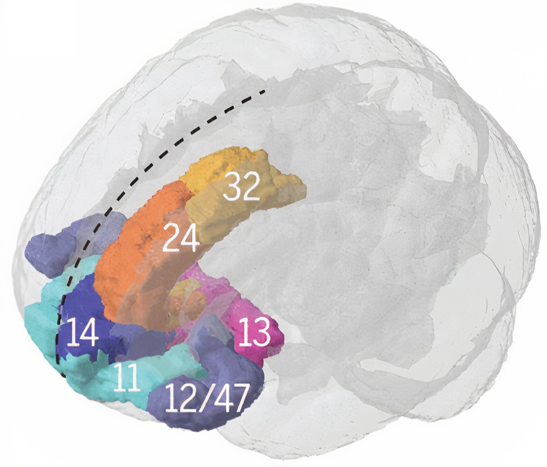
.jpg)
Brodmann areas
The insular cortex is associated with Brodmann areas 13, 14, and 52.Location
The insular cortex is located deep within the lateral sulcus, separating the frontal and parietal lobes from the temporal lobe.Connections
The insular cortex has widespread connections with various brain regions, including the prefrontal cortex, parietal cortex, temporal cortex, and limbic structures (Augustine, 1996).Participation in brain networks
The insular cortex is involved in multiple brain networks, including the salience network, and is responsible for detecting and responding to salient stimuli (Menon & Uddin, 2010).Functions
The insular cortex involves various functions, including interoception, emotional processing, pain perception, and cognitive control (Craig, 2009).Brodmann Area Involvement in Clinical Disorders
The clinical correlations between psychiatric and neurological disorders and specific Brodmann areas provide valuable insights into the neural mechanisms of these conditions. Understanding the abnormalities in these regions can offer critical information for diagnosis, treatment, and the development of targeted therapies. This section explores the associations between specific Brodmann areas and disorders including ADHD, autism spectrum disorder, bipolar disorder, major depressive disorder, panic disorder, PTSD, schizophrenia, and substance use disorder.
Attention-Deficit Hyperactivity Disorder (ADHD)
ADHD is associated with abnormalities in multiple Brodmann areas, including BAs 44/45 (Broca's area), 8/9, 10, 11, 46 (frontal regions), 7, 39, 40 (parietal regions), 4 (motor cortex), 30 (cingulate gyrus), 21, 38 (temporal regions), and 6 (premotor cortex). These abnormalities contribute to the diverse cognitive, attentional, and motor deficits observed in individuals with ADHD.
In individuals with ADHD, Broca's area (BA 44 and 45) exhibits functional disturbances, particularly a lower fractional amplitude of low-frequency fluctuations (fALFF). This dysfunction is linked to language deficits, with different ADHD subtypes affecting distinct parts of Broca's area: Area 44 in the inattentive subtype and Area 45 in the combined subtype (Pikusa & Jończyk, 2015; Silk et al., 2005).
In the frontal regions, children with ADHD display grey matter deficits in the right superior frontal gyrus (BA 8 and 9), suggesting disruptions in attentional networks (Overmeyer et al., 2001). Adolescents with ADHD show decreased activation in the middle frontal gyrus (Area 10) and the dorsolateral prefrontal cortex (Area 46), indicating widespread frontal lobe dysfunction (Silk et al., 2005). Abnormalities in the ventromedial orbitofrontal cortex (Area 11) are linked to motivational deficits in adults with ADHD (Farré-Colomés et al., 2021).
The parietal regions, including the superior parietal lobe (Area 7) and the inferior parietal lobule (Areas 39 and 40), also show decreased activation in ADHD, implicating these areas in attentional and action-attentional systems (Silk et al., 2005). Children with ADHD exhibit decreased activation in the primary motor cortex (Area 4) during motor tasks, suggesting anomalies in motor development (Gaddis et al., 2015; Gilbert et al., 2011, 2019).
The right posterior cingulate gyrus (Area 30) shows grey matter deficits in children with ADHD, which may contribute to difficulties in attentional control (Overmeyer et al., 2001). Structural alterations are also observed in the temporal regions, with lower cortical thickness in the fusiform gyrus and temporal pole (Areas 21 and 38) in children with ADHD (Hoogman et al., 2019; Karalok et al., 2019; Lake et al., 2019; McLaughlin et al., 2014). Increased connectivity in the premotor cortex (Area 6) may be associated with challenges in motor planning and execution (Hoshi & Tanji, 2007; Oldehinkel et al., 2016; Sörös et al., 2019; Suskauer et al., 2008).
Autism Spectrum Disorder (ASD)
Research indicates that abnormalities in Brodmann areas 24, 44, 45, and 10 are associated with Autism Spectrum Disorder. These abnormalities include changes in neuron size and density, gray matter volume, and functional connectivity, which are linked to the social and communication deficits characteristic of ASD. In the anterior cingulate cortex (BA 24), there is a notable reduction in cell size and cell packing density, specifically in areas 24b and 24c, along with elevated levels of glial fibrillary acidic protein (GFAP) in the white matter, indicating increased astrocyte activation. During verbal memory tasks, decreased glucose metabolism is observed in BA24, and dysregulated DNA methylation patterns in this area impact genes related to immune functions and synaptic membranes.
In the inferior frontal cortex (BA 44 and 45), there is a decrease in the size of pyramidal neurons, which play crucial roles in language processing and social behaviors (Jacot-Descombes et al., 2012). Reduced gray matter volume in the pars opercularis (BA44) and pars triangularis (BA45) is linked to difficulties in social communication (Yamasaki et al., 2010).
The anterior prefrontal cortex (BA 10) exhibits abnormal functional connectivity between the right and left hemispheres, correlating with the severity of social deficits (Kikuchi et al., 2013). Similar to BA24, BA10 also shows dysregulated DNA methylation affecting genes involved in immune response and synaptic function (Nardone et al., 2014).
Other areas affected include the medial/cingulate regions (BAs 32, 24, and 25), where lower glucose metabolism is observed during verbal memory tasks (Deery et al., 2022; Hazlett et al., 2004, 2010). There is also a reduction in white matter volumes in the posterior frontal lobe and along the cingulate arch (Aalst et al., 2021; Gardener et al., 2016).
Bipolar Disorder (BD)
Research indicates that abnormalities in several Brodmann areas, including BAs 9, 24, 38, 41, 42, 46, and 10, are associated with bipolar disorder. These abnormalities span structural changes such as reduced glial numbers and cortical volume, as well as functional and molecular disruptions including altered gene expression and DNA methylation. In BA 9 (part of the dorsolateral prefrontal cortex), abnormalities in DNA methylation and gene expression indicate its role in the pathophysiology of BD, and this area shows decreased numbers and density of glial cells, suggesting disruptions in neurochemical regulation.
The subgenual prefrontal cortex (BA 24) exhibits reduced volume and fewer glial cells, associated with familial forms of BD. This area is significant for mood regulation and reflects neurobiological changes linked to the disorder (Öngür et al., 1998; Scarr et al., 2019).
In the temporal pole (BA 38), notable differences in DNA methylation are associated with BD, indicating involvement in the molecular mechanisms underlying the disorder (Ho et al., 2019). In the primary auditory cortex (BA 41 and 42), decreased BOLD signals in response to auditory stimuli have been identified as potential biomarkers for BD (Okamoto et al., 2022).
BA 46, another region of the dorsolateral prefrontal cortex, shows altered gene expression with specific genes exhibiting differential expression in BD, suggesting disruptions in neuronal network functions (Nakatani et al., 2006; Vizueta et al., 2012). The prefrontal cortex (BA 10) also presents changes in gene expression in BD, highlighting complex regional-specific changes in cortical gene expression (Scarr et al., 2019; Vizueta et al., 2012).
Major Depressive Disorder (MDD)
Research consistently implicates several Brodmann areas in the pathology of major depressive disorder, including the anterior cingulate cortex (BA 24 and BA 32), dorsolateral prefrontal cortex (BA 9 and BA 46), ventromedial prefrontal cortex (BA 10), orbitofrontal cortex (BA 13 and BA 47/12), and subgenual cingulate cortex (BA 25). In the anterior cingulate cortex, structural abnormalities and reduced volume in the subgenual part of BA 24 are linked to familial forms of MDD, and elevated levels of tumor necrosis factor (TNF) have been observed in BA 24 among MDD patients, suggesting the involvement of pro-inflammatory pathways. Abnormalities in BA 32 have been associated with antidepressant treatment.
In the dorsolateral prefrontal cortex (BA 9 and 46), both structural and functional abnormalities have been connected to MDD. In BA 9, changes in connectivity and increased fractional amplitude of low-frequency fluctuation (fALFF) are noted (Gao et al., 2021; Lai & Wu, 2015; Ye et al., 2012; Vasic et al. 2008; Zhukovsky et al., 2020). Elevated TNF levels in BA 46 indicate a role in the disorder's pathophysiology (Dean et al., 2010).
The ventromedial prefrontal cortex (BA 10) shows abnormal functional connectivity linked to emotional regulation deficits in MDD patients (Almeida et al., 2011; Johnstone et al., 2007; Wackerhagen et al., 2017; Young et al., 2016). Epigenetic modifications, such as methylation changes, have been identified in BA 9 and BA 38 (Ho et al., 2019).
In the orbitofrontal cortex (BA 13 and BA 47/12), reduced functional connectivity in BA 13 is associated with depressive symptoms, while increased functional connectivity in BA 47/12 correlates with negative self-perception in MDD (Cheng et al., 2016). The subgenual cingulate cortex (BA 25) exhibits reduced functional connectivity with regions involved in emotional regulation (Peng et al., 2020), and methylation changes in BA 25 have been consistently found in independent brain samples (Åberg et al., 2018).
Panic Disorder
Research indicates that abnormalities in Brodmann areas 11, 25, 32, and 15, as well as regions within the prefrontal cortex and parahippocampal gyrus, are associated with panic disorder. During panic attacks, there is a noticeable decrease in regional cerebral blood flow (rCBF) in the right orbitofrontal cortex (BA 11) and the prelimbic cortex (BA 25). This reduction is also observed in the anterior cingulate cortex (BA 32), which exhibits mixed patterns of hyperactivation and hypoactivation during emotional processing.
The parahippocampal gyrus in panic disorder patients shows significant abnormal asymmetry in cerebral blood flow. The anterior temporal cortex (BA 15) experiences decreased rCBF during panic attacks (Hasler et al., 2007). Key regions within the prefronto-limbic network, particularly areas within the PFC, show selective deficits in emotional processing among panic disorder patients (Hasler et al., 2007; Shang et al., 2014). Dysfunctional communication within frontotemporal structures is indicated by decreased coherence imaging values (Shang et al., 2014; Speer et al., 2003).
Increased resting-state functional connectivity between the thalamus and insula suggests excessive sensitivity to external information in panic disorder patients, highlighting a potential neural mechanism underlying their heightened responsiveness to stimuli (Feldker et al., 2016, 2019; Zhou et al., 2022).
Post-Traumatic Stress Disorder (PTSD)
PTSD is associated with abnormalities in the anterior cingulate cortex (BA 32 and BA 24), medial prefrontal cortex (BA 10 and BA 11), dorsolateral prefrontal cortex (BA 46), insula (BA 13), orbitofrontal cortex (BA 25), and sensorimotor areas (BA 4/6). These regions are involved in emotional regulation, memory processing, and response to trauma-related stimuli. In individuals with PTSD, the DLPFC (BA 46) shows decreased activity and signs of mitochondrial dysfunction, and the anterior cingulate gyrus (BA 32) exhibits less activation compared to those without PTSD.
The medial prefrontal cortex (BA 10 and 11) shows reduced activation in PTSD subjects (Etkin & Wager, 2007; Herringa et al., 2012; Manthey et al., 2021; Sartory et al., 2013). The activation levels in BA 10 correlate with PTSD symptoms during threat processing. The insula (BA 13) demonstrates increased delta slow waves linked to worsening PTSD symptoms over time (Harricharan et al., 2019; Herringa et al., 2012; Rabinak et al., 2011), and differences in functional connectivity in the insula are observed between PTSD and non-PTSD individuals (Rabinak et al., 2011; Sripada et al., 2012; Zhang et al., 2016).
During symptom provocation in PTSD patients, there is increased regional cerebral blood flow (rCBF) in the right sensorimotor areas (BA 4 and 6), while lowered rCBF is observed in the right retrosplenial cortex (BA 26, 29, and 30) (Pissiota et al., 2000). Structural and functional changes in the hippocampus and amygdala are consistently observed in PTSD, highlighting their crucial roles in memory and emotional processing (Chen et al., 2018; Hull, 2002).
Schizophrenia
Abnormalities in frontal (BAs 4, 6, 8, 9, 10, 44, 46, 47), temporal (BAs 20, 21, 22, 37, 39, 42), and cingulate (BAs 24, 25, 29, 30, 31) cortices are associated with schizophrenia. These include disrupted intercorrelations, altered receptor binding, gene expression changes, and structural deficits. Significant reductions in gray matter volume have been observed in the left anterior hippocampus-amygdala, left parahippocampal gyrus, and left superior temporal gyrus, with correlations between the volume of the left posterior superior temporal gyrus and the severity of thought disorders.
Disruptions in D2 dopamine receptor patterns have been identified in the perirhinal, superior, and inferior temporal cortices (BA 20, 22, 37, 39, and 42). These disruptions are distinct to the temporal lobe and are not attributable to long-term antipsychotic treatment, suggesting a specific role in auditory hallucinations and other positive symptoms (Goldsmith et al., 1997; Joyce et al., 1997).
In schizotypal personality disorder, lower fractional anisotropy (FA) in the left temporal lobe (BA 20, 21, and 22) indicates alterations in white matter microstructure (Chan et al., 2018; Ellison-Wright & Bullmore, 2009; Hazlett et al., 2011; Lee et al., 2016; Szeszko et al., 2008). Schizophrenia is also associated with a thinner cortex and reduced surface area in the temporal lobe, with the most significant cortical thickness reductions at the temporal pole (Kaur et al., 2020; McCarley et al., 1999; Shenton et al., 1992). Progressive volumetric changes over time, particularly decreased temporal white matter volume, indicate ongoing structural abnormalities (Kaur et al., 2020; Mathew et al., 2014; Olabi et al., 2011).
Enhanced functional connectivity between the left DLPFC and the left mid-posterior temporal lobe has been identified in patients experiencing their first episode of schizophrenia (Zhou et al., 2007), a connectivity pattern that may contribute to the cognitive and perceptual disturbances characteristic of the disorder.
Substance Use Disorder (SUD)
Abnormalities in specific Brodmann areas, particularly BA 10, BA 24, BA 30, BA 18, BA 21/22, and BA 19, are associated with substance use disorder. These areas are involved in critical functions such as self-reflection, emotional regulation, memory, and executive function, which are often impaired in individuals with SUD.
In adolescents with SUD, there is reduced activity in BA 10 (superior, medial, and middle frontal gyrus), suggesting difficulties in self-referential evaluation and future planning (Dalwani et al., 2014). The anterior cingulate cortex (BA 24) shows abnormal activity, impacting emotional behavior and executive function in individuals with SUD.
Conclusion
The examination of specific Brodmann areas reveals significant correlations with various psychiatric and neurological disorders, underscoring the importance of these brain regions in disease pathology. ADHD features disruptions in regions related to attention and executive function. Autism spectrum disorder is characterized by changes in areas affecting social and communication skills. Bipolar disorder involves alterations in areas associated with mood regulation.
Panic disorder and PTSD both involve the prefronto-limbic network, impacting emotional and stress responses. Schizophrenia is associated with widespread disruptions affecting cognition and perception, and substance use disorder involves regions critical for executive function and emotional regulation. For biofeedback practitioners, these Brodmann area findings provide a neuroanatomical framework for understanding the EEG patterns and connectivity abnormalities observed in clinical assessment.
Check Your Understanding
- Why do source localization reports name Brodmann areas rather than electrode sites?
- Which Brodmann area lies beneath F3 and F4, and what functions does it support?
- Which areas underlie P3 and P4, and why does that matter for reading and spatial attention?
- What limits the precision of the translation between a 10-20 electrode site and a Brodmann area?
- How would you explain to a referring clinician why a report mentioning Brodmann area 46 is relevant to a complaint about working memory?
Cutting-Edge Topics in qEEG Research
Beyond Brodmann: Multimodal Parcellation
Brodmann had one kind of evidence, the stained section, and one observer, himself. Glasser and colleagues (2016) parcellated the human cortex using four kinds of evidence at once: cortical thickness and myelin content, task-based activation, resting-state functional connectivity, and topographic organization. Their map contains 180 areas per hemisphere, roughly three and a half times Brodmann's count, and it was validated on a separate group of participants. Many of Brodmann's borders survived the exercise intact, which is a striking result for work done with a microscope in 1909.
Probabilistic Atlases and the Problem of Individual Variability
Brodmann drew borders as lines, but real borders move from brain to brain. Amunts and Zilles (2015) argued that the honest representation of a cortical area is a probability map showing how likely each voxel is to belong to that area across a sample of brains. The Julich-Brain atlas implements exactly that, mapping cytoarchitectonic areas in a large postmortem sample and expressing each one as a three-dimensional probability distribution (Amunts et al., 2020). For source localization this matters directly, since labeling a current-density maximum as area 46 in an individual client is a probabilistic statement rather than a certainty.
Brodmann-Guided Source Neurofeedback
Once source estimates are expressed in Brodmann coordinates, they can be used as training targets rather than only as findings. Methods such as sLORETA compute current density throughout the cortical volume, which allows a feedback signal to be derived from a specified region rather than from a single electrode (Pascual-Marqui, 2002). The appeal is obvious: a clinician who suspects dorsolateral prefrontal underactivation can target area 46 rather than approximating it with F3. The corresponding caution is equally obvious, since the spatial resolution of any scalp-based inverse solution is limited and adjacent areas are not independently addressable.
Cytoarchitecture as a Predictor of Electrophysiology
An underexplored question is how directly cellular architecture predicts the rhythms you record. The thick layer IV that marks primary sensory cortex, including the striate band of area 17, receives dense thalamic input, and thalamocortical loops are the engine of the alpha rhythm (Hubel & Wiesel, 1968). Areas with sparse layer IV and prominent layer III, such as association cortex, show different spectral signatures. Linking Brodmann's histology to band-specific EEG features would give the map a second life as an electrophysiological prediction rather than only an anatomical description.
Assignment
Now that you have completed this unit, describe the important functions of the dorsolateral prefrontal cortex. Name the Brodmann area involved, identify the 10-20 electrode sites that sit above it, and explain in your own words what a client with dorsolateral prefrontal dysfunction might report in daily life. Close by stating one reason a qEEG finding at that location should be interpreted cautiously rather than as a diagnosis.
Glossary
affective network: a network including the anterior cingulate, entorhinal, and temporal cortices, posterior parietal cortex, globus pallidus, substantia nigra pars reticulata, and mediodorsal thalamus that monitors performance and signals when greater executive control is needed.
amygdala: a group of medial temporal-lobe nuclei involved in salience evaluation, associative learning, affective memory, and coordination of defensive and autonomic responses.
angular gyrus: a region of the parietal lobe near the junction of the temporal and occipital lobes that corresponds to Brodmann area 39. It plays a role in language processing, attention, spatial cognition, and the integration of sensory information.
anterior cingulate cortex (ACC): medial frontal cortex involved in cognitive control, motivation, affect, pain processing, and autonomic regulation, with functionally heterogeneous subdivisions.
anterior prefrontal cortex (aPFC): the most anterior region of the prefrontal cortex, corresponding to Brodmann area 10. It supports complex cognitive processes such as planning, decision-making, working memory, prospective memory, and abstract reasoning.
auditory cortex: the region of the temporal lobe that processes auditory information within the dorsal and ventral streams, supporting sound localization, pitch discrimination, and speech comprehension.
Brodmann area (BA): a numbered cytoarchitectural zone of the cerebral cortex mapped by Brodmann from Nissl staining; the numbering runs to 52, but areas 12-16 and 48-51 were defined in non-human species.
central autonomic network (CAN): interconnected forebrain, hypothalamic, brainstem, and spinal regions that integrate internal-state information and regulate autonomic output.
central executive network: a frontoparietal control network anchored in the dorsolateral prefrontal cortex and posterior parietal cortex that maintains and manipulates information for goal-directed behavior.
cingulate cortex: a limbic region on the medial surface of the frontal and parietal lobes, comprising anterior (Brodmann areas 24, 32, 33) and posterior (areas 23, 29, 30, 31) divisions serving emotion, memory, attention, and cognitive control.
cytoarchitecture: the microscopic appearance and organization of neurons within the cortex, including cell types, cell densities, and the arrangement of cortical layers.
default mode network (DMN): a cortical network of sites located in frontal, temporal, and parietal regions that is most active during introspection and daydreaming and relatively inactive when pursuing external goals.
dorsal anterior cingulate cortex (dACC): the dorsal subdivision of the anterior cingulate cortex, comprising Brodmann areas 24 and 32. It plays a role in cognitive control, decision-making, and conflict monitoring.
dorsal entorhinal cortex (DEC): a medial temporal lobe region often associated with Brodmann areas 28 and 34, although the entorhinal cortex is allocortex and does not map neatly onto Brodmann's cytoarchitectonic divisions. It is involved in spatial memory and navigation.
dorsal posterior cingulate cortex (dPCC): the dorsal part of the posterior cingulate cortex, composed primarily of Brodmann areas 23 and 31. It is involved in self-referential thought, memory, and spatial awareness.
dorsolateral prefrontal cortex: lateral prefrontal cortex involved in working memory, cognitive control, planning, rule use, and goal-directed behavior; simple left-positive and right-negative mappings are inadequate.
ectosplenial cerebral cortex: a narrow band of retrosplenial cortex in the isthmus of the cingulate gyrus corresponding to Brodmann area 26, involved in spatial memory, navigation, and contextual processing.
frontal eye field (FEF): a premotor region in the caudal middle frontal gyrus at its junction with the precentral gyrus, corresponding to Brodmann area 8, that controls voluntary eye movements and directs gaze toward attended targets.
functional networks: sets of brain regions whose activity is correlated over time, in contrast to structural networks defined by axonal projections and pathways.
fusiform gyrus: a region on the ventral surface of the temporal and occipital lobes associated with Brodmann areas 37 and 19. It is involved in face recognition, object recognition, and the processing of color and visual form.
inferior temporal gyrus (ITG): a region of the inferior temporal lobe, primarily Brodmann areas 20 and 37, that supports visual object recognition and semantic memory.
insular cortex (insula): a cortical region buried within the lateral sulcus, comprising Brodmann areas 13, 14, 15, 16, and parts of area 52, that supports interoception, self-awareness, emotion, pain perception, and taste and serves as an integrative hub for the salience network.
middle temporal gyrus (MTG): a temporal lobe convolution between the superior and inferior temporal gyri, corresponding chiefly to Brodmann area 21 and extending posteriorly into area 37, that supports language processing, semantic memory, and visual motion processing.
Nissl stain: a histological stain that binds to the rough endoplasmic reticulum of neurons, making cell bodies visible so that cortical layering and cell density can be examined.
orbitofrontal cortex: prefrontal cortex above the orbits involved in valuation, outcome updating, emotion, sensory integration, and context-appropriate behavior.
parainsular area: a region at the junction of the temporal lobe and insula that is associated with Brodmann areas 13, 14, and 52. It participates in auditory and somatosensory integration and in processing pain and temperature sensations.
pars opercularis: the opercular part of the inferior frontal gyrus, corresponding to Brodmann area 44. It plays a role in language production and forms part of Broca's area.
pars orbitalis: the orbital part of the inferior frontal gyrus, corresponding to Brodmann area 47. It is involved in language processing, social cognition, and emotional regulation.
pars triangularis: the triangular part of the inferior frontal gyrus, corresponding to Brodmann area 45. It is involved in language processing and forms part of Broca's area.
perirhinal cortex (PRC): a medial temporal lobe region comprising Brodmann areas 35 and 36 that supports object recognition, associative memory, and contextual processing.
prefrontal cortex (PFC): the anterior association cortex of the frontal lobes involved in executive control, planning, working memory, valuation, social cognition, and emotion regulation.
primary gustatory cortex (PGC): a region associated with Brodmann area 43, which lies at the opercular part of the inferior frontal gyrus and extends into the insular cortex (Brodmann area 13), responsible for processing taste information.
primary motor cortex: cortex in the precentral gyrus that contributes substantially to execution and control of voluntary movement.
primary somatosensory cortex (S1): cortex in the postcentral gyrus that receives organized thalamic somatosensory input and represents touch, proprioception, and nociceptive features.
primary visual cortex (V1): the occipital lobe region located in the calcarine sulcus (BA 17, also called striate cortex) that receives most visual information from the lateral geniculate nucleus of the thalamus and performs the initial processing of visual information received from the retinas.
pyriform cortex: the ventral temporal lobe region that processes olfactory input, also called primary olfactory cortex and conventionally placed at Brodmann area 27 although it is allocortex.
resting-state functional connectivity (RSFC): a neuroimaging method that identifies brain networks whose regions show synchronous activity when a person is not performing an explicit task.
retinotopic organization: the mapping of spatial information from the visual field onto the surface of the visual cortex, so that neighboring points in the visual scene are represented by neighboring points in cortex.
retrosplenial cingulate cortex: a region in the posterior part of the cingulate cortex comprising Brodmann areas 29 and 30, involved in spatial memory, navigation, and contextual processing.
retrosubicular area: a small medial temporal lobe region of the hippocampal formation designated Brodmann area 48, involved in spatial navigation and memory.
salience network: a large-scale network centered on anterior insula and dorsal anterior cingulate cortex that detects behaviorally relevant events and coordinates network switching.
secondary visual cortex (V2): a region of the occipital lobe adjacent to the primary visual cortex, corresponding to Brodmann area 18, that processes shape, color, and spatial orientation.
sLORETA: standardized low-resolution brain electromagnetic tomography, a version of LORETA that standardizes the current density estimate to achieve zero localization error under ideal conditions (Pascual-Marqui, 2002).
somatosensory association cortex (SAC): a posterior parietal region encompassing Brodmann areas 5 and 7 that integrates and interprets somatosensory information such as touch, pain, temperature, and proprioception.
somatotopic organization: the mapping of body parts onto the cortical surface, in which the amount of cortex devoted to a region reflects the precision required rather than the physical size of that region.
striate cortex: the primary visual cortex (BA 17), responsible for processing basic visual information such as orientation, spatial frequency, and color.
subgenual ventromedial prefrontal cortex (subgenual vmPFC): the ventromedial prefrontal region beneath the genu of the corpus callosum, comprising Brodmann areas 25 and 24b, involved in emotion regulation, decision-making, and social cognition.
superior temporal gyrus (STG): the uppermost gyrus of the temporal lobe, spanning Brodmann areas 22, 41, and 42, which supports auditory processing, language comprehension, and social cognition; its posterior part contains Wernicke's area.
supplementary motor cortex (SMA): a region on the medial aspect of the superior frontal gyrus corresponding to Brodmann area 6, involved in planning and coordinating complex movements and in motor learning.
supramarginal gyrus: a part of the inferior parietal lobule corresponding to Brodmann area 40, involved in phonological processing, language perception, attention, spatial cognition, and the integration of sensory information.
temporopolar area: the most anterior part of the temporal lobe, associated with Brodmann area 38. It is involved in olfactory processing, social cognition, and semantic memory.
ventral anterior cingulate cortex (vACC): the ventral subdivision of the anterior cingulate cortex, comprising Brodmann areas 24 and 25 and, in some parcellations, area 33. It is involved in emotional regulation, attention, and pain processing.
ventral entorhinal cortex (vEC): a medial temporal lobe region associated with Brodmann area 28, although the entorhinal cortex is allocortex and is not readily defined by Brodmann areas. It is involved in object recognition, memory, and contextual processing.
ventral posterior cingulate cortex (vPCC): the ventral part of the posterior cingulate cortex, comprising primarily Brodmann area 23. It supports self-referential thought, episodic memory retrieval, and emotional processing.
visual association cortex: a set of occipital and temporal lobe regions comprising Brodmann areas 18, 19, 21, 22, and 37 that includes areas V3, V4, and V5. It is responsible for higher-level visual processing, including object recognition, motion perception, and spatial awareness.
Wernicke's aphasia: a language disorder following damage to Wernicke's area, characterized by fluent but nonsensical speech and impaired comprehension of language.
Wernicke's area: the area of the temporoparietal cortex (BA 22) of the dominant hemisphere specialized for speech comprehension. Damage can result in an inability to understand the meaning of speech and construct intelligible sentences.
Test Yourself on ClassMarker
Click the button below to take a 10-question exam over this entire unit. There is no password.
Review Flash Cards on Quizlet
Click the button below to review our chapter flash cards.
References
Amunts, K., Mohlberg, H., Bludau, S., & Zilles, K. (2020). Julich-Brain: A 3D probabilistic atlas of the human brain's cytoarchitecture. Science, 369(6506), 988-992. https://doi.org/10.1126/science.abb4588
Amunts, K., & Zilles, K. (2015). Architectonic mapping of the human brain beyond Brodmann. Neuron, 88(6), 1086-1107. https://doi.org/10.1016/j.neuron.2015.12.001
Brodmann, K. (1909). Vergleichende Lokalisationslehre der Großhirnrinde in ihren Prinzipien dargestellt auf Grund des Zellenbaues. Barth.
Geschwind, N. (1970). The organization of language and the brain. Science, 170(3961), 940-944. https://doi.org/10.1126/science.170.3961.940
Glasser, M. F., Coalson, T. S., Robinson, E. C., Hacker, C. D., Harwell, J., Yacoub, E., Ugurbil, K., Andersson, J., Beckmann, C. F., Jenkinson, M., Smith, S. M., & Van Essen, D. C. (2016). A multi-modal parcellation of human cerebral cortex. Nature, 536(7615), 171-178. https://doi.org/10.1038/nature18933
Hubel, D. H., & Wiesel, T. N. (1968). Receptive fields and functional architecture of monkey striate cortex. Journal of Physiology, 195(1), 215-243. https://doi.org/10.1113/jphysiol.1968.sp008455
Kaas, J. H. (1993). The organization of somatosensory cortex in primates. In A. D. Keller & J. H. Asanuma (Eds.), Cerebral cortex (pp. 195-245). Springer.
Miller, E. K., & Cohen, J. D. (2001). An integrative theory of prefrontal cortex function. Annual Review of Neuroscience, 24, 167-202. https://doi.org/10.1146/annurev.neuro.24.1.167
Pascual-Marqui, R. D. (2002). Standardized low-resolution brain electromagnetic tomography (sLORETA): Technical details. Methods and Findings in Experimental and Clinical Pharmacology, 24(Suppl D), 5-12.
Penfield, W., & Boldrey, E. (1937). Somatic motor and sensory representation in the cerebral cortex of man as studied by electrical stimulation. Brain, 60(4), 389-443. https://doi.org/10.1093/brain/60.4.389
Rizzolatti, G., Fogassi, L., & Gallese, V. (2002). Motor and cognitive functions of the ventral premotor cortex. Current Opinion in Neurobiology, 12(2), 149-154. https://doi.org/10.1016/S0959-4388(02)00308-2
Return to Top