
Behavioral Correlates to Brain Regions and Networks
What You Will Learn in This Chapter
You know that F3 sits over the left dorsolateral prefrontal cortex. But what does that region actually do, and what happens to your client when it is underactive? This chapter connects scalp locations to cortical anatomy, and cortical anatomy to behavior.
You will tour the five cortical lobes and the insula, the white matter bundles that link them, and the subcortical structures that feed them: thalamus, basal ganglia, hippocampus, amygdala, and septal nuclei. Each region is presented with its functions and the clinical concerns that follow when it malfunctions.
The chapter closes with a deep dive through the Brodmann area profiles. Each profile gives you the same six facts: which Brodmann areas are involved, where they sit, what they connect to, which networks they join, what they do, and their role in clinical disorders. A final section runs the same material in reverse, from a diagnosis to the areas implicated in it, for ADHD, autism spectrum disorder, bipolar disorder, major depressive disorder, panic disorder, PTSD, schizophrenia, and substance use disorder. Treat both as a reference you return to when you are planning a montage.
IQCB Blueprint Coverage: This unit addresses Behavioral Correlates of Brain Regions (II. Neuroscience), Cortical and Subcortical Anatomy (II. Neuroscience), and Brodmann Areas and Their Functions (II. Neuroscience).
Learning Objectives
After completing this section, you will be able to:
Identify the five cortical lobes and the insula, and name the electrode sites that overlie each.
Describe the principal functions of the primary motor cortex, motor association cortex, Broca's area, and the prefrontal subdivisions.
Contrast left and right frontal lobe functions and the clinical concerns associated with each.
Distinguish association, projection, and commissural fiber bundles and give an example of each.
Describe the functions of the thalamus, basal ganglia, hippocampus, amygdala, and septal nuclei.
Explain how thalamocortical and corticothalamic feedback loops generate EEG rhythms.
Explain what Brodmann areas represent, and why cytoarchitecture alone does not capture functional organization.
Look up any Brodmann area and state its location, connections, network membership, functions, and clinical associations.
Name the Brodmann areas implicated in ADHD, autism spectrum disorder, bipolar disorder, major depressive disorder, panic disorder, PTSD, schizophrenia, and substance use disorder, and explain why those correlations cannot stand alone as diagnostic evidence.
Translate a client's presenting problem into candidate cortical regions and the networks that serve them.
We will review Cortical Lobes, Subcortical Structures, and A Deep Dive Into Brodmann Areas.
Listen to the Full-Length Lecture: Cortical Lobes
Cortical Lobes
The cortical lobes are named for the overlying bones of the skull (Breedlove & Watson, 2023).
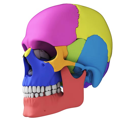
Graphic © Sebastian Kaulitzki/Shutterstock.com.
The cortex is required for executive functions like attention, planning, and problem-solving.
Without a cerebral cortex, a person would be blind, deaf, dumb, and unable to initiate voluntary movement (Bear, Connors, & Paradiso, 2020, p. 205).
The five major cortical regions include the frontal, parietal, temporal, and occipital lobes, and the insula (not shown).
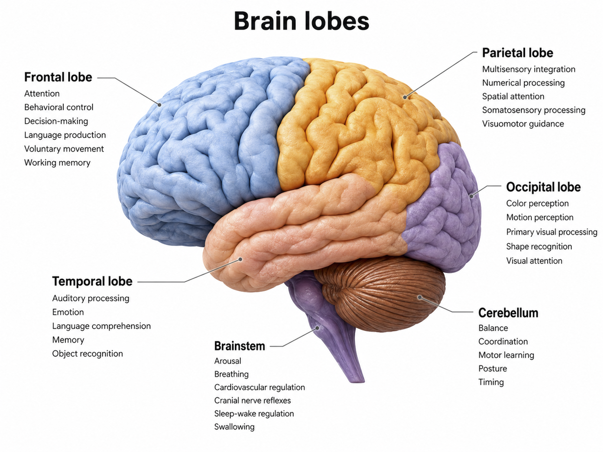
Cortical lobes graphic adapted from © Madrock24/Shutterstock.com.
Frontal Lobes
The frontal lobes (Fp1, Fp2, F7, F3, Fz, F8, F4) consist of the cortex anterior to the central sulcus and include the primary motor cortex, motor association cortex, Broca's area, and prefrontal cortex. These structures collectively support everything from fine motor control to the highest-level executive functions, which is why frontal sites carry so much of the interpretive weight in a qEEG.
Left Hemisphere
Essential functions include working memory, concentration, planning, and positive emotion. The main clinical concern is Major Depressive Disorder (MDD).
Right Hemisphere
Critical functions include declarative memory, social awareness, and negative emotions. Principal clinical concerns include Generalized Anxiety Disorder (GAD), fear, and impaired executive functioning.
Frontal lobe damage may result in impaired flexibility and problem solving, increased risk-taking, changes in social behavior, an inability to use external cues, and deficits in emotional self-regulation.
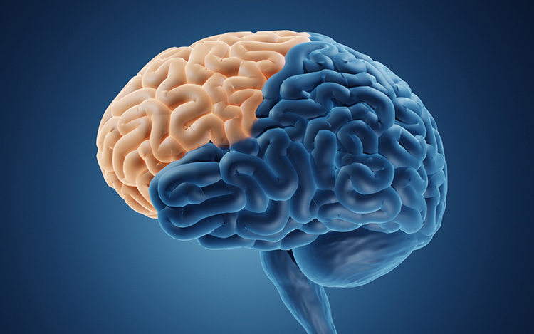
Graphic © ART-ur/Shutterstock.com.
The primary motor cortex is located in the precentral gyrus (Brodmann area 4, BA 4). It organizes the opposite side of the body's muscles and movements required for fine motor coordination in tasks like writing. Lesions can result in loss of motor control, including rigid paralysis. The graphic below, which shows the motor and sensory homunculi, was retrieved from the nccpbwikiproject.
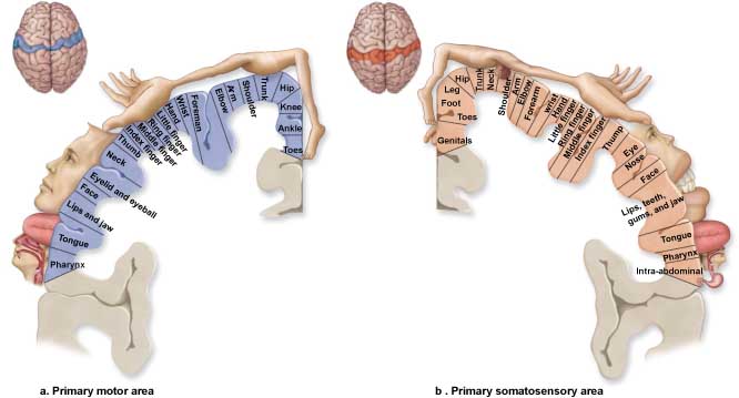
The motor association cortex (premotor cortex) is rostral to the primary motor cortex (BA 6) and helps program and execute movements. Think of the motor association cortex as the piano player and the primary motor cortex as the keyboard (Carlson & Birkett, 2017). The primary and motor association cortex collectively appear to map behaviors rather than specific muscles or movements (Breedlove & Watson, 2023).
Broca's area, which is located in the inferior frontal gyrus (BA 44 and 45) of the dominant hemisphere (F7-T3 in the left hemisphere), is concerned with speech production, grammar, language comprehension, and sequencing (Caplan, 2006). Lesions to Broca's area can produce Broca's aphasia, an inability to produce fluent speech despite relatively intact comprehension. Broca's area receives input from Wernicke's area via the arcuate fasciculus (Breedlove & Watson, 2023).
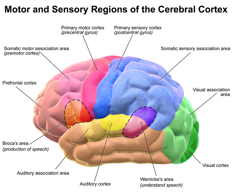
Graphic courtesy of Blausen.com staff "Blausen gallery 2014," Wikiversity Journal of Medicine.
The prefrontal cortex (PFC) (BA 9, 10, 11, 12, 25, 32, 44, 45, 46, 47) is rostral to the motor association area. The PFC participates in executive functions, including attention, working memory, outcome prediction for current and hypothetical actions, goal-directed behavior, problem-solving, planning, and the ability to suppress actions that could lead to unwanted outcomes (Diamond, 2013). The PFC integrates emotion and reward in decision-making (Fuster, 2015).
Important subdivisions of the PFC include the orbitofrontal cortex, ventromedial PFC, and dorsolateral PFC.
The orbitofrontal cortex (OFC) comprises Brodmann areas 10, 11, and 47 in Brodmann's human map, subdivided in the Walker scheme as areas 11, 13, 14, and 47/12 (Kringelbach, 2005). Areas 9 and 46 belong to the dorsolateral prefrontal cortex, not the OFC. The OFC aids planning by evaluating the consequences (rewards and punishments) of our actions and helps generate the motivation to ingest drugs. The profound personality changes suffered by Phineas Gage resulted from damage to this subdivision and the ventromedial PFC. The OFC adjusts decision-making based on the stakes involved, enabling us to switch between significant (investments) and trivial (snacks) choices. It compares current options with recent ones, while the anterior cingulate cortex registers our predictions and prediction errors (Kennerley et al., 2011).
The ventromedial prefrontal cortex (VMPFC) corresponds to the ventromedial reward network (Öngür & Price, 2000) and includes BA 10, 25, 32, and parts of 11 and 12. The VMPFC is implicated in decisions where outcomes are uncertain and moral values must be applied to real situations. Patients with VMPFC damage choose options that lead to immediate reward regardless of future cost and do not learn from their mistakes.
Since they have difficulty reading social cues, they may not recognize deception, irony, or sarcasm (Zald & Andreotti, 2010) and may struggle to control emotional reactions in social situations, particularly anger and violence (Carlson & Birkett, 2017).
The dorsolateral prefrontal cortex (DLPFC) is located in the middle frontal gyrus and includes BA 9 and 46. The DLPFC shares responsibility with cortical and subcortical networks for executive functions like abstract reasoning, cognitive flexibility, decision-making, inhibition, planning, and working memory (Miller & Cummings, 2007). It exercises the highest cortical level of motor control (Hale & Fiorello, 2004).
The left DLPFC is concerned with approach behavior and positive affect. It helps us select positive goals and organizes and implements behavior to achieve them. The right DLPFC organizes withdrawal-related behavior and negative affect and mediates threat-related vigilance. It plays a role in working memory for object location. In unipolar depression and premenstrual dysphoric disorder, the right DLPFC may be more active than the left, producing the alpha asymmetry pattern that is among the most frequently reported frontal findings in the qEEG literature. Read that pattern in context: an asymmetry index is a comparison between two sites, so an artifact, a drowsy epoch, or an unequal impedance at F3 and F4 can manufacture one.
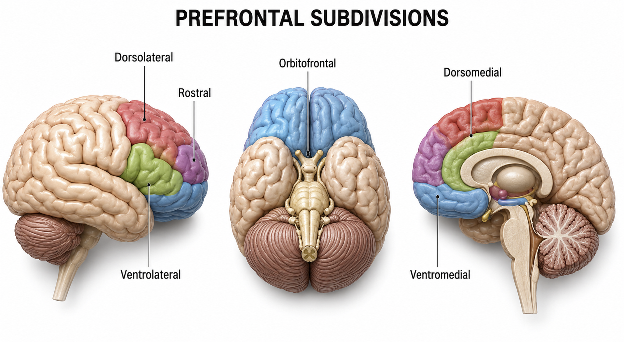
Prefrontal cortex subdivision graphic redrawn for the Tutor series.
Anterior Cingulate Cortex (ACC)
The cingulate cortex has reciprocal connections with the parahippocampal gyri, integrates limbic functions, and is part of the salience network. Cingulate cortical functions include nurturing, grooming, play, and organizing and managing input/output functions.
The anterior cingulate cortex (ACC) (Fpz and Fz) lies above the corpus callosum (BA 24, 32, 33). The dorsal ACC connects to both the PFC and parietal cortex. The ACC plays a vital role in attention, is activated during working memory, and mediates both emotional and physical pain. It has cognitive (dorsal anterior cingulate) and affective (ventral anterior cingulate) conflict-monitoring components. Because the ACC lies on the medial surface, no scalp site records it directly; a midline finding at Fz or Cz reflects the ACC only through volume conduction and source estimation.
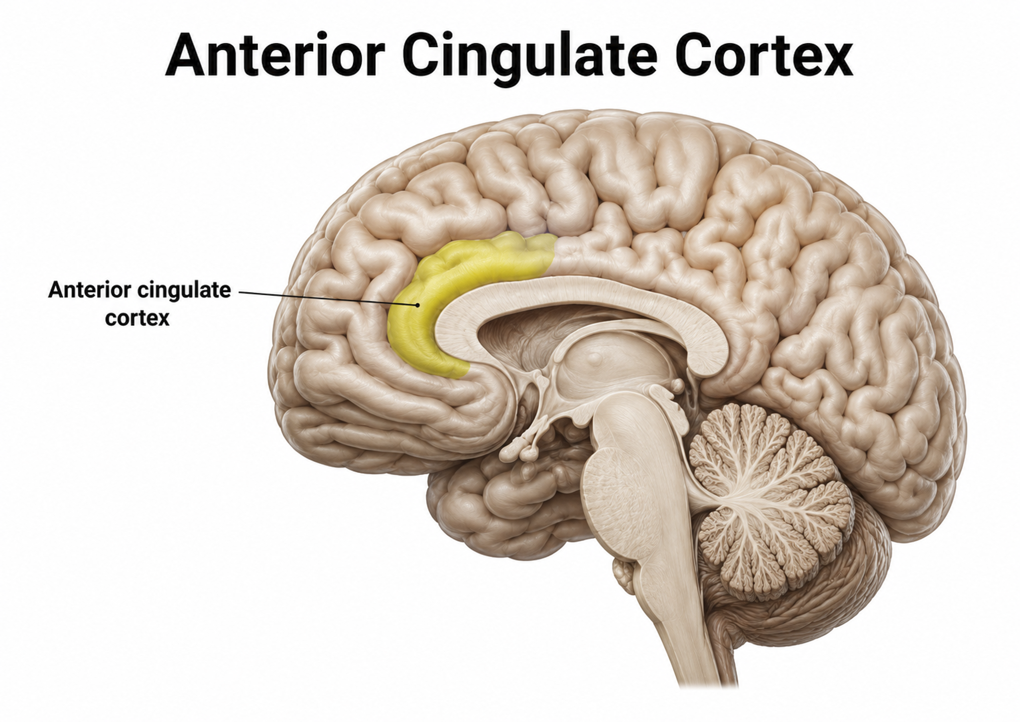
Graphic courtesy of Geoff B. Hall in Wikimedia Commons.

The Stroop test illustrates a cognitive monitoring task where color and name information conflict, while discrepancies between facial and vocal cues present an affective conflict. The anterior cingulate recruits other brain areas to resolve these conflicts.
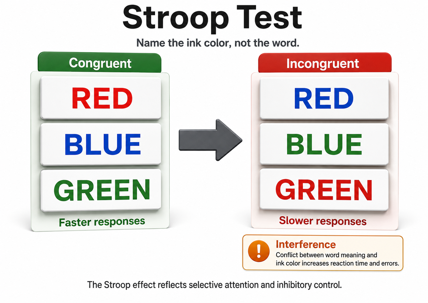
The Stroop test pits the color of the ink against the word it spells.
The anterior cingulate gyrus helps us allocate attention: focusing on a target, disengaging, perceiving options, and making adaptive choices. The anterior cingulate gyrus, prefrontal cortex, and caudate function abnormally in children diagnosed with ADHD during selective attention tasks, and fMRI evaluation showed that these structures can be trained toward normal activity (Beauregard & Levesque, 2006). That finding matters for assessment: an attentional complaint should send you looking at midline and frontal sites together rather than at a single electrode.
The anterior cingulate gyrus is also involved in motivation and the perception of emotional and physical pain. Eisenberger, Lieberman, and Williams (2003) used fMRI to study the brains of subjects who believed that two companions in Cyberball, an online ball-tossing game, had suddenly begun excluding them; their emotional distress activated the anterior cingulate cortex, the same region that evaluates physical pain. deCharms and colleagues (2005) provided real-time fMRI feedback from the rostral anterior cingulate, and subjects learned to increase and decrease its activation, with corresponding increases and decreases in the rated intensity of a noxious thermal stimulus.
Lesions to the cingulate can produce akinetic mutism, a state of markedly reduced spontaneous movement and speech despite preserved wakefulness, in which orienting responses are absent or greatly diminished. Cingulate malfunction can also result in addictive behaviors (alcohol or drug abuse, eating disorders, chronic pain), obsessive-compulsive disorder and OCD spectrum disorders, and “road rage.”
Parahippocampal Gyri
The parahippocampal gyri are located within the medial temporal lobe. They form spatial and nonspatial contextual associations, which serve as building blocks for contextual processing, episodic memory, navigation, and scene processing (Aminoff, Kveraga, & Bar, 2013). They may also play a role in emotional responsiveness.
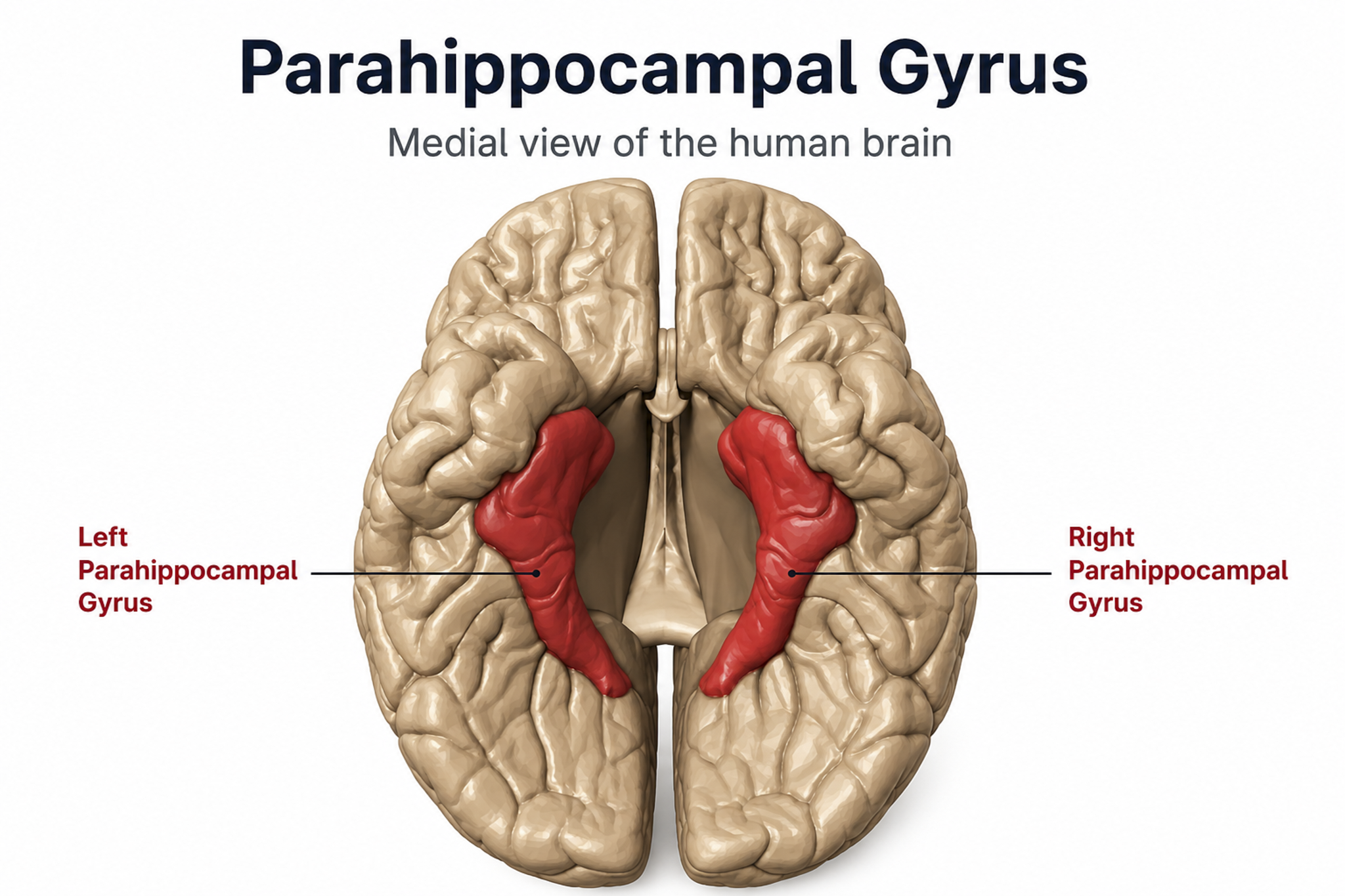
Polygon data were generated by Database Center for Life Science (DBCLS). Creative Commons Attribution-Share Alike 2.1 jp.
Parietal Lobes
The parietal lobes (Pz, P3, P4) are posterior to the frontal lobes (BA 1, 2, 3, 5, 7, 39, 40) and are divided into the primary somatosensory cortex and secondary somatosensory cortex. Their main function is to process somatosensory information like pain and touch.
Left Hemisphere
Major functions include attention, association, complex grammar, math, object names, problem-solving, and somatosensation.
Right Hemisphere
Major functions include body boundary awareness, geometry, guiding reaching with the hands, somatosensation, spatial awareness, and spatial perception (Demos, 2019).
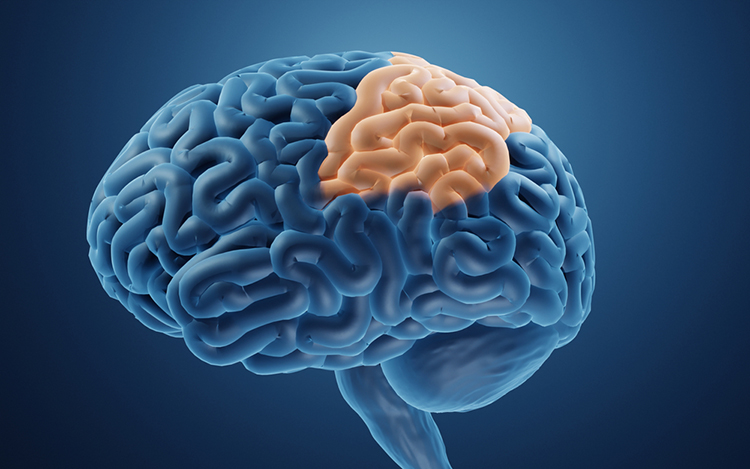
Graphic © ART-ur/Shutterstock.com.
The primary somatosensory cortex (S1) is located in the parietal lobe's postcentral gyrus posterior to the central sulcus (BA 3, 1, and 2). S1 maps touch and pain information from the opposite side of the body. The secondary somatosensory cortex (S2), which lies in the parietal operculum (BA 40 and 43), receives projections from it and maps touch and pain from both sides of the body (Breedlove & Watson, 2023).
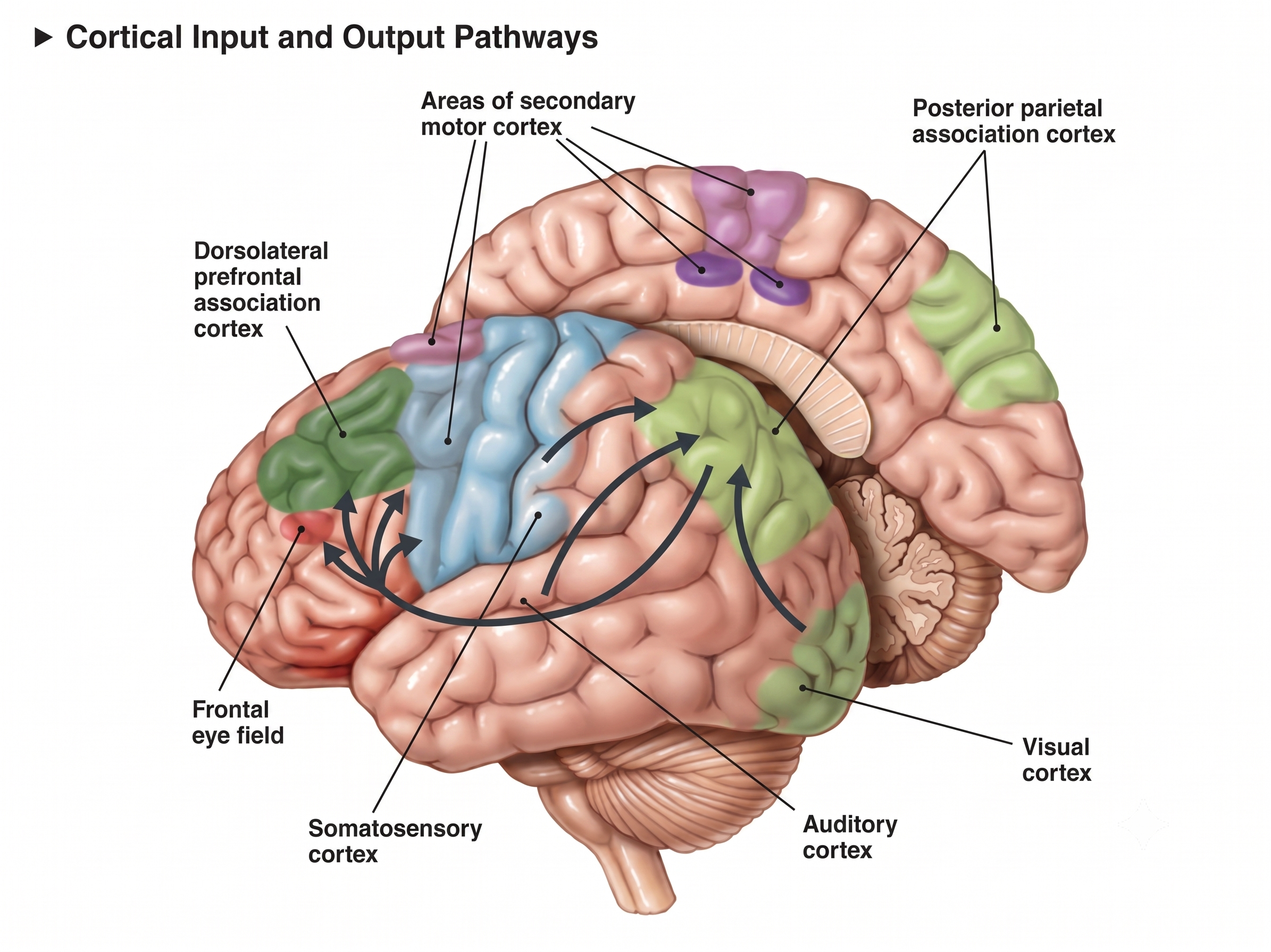
Graphic adapted from Paskari via Wikimedia Commons.
The parietal cortex monitors our preparation for a movement and is responsible for our subjective feeling of intending to move (Sirigu et al., 2004).
The angular gyrus, located near the superior temporal lobe (BA 39), is involved in reading, math, and copying writing.
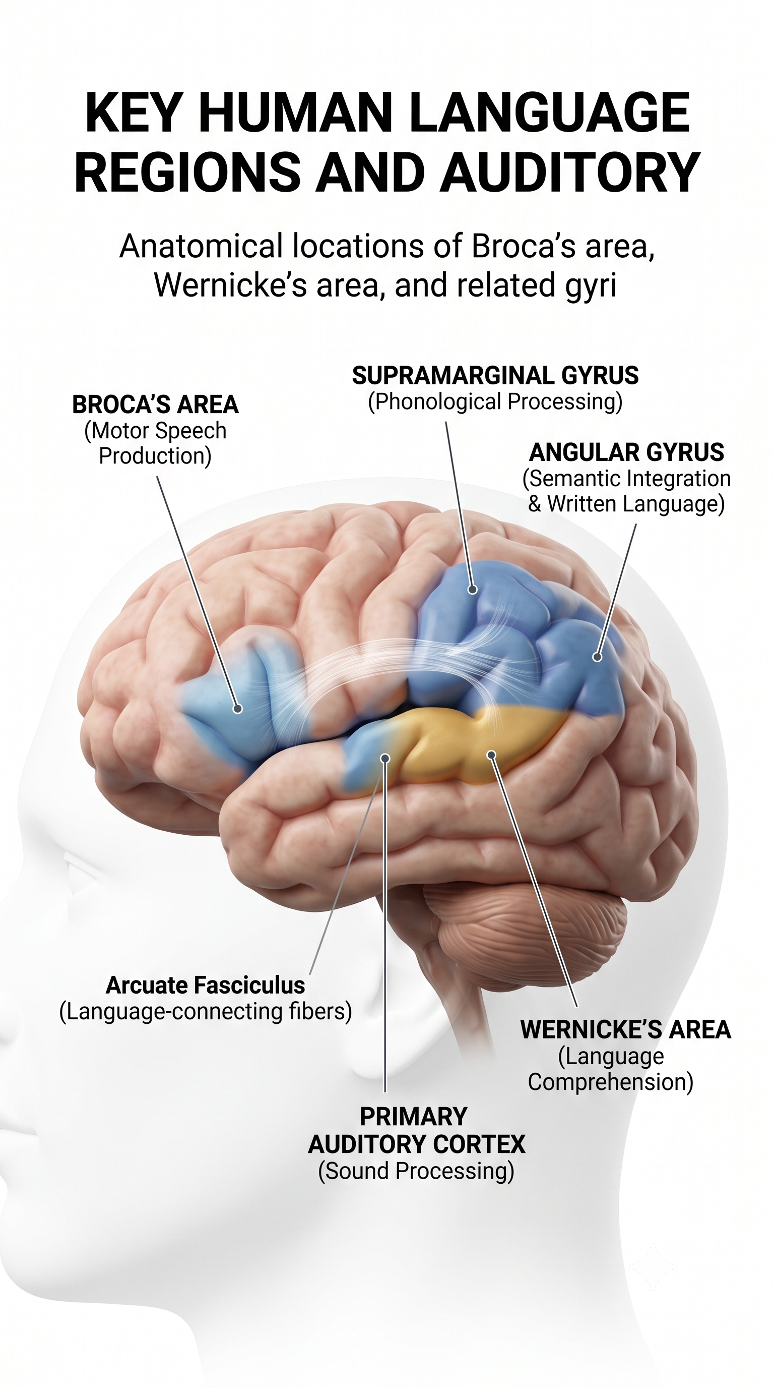
The cortical language areas, including the angular gyrus.
The supramarginal gyrus (BA 40) forms part of the inferior parietal lobule and, like the neighboring angular gyrus (BA 39), functions as a convergence zone where somatosensory, auditory, and visual information are integrated. Positioned at the posterior end of the lateral sulcus, it draws on this multimodal input to support several higher-order functions.
In written language, it contributes to phonological processing during reading and writing, helping map the sounds of speech onto their corresponding letters and words. Its role in verbal working memory reflects its involvement in the phonological store, the component of the phonological loop that briefly holds speech-based information for rehearsal. The right supramarginal gyrus also supports social cognition by helping a person distinguish their own emotional state from that of others, a process central to empathy.
Both the angular gyrus and the supramarginal gyrus lie in the vicinity of the P3 electrode site, so a P3 finding rarely belongs to one of them alone.
Temporal Lobes
The temporal lobes are separated from the rest of the cortical lobes by the Sylvian fissure and lie beneath the temporal bone at the sides of the head, below the T3 and T5 electrode sites on the left and the T4 and T6 sites on the right. They contain numerous Brodmann areas, both laterally (20, 21, 22, 37, 38, 41, and 42) and medially (20, 27, 28, 34, 35, 36, 37, and 38); several of these areas wrap around from the lateral to the medial cortical surface. The temporal lobes process hearing, smell, and taste information and help us understand spoken language and recognize visual objects and faces (Breedlove & Watson, 2023).
Their medial cortex is involved in emotional experience and new memory formation. In the language-dominant hemisphere, usually the left, Wernicke's area lies beneath electrode sites C3, T3, and T5 and encompasses posterior regions of Brodmann areas 22, 41, and 42.
Wernicke's area, located in the temporoparietal cortex (BA 22) of the dominant hemisphere, is specialized for the comprehension of spoken and written language. Damage can result in an inability to understand the meaning of speech and to construct meaningful sentences, a condition known as Wernicke's aphasia, in which speech output remains fluent but is empty of meaning and carries paraphasias and neologisms.
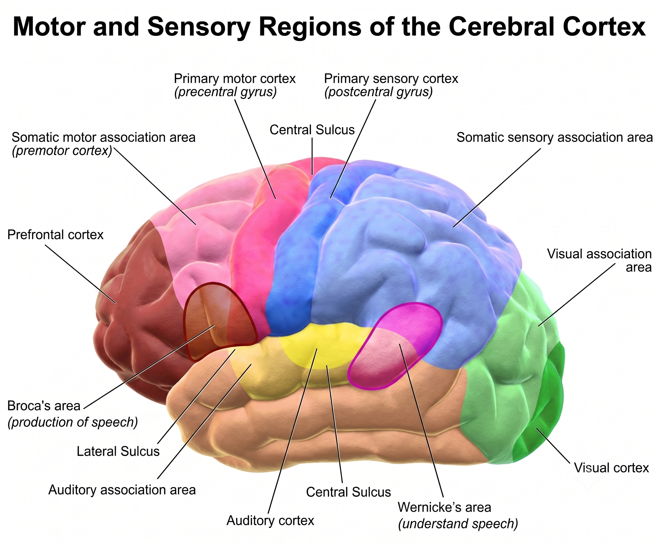
Graphic adapted from Blausen.com in the Wikiversity Journal of Medicine.
Left Hemisphere
Major functions include affect, declarative memories, language comprehension, perception of movement, reading, and word recognition.
Right Hemisphere
Important functions include face and object recognition, music, and social cues (Demos, 2019).
Occipital Lobes
The occipital lobes (Oz, O1, O2) are posterior to the parietal lobes. The primary visual cortex (V1) is located within the calcarine sulcus (BA 17). The occipital lobes process visual information from the eyes in collaboration with the frontal, parietal, and temporal lobes.
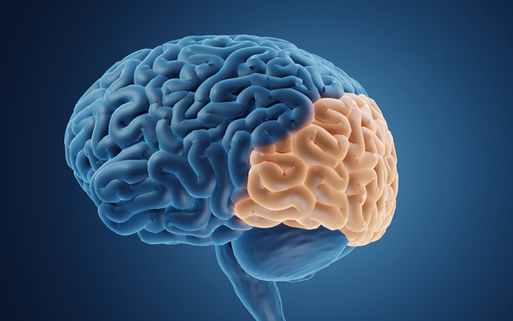
Graphic © ART-ur/Shutterstock.com.
Their primary functions are visual, including the analysis of orientation, color, spatial frequency, illusory contours, and complex patterns like concentric and radial stimuli (Breedlove & Watson, 2023).
The occipital lobes are the primary source of the posterior dominant rhythm, which makes O1, O2, and Oz the sites where eyes-closed alpha is largest and where an eyes-open alpha blocking response is easiest to confirm.
Insular Cortex
The insular cortex lies buried deep within the lateral sulcus, concealed by the frontal, parietal, and temporal opercula, and corresponds to Brodmann area 13. It supports interoception (awareness of internal bodily states), autonomic regulation, emotional processing, and social cognition, and serves as a core hub of the salience network. It detects salient events via afferent pathways and switches between other large-scale networks when such events are identified, affecting attention and working memory. No scalp electrode sits over the insula, so it enters a qEEG only through source estimation and through the networks it anchors.
The anterior and posterior insulae interact to regulate autonomic responses to salient stimuli, and interactive communication between the insula and anterior cingulate cortex facilitates motor control (Menon & Uddin, 2010). The right insula mediates awareness of our body, empathy, and understanding others’ points of view (Khazan, 2019).
Increased heart rate variability strengthens the connectivity between the ACC and the insula for empathy and the ability to understand others’ emotions, feel gratitude, socially connect, understand our own psychophysiological states, and restore nervous system balance. Mindfulness meditation increases insula gray matter and activation. That finding is worth noting at assessment, because a client’s contemplative practice history may itself be reflected in the measures you are interpreting.
The insula functions as an integrative and organizational hub for the salience network, integrating interoceptive awareness, emotional experience, and external perception to facilitate our global perception of the world and our relationship with it. The insula directs specific networks in the processing of salient stimuli and in generating appropriate behavioral responses (Wiebking & Northoff, 2014).
The insula is the primary taste cortex and is activated when you see something that disgusts you or see another person’s expression of disgust. Pictures of lovers also activate the anterior insula, as opposed to friends. In neuroeconomic studies, anterior insula activation predicted risk-avoidant financial strategies (choosing bonds instead of stocks), and in the Prisoner’s Dilemma game, mutually cooperative decisions also activated this region.
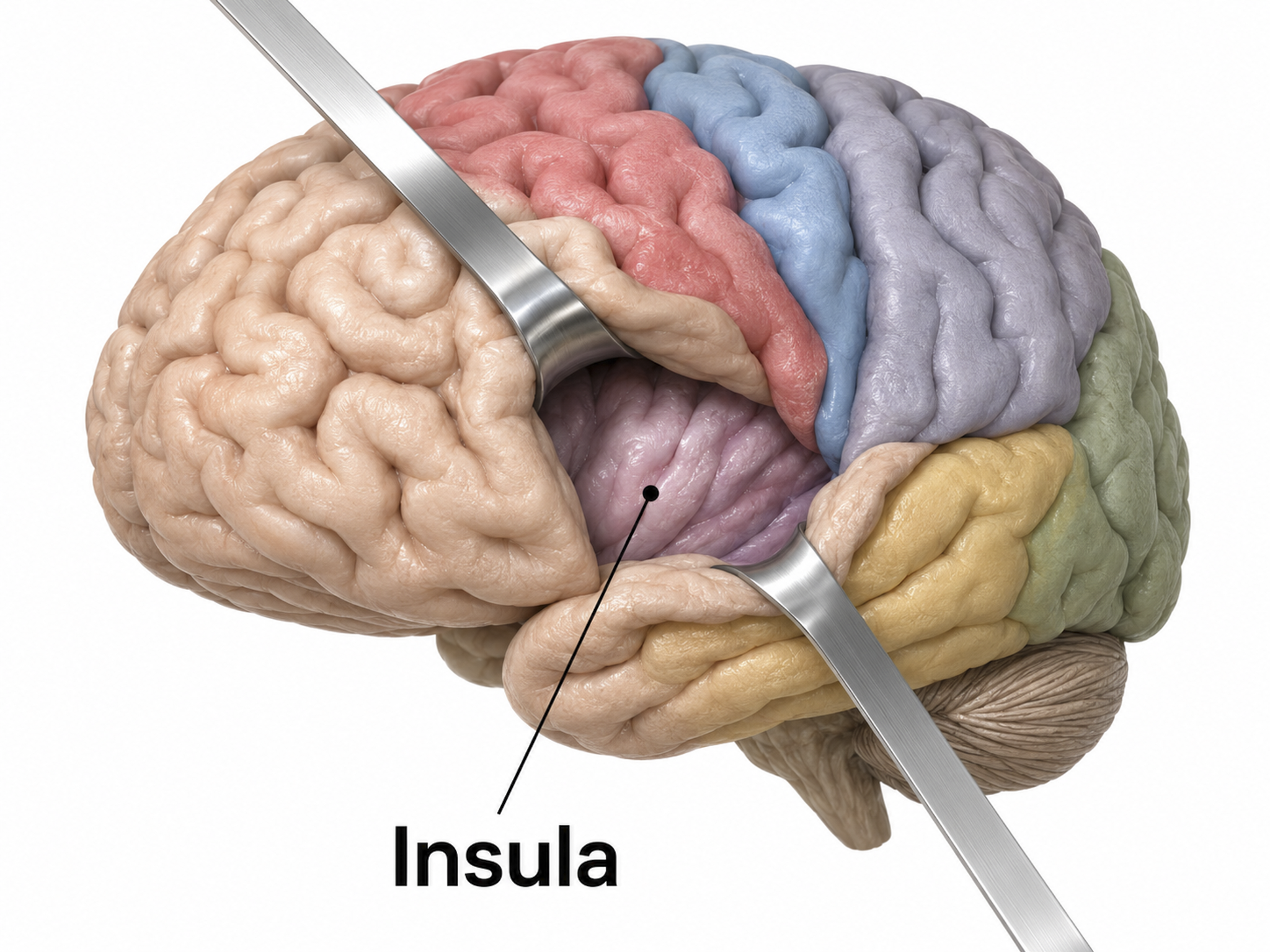
Insula graphic redrawn for the Tutor series.
Antonio Damasio has proposed that this region helps map visceral states associated with emotional experience and generate conscious feelings, providing the basis for somatic markers, gut feelings like the discomfort produced by a risky decision.
The insular cortex has been implicated in the experience of pain and basic emotions including anger, disgust, fear, happiness, and sadness. It receives reports of internal states like hunger and drug craving, motivating individuals to engage in consummatory behavior. The insular cortex plays a crucial role in craving and impulse control. It is stimulated by drug-related cues and may activate memories of pleasurable drug-related experiences. Smokers whose strokes damaged the insular cortex have been found far more likely than smokers with damage elsewhere to quit easily, immediately, and without relapse, although not all of them quit.
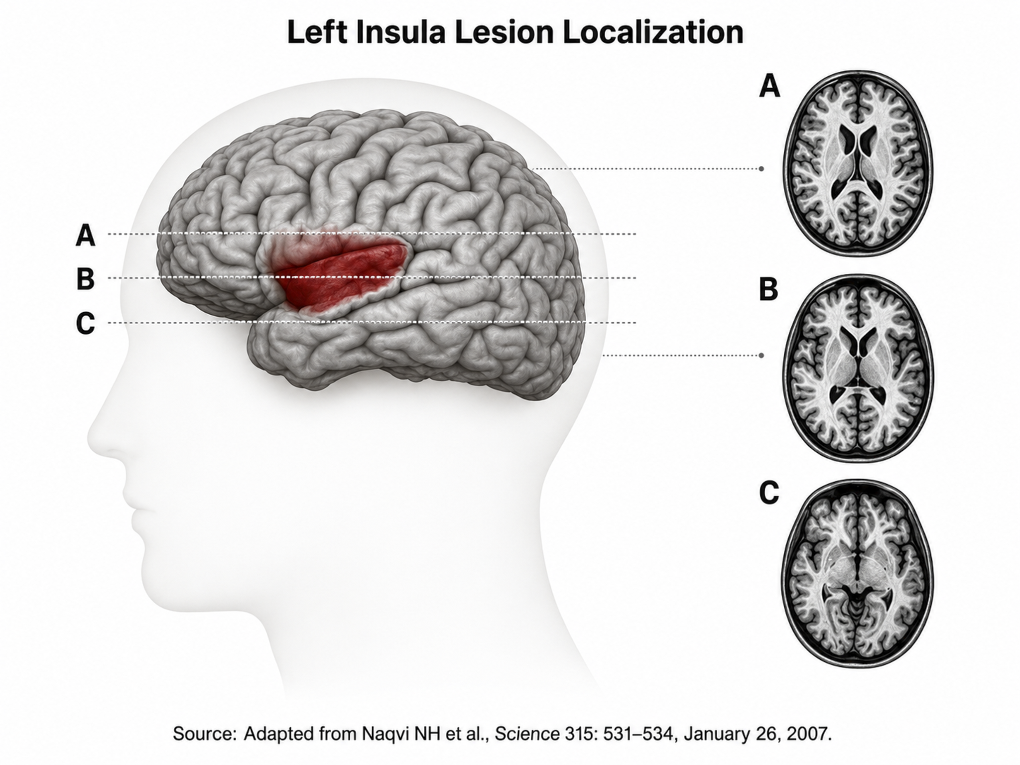
National Institute of Drug Abuse graphic retrieved from Wikimedia Commons and adapted.
Cortical and Subcortical Connections
Listen to the Full-Length Lecture: Cortical and Subcortical Connections
Neocortical zones are connected with cortical and subcortical regions by specialized fiber tracts: association bundles, projection fibers, and commissural bundles. A meta-networking model proposes that the dynamic interaction of "distributed but relatively specialized networks" mediates brain functions like language (Herbet & Duffau, 2020, p. 1181). These tracts are the anatomical substrate of the coherence and phase measures you compute between electrode pairs, which is why a connectivity finding is a claim about white matter as much as about cortex.
We adapted the figure below from Do Tran et al. (2025), which shows the major white matter tracts of the brain.
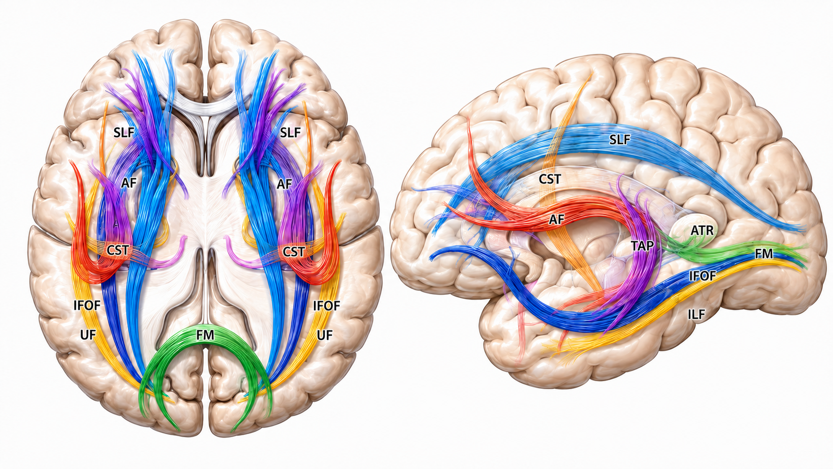
Artistic view of the main white matter tracts in axial view (left panel) and sagittal view (right panel): AF arcuate fasciculus, ATR anterior thalamic radiations, CST corticospinal tract, FM forceps major, IFOF inferior fronto-occipital fasciculus, ILF inferior longitudinal fasciculus, SLF superior longitudinal fasciculus, TAP tapetum, UF uncinate fasciculus. Pencil drawing performed by Nathan Beucler, MD.
Association Bundles
Association bundles link cortical regions located in the same hemisphere. Short association fibers, called U-fibers, arch beneath a sulcus to join adjacent gyri, while long association fasciculi connect widely separated regions. The long fasciculi include the arcuate fasciculus (AF), frontal aslant tract (FAT), inferior fronto-occipital fasciculus (IFOF), inferior longitudinal fascicle (ILF), middle longitudinal fasciculus (MdLF), superior longitudinal fasciculus (SLF), and uncinate fasciculus (UF).
Projection Fibers
Projection fibers connect the cortex with structures deep in the brain, the brainstem, and the spinal cord. They include the frontostriatal tract (FST), which connects the premotor cortex with the caudate nucleus and putamen, as well as the thalamocortical, optic, and pyramidal tracts.
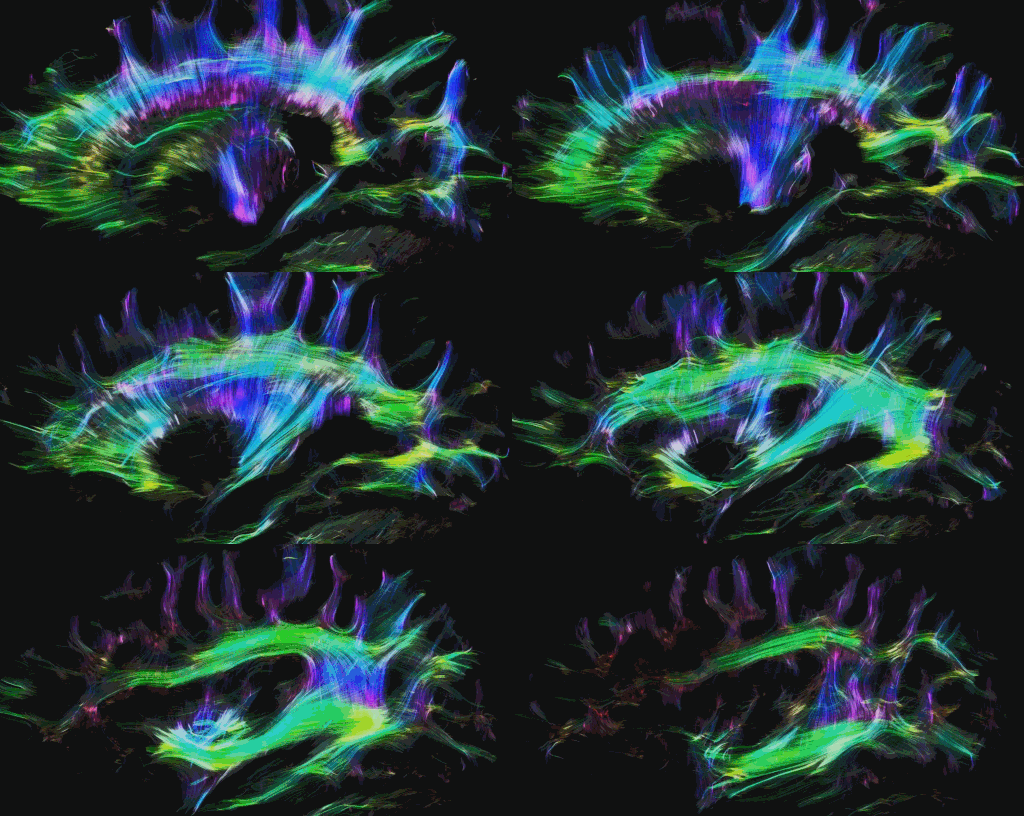
Tractography animation Alfred Anwander, CC BY-SA 4.0, via Wikimedia Commons.

Commissural Bundles
The left and right hemispheres communicate using three commissures, or axon tracts. The corpus callosum is the largest tract and connects the left and right frontal, parietal, and occipital lobes. Certain conditions, such as prenatal exposure to alcohol and other drugs, may result in agenesis of the corpus callosum, in which part or all of this fiber bundle is missing. Agenesis is worth knowing at assessment, because a client whose callosum is partly absent cannot produce the interhemispheric coherence values a normative database expects.
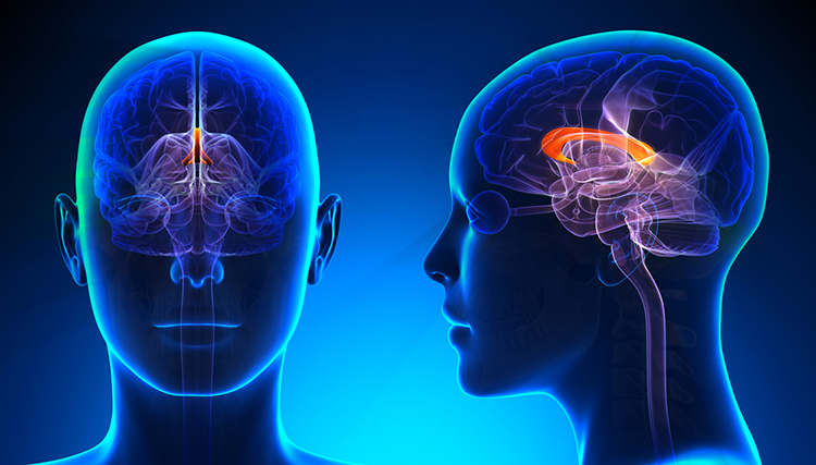
Graphic © decade3d - anatomy online/Shutterstock.com.
The anterior commissure, shown above the third ventricle at the bottom of the diagram, is considerably smaller than the corpus callosum and connects the left and right temporal lobes and the hippocampus and amygdala.
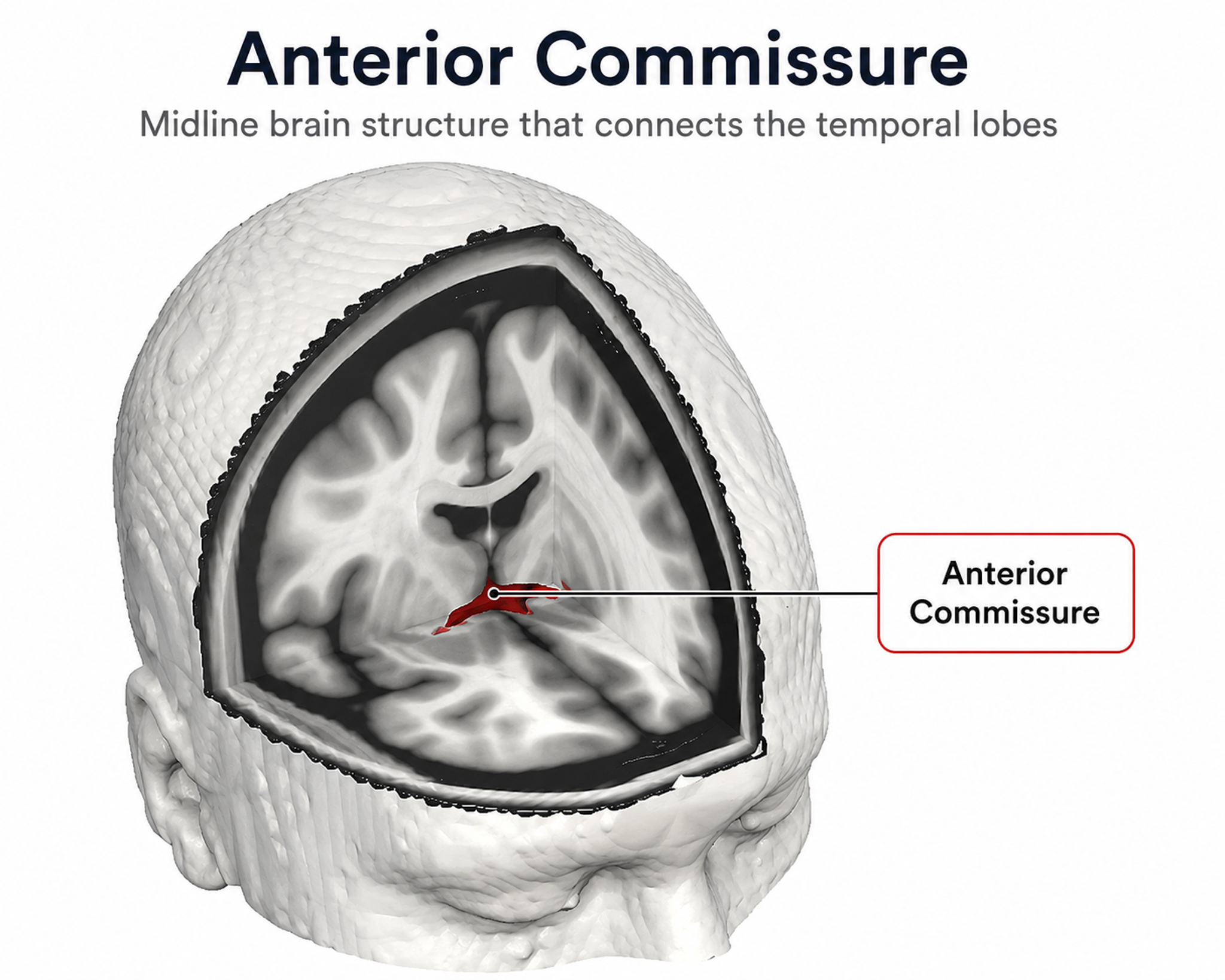
Anterior commissure graphic adapted from Winter, T. J. & Franz, E. A., CC BY 3.0, via Wikimedia Commons.

The posterior commissure, located below the corpus callosum, connects the right and left diencephalon and mesencephalon (Breedlove & Watson, 2023).
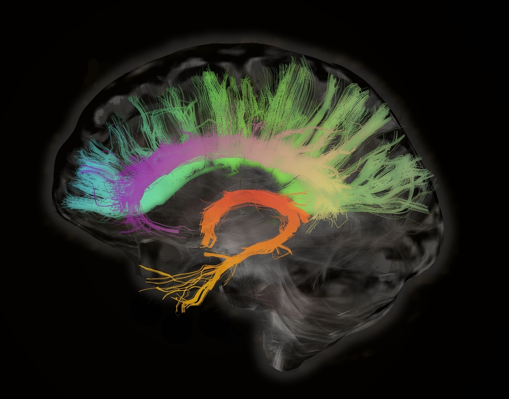
Corpus callosum graphic © Image Trading Source Ltd/Shutterstock.com.
The cortical lobes are named for the overlying skull bones, and each maps onto familiar electrode sites. The frontal lobes house the primary motor cortex, motor association cortex, Broca's area, and prefrontal cortex, with the left side serving approach behavior and positive affect and the right side serving withdrawal and negative affect. The parietal lobes process somatosensory information, the temporal lobes handle hearing and object recognition, and the occipital lobes process vision. The insula, hidden in the lateral sulcus, anchors the salience network and mediates interoceptive awareness. Association, projection, and commissural bundles connect these regions to each other, to subcortical structures, and across the midline.
Check Your Understanding
- Which electrode sites overlie the frontal lobes, and which structures do those lobes contain?
- How do left and right frontal lobe functions differ, and which clinical concern is associated with each side?
- What is the relationship between the motor association cortex and the primary motor cortex, using the piano analogy from the text?
- How do association, projection, and commissural bundles differ in what they connect?
- Why is the insula relevant to biofeedback even though it cannot be recorded directly from the scalp?
Subcortical Structures
Subcortical regions, including the thalamus, basal ganglia, and limbic system (for example, the amygdala, hippocampus, and hypothalamus), regulate fundamental functions like sensory processing, movement, and emotion. The cerebellum is sometimes included among them. These regions also contribute to the regulation of various qualities of thinking and emotion, and several of them shape the rhythms you record at the scalp even though no electrode sits over them.
Thalamus
The thalamus consists of specialized nuclei that process and relay data to and from the telencephalon (cerebral cortex, basal ganglia, and limbic system). The thalamus analyzes all sensory data except olfaction before distributing this information to the cortex via thalamocortical afferent fibers (Breedlove & Watson, 2023). The cortex also sends information to the thalamus to adjust its information processing via corticothalamic fibers.
This two-way conversation creates feedback loops crucial for generating several EEG rhythms. The thalamus helps regulate arousal, sleep, and wakefulness (Steriade & Llinás, 1988) and, through its functional connection to the hippocampus, plays a crucial role in episodic memory (Aggleton et al., 2010).
The thalamus contributes to SCPs, delta, theta, alpha, SMR activity, and beta-gamma activity (Thompson & Thompson, 2015). That range makes the thalamus the single most important subcortical structure for understanding where the rhythms in your recording originate.
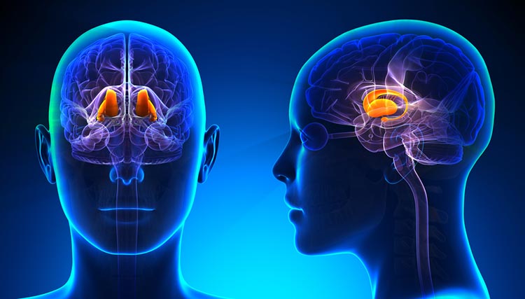
Thalamus graphic © decade3d - anatomy online/Shutterstock.com.
Basal Ganglia
The basal ganglia (comprising the caudate nucleus, putamen, globus pallidus, subthalamic nucleus, and substantia nigra) modulate movement. The basal ganglia, prefrontal cortex, cingulate cortex, and parietal cortex are involved in self-awareness, attention, and emotional regulation. Researchers have identified several distinct prefrontal-basal ganglia-thalamic loops that are implicated in a range of psychiatric conditions.
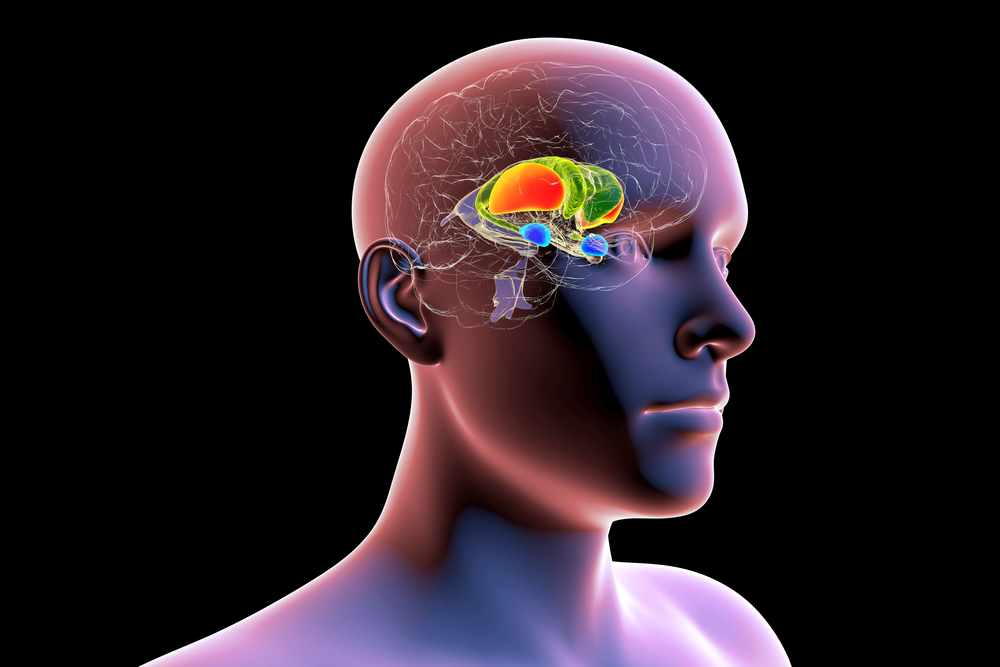
The dorsal striatum consists of the caudate nucleus (green) and the putamen (orange). The amygdala is colored blue. Graphic © Kateryna Kon/Shutterstock.com.
Limbic System
The limbic system is a widespread network of nuclei involved in emotion, motivation, learning, memory, and navigation. Three important limbic structures are the hippocampus, amygdala, and septal nuclei, along with the hypothalamus, anterior thalamus, and cingulate gyrus (Breedlove & Watson, 2023).
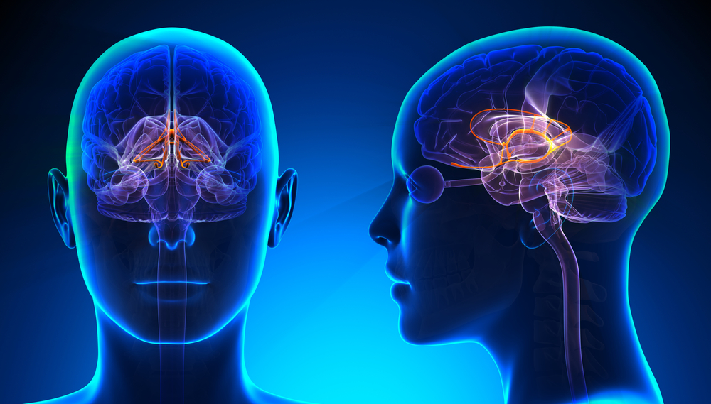
Limbic system graphic © decade3d - anatomy online/Shutterstock.com.
Hippocampus
The hippocampus is a seahorse-shaped limbic structure required to form declarative memories. It plays a vital role in emotion, navigation, spatial memory, and dampening the endocrine stress response. The hippocampus simultaneously integrates emotional, auditory, and visuospatial information to create episodic memories and contains leukocyte receptors, making it part of the feedback loop for immune system regulation. Hippocampal neurons and networks that include it are important sources of the theta rhythm (Amzica & Lopes da Silva, 2018).
Hippocampal Brainwaves Travel in Two Directions
The old-school view was that hippocampal brainwaves travel in one direction. This model could not explain how the hippocampus integrates information from interconnected specialized systems. Based on recordings from human participants undergoing brain surgery, the new-school view is that brainwaves travel through the hippocampus in both directions: from the back to the front and from the front to the back (Kleen et al., 2021). Moreover, cognitive activity differentially influences the direction of movement for low (e.g., 1.9 Hz) and high (e.g., 13.8 Hz) frequency waveforms.
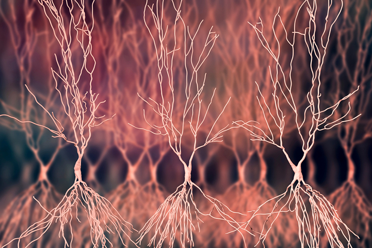
Human hippocampal neuron graphic © Kateryna Kon/Shutterstock.com.
Watch Sam Kean's TED-Ed Talk, What Happens When You Remove the Hippocampus.
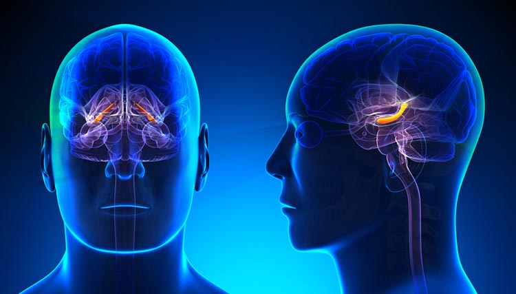
Hippocampus graphic © decade3d - anatomy online/Shutterstock.com.
Amygdala
The amygdala is a collection of nuclei located deep within the medial temporal lobes at the end of the hippocampus. It is an essential limbic structure that includes, among others, the lateral nucleus and the central nucleus.
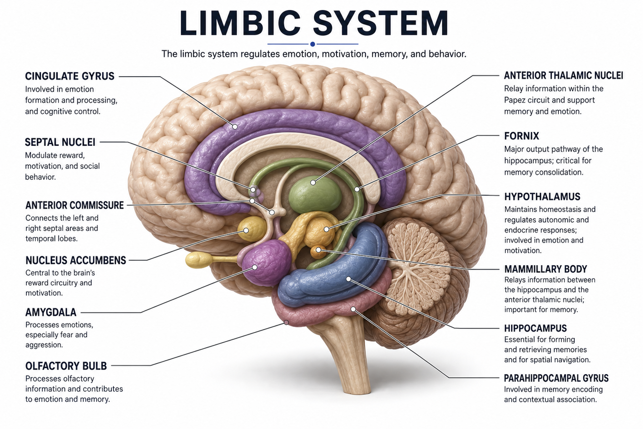
The amygdala shown among the limbic structures of the medial temporal lobe.
The lateral nucleus processes sensory information and distributes it throughout the amygdala. The central nucleus orchestrates the nervous system's response to important stimuli by activating circuits in the brainstem (autonomic arousal), the basal ganglia, and periaqueductal gray (defensive behavior). The amygdala plays a crucial role in learning about the consequences of our actions and creating declarative memories of events with emotional significance (Breedlove & Watson, 2023). Its role in fear conditioning is worth holding in mind when a referral question concerns PTSD or an anxiety disorder, because the behavioral picture will not have a scalp signature of its own.
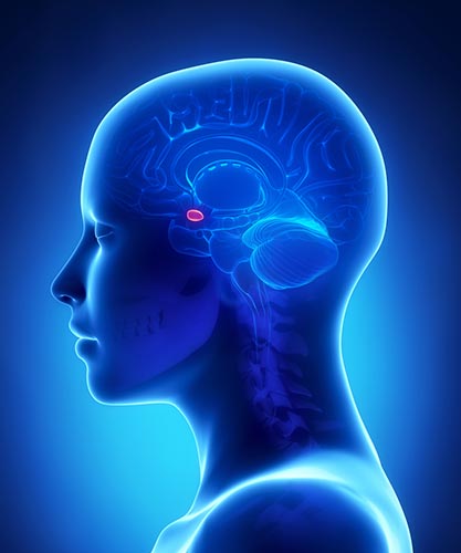
Amygdala graphic © decade3d - anatomy online/Shutterstock.com.
Septal Nuclei
The septal nuclei are a limbic structure containing several nuclei involved in emotion, control of aggressive behavior, reward, and addiction (Breedlove & Watson, 2023). The septohippocampal system contributes to the theta rhythm (Amzica & Lopes da Silva, 2018).
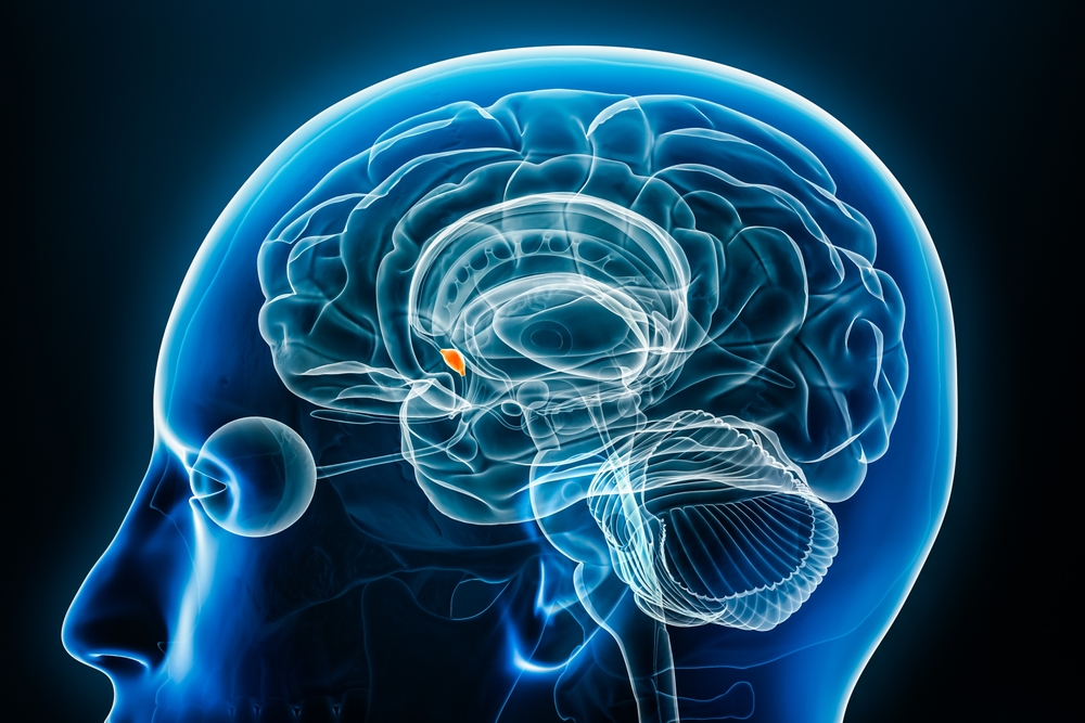
Septal nuclei graphic © MattL_Images/Shutterstock.com.
The cortical lobes and subcortical structures work together through extensive reciprocal connections to support cognition, emotion, movement, and memory. Recognizing which structures contribute to specific EEG patterns, and which clinical disorders involve dysfunction in these regions, is the foundation of a defensible qEEG interpretation.
A referral arrives for a 44-year-old client with a two-year history of low mood. The eyes-closed record shows higher alpha over F3 than over F4. The frontal alpha asymmetry literature would read greater left frontal alpha as reduced approach-related activation, consistent with the right dorsolateral prefrontal cortex being relatively more active than the left. Before that reading goes into a report, check what else could produce it: unequal impedance at the two sites, a drowsy or eye-movement-contaminated epoch, an unrepresentative sample of the record, or a reference that loads the two hemispheres differently. Confirm the asymmetry across artifact-free epochs and against the client's history and symptoms before you let a single index carry the interpretation.
Subcortical structures supply and shape the activity we record at the scalp. The thalamus relays all sensory data except olfaction to the cortex, and its two-way conversation with the cortex generates several EEG rhythms while regulating arousal, sleep, and wakefulness. The basal ganglia modulate movement and contribute to attention and emotional regulation. Within the limbic system, the hippocampus forms declarative memories and dampens the endocrine stress response, the amygdala evaluates salience and drives defensive responses through its lateral and central nuclei, and the septal nuclei contribute to the theta rhythm through the septohippocampal system.
Check Your Understanding
- How do thalamocortical and corticothalamic fibers together produce EEG rhythms?
- Which sensory modality bypasses the thalamus, and why does that matter anatomically?
- What roles do the lateral and central nuclei of the amygdala play in responding to a salient stimulus?
- How does the septohippocampal system relate to the theta rhythm you observe in a recording?
- Beyond memory, what other functions does the hippocampus serve?
Listen to the Full-Length Lecture: A Deep Dive into Brodmann Areas
A Deep Dive into Brodmann Areas
Brodmann areas are regions of the cerebral cortex defined by their distinct cytoarchitectonic characteristics, meaning the organization, density, and layering of neurons. They were first delineated by the German neurologist Korbinian Brodmann in the early twentieth century (Brodmann, 1909). Using Nissl staining, Brodmann examined cortical cell organization across different brain regions in multiple mammalian species, including humans.
Brodmann compared the human cortex with that of a range of other mammals, including primates, rodents, and marsupials, publishing maps for humans and eight other species and enabling comparative conclusions about cortical organization (Garey, 1994). This produced a map whose areas are numbered 1 through 52, each characterized by unique cytoarchitectonic features. Roughly 43 to 47 of those areas were delineated in the human cortex; the remaining numbers were defined only in non-human species, which is why numbers such as 12 through 16 and 48 through 51 are absent from most human maps (Zilles & Amunts, 2010).
Although refinements have been made since Brodmann's initial classification, many areas retain their original numbering, and qEEG practitioners continue to use these divisions when localizing findings and writing reports.
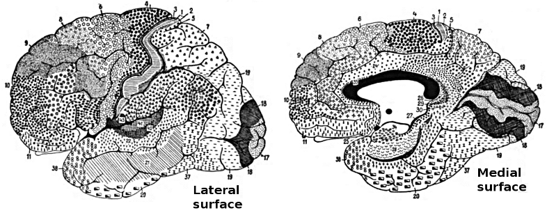
Lateral and medial views of the Brodmann areas.
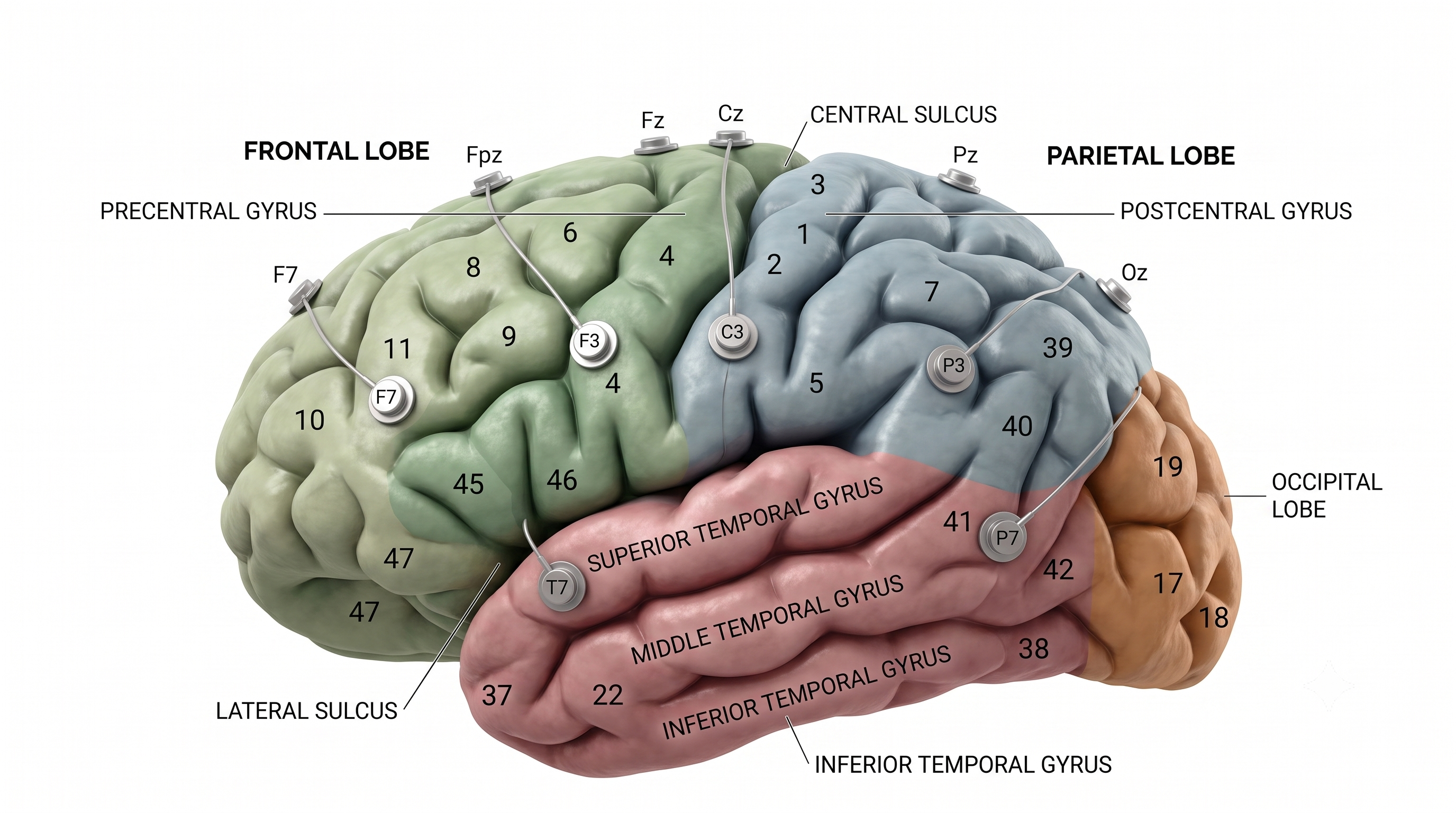
This figure superimposes the 10-20 System electrode sites on the Brodmann areas of the brain's lateral convexity.
The Importance of Brodmann Areas
The identification of Brodmann areas has facilitated the investigation of functional specialization within the cortex. Most cortical functions involve the networked activity of multiple Brodmann areas. Several Brodmann areas are now associated with specific functions, such as primary sensory and motor areas, as well as higher cognitive functions like language processing and decision-making (Glasser et al., 2016). For instance, Brodmann area 4 corresponds to the primary motor cortex, area 17 to the primary visual cortex, and areas 44 and 45 to Broca's area, which is crucial for speech production.
The Brodmann map remains a widely used anatomical reference in both clinical and research neuroanatomy. Area 4 corresponds to the primary motor cortex in the precentral gyrus and is critical for voluntary motor control (Penfield & Boldrey, 1937). Area 17, the primary visual cortex in the occipital lobe, is the initial cortical recipient of visual input from the lateral geniculate nucleus (Zeki, 1993). Naming areas this way lets a report describe a finding in terms every reader can locate.
Language processing illustrates how Brodmann areas map onto clinical function. Broca's area (BA 44 and 45) in the dominant hemisphere's inferior frontal gyrus supports speech production and grammar, while Wernicke's area (BA 22) in the temporal cortex supports speech perception and comprehension. Damage to either area produces distinct aphasia syndromes that can be identified through neuropsychological testing and correlated with qEEG findings. The arcuate fasciculus connecting these regions is a critical white matter tract whose integrity affects language network coherence.
Advances in neuroimaging, including fMRI and PET, have refined the functional mapping of Brodmann areas. Probabilistic cytoarchitectonic atlases, such as those developed by the Jülich Brain Atlas project, supplement Brodmann's static classification with data derived from multiple brains, allowing for population-level variability in cortical organization (Eickhoff et al., 2005). This evolution reflects the growing recognition that individual differences in cortical morphology and function require more dynamic and data-driven mapping approaches (Amunts & Zilles, 2015).
That variability has a practical consequence for you. Brodmann areas remain the standard language for communicating about cortical regions, but a given area may not occupy the same cortical territory in every client, so a source-localized finding names a likely region rather than a certainty.
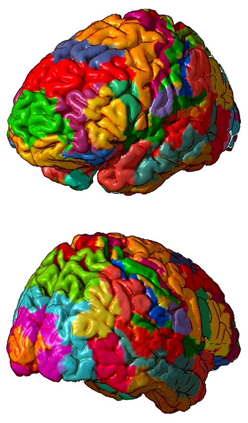
Brodmann maps contributed by Mark Dow, Research Assistant at the Brain Development Lab, University of Oregon, to Wikimedia Commons.
While Brodmann areas provide a valuable framework for understanding cortical organization, they do not capture the full complexity of the brain's functional architecture. Advances in neuroimaging techniques have identified additional areas and functional networks, highlighting the intricate organization of the cortex beyond Brodmann's classification (Glasser et al., 2016).
Areas 3, 1, and 2: Primary Somatosensory Cortex (S1)
The primary somatosensory cortex (S1) is a critical region for processing somatosensory information in the brain. It is involved in processing touch, proprioception, and temperature. These four cortical areas contain separate somatotopic maps (Purves, 2018).
.jpg)
Graphic © Big8/Shutterstock.com.
Brodmann areas. The S1 is located in the postcentral gyrus, mainly in Brodmann areas 3, 1, and 2. These areas have distinct functions; Brodmann area 3 receives and processes cutaneous and proprioceptive inputs, area 1 processes tactile stimuli, and area 2 integrates proprioceptive and tactile inputs (Kaas, 2008).
Location. The S1 is located in the parietal lobe, immediately posterior to the central sulcus. It is bordered by the primary motor cortex (M1) anteriorly and the secondary somatosensory cortex (S2) posteriorly. The closest sites are C3 and C4, which overlie the central sulcus (Jasper, 1958).
Connections. The S1 strongly connects with other cortical and subcortical areas, including the M1, premotor cortex, supplementary motor area, posterior parietal cortex, and thalamus (Lemon, 2008). These connections are essential for sensorimotor integration and control.
Participation in brain networks. The S1 is a critical node in the somatosensory network, which includes other areas like the S2, insular cortex, and parietal operculum. It also participates in the sensorimotor network, interacting with the motor and premotor cortices (Sepulcre, 2012).
Functions. The S1 is crucial for processing somatosensory information like touch, proprioception, and temperature. It plays a significant role in perceiving object features, body awareness, and sensorimotor integration.
Role in clinical disorders. The altered functioning of the S1 has been implicated in various clinical conditions, including neuropathic pain (Baliki et al., 2011), phantom limb pain (Makin et al., 2013), and stroke-related sensory deficits (Carey et al., 2002).
Area 4: Primary Motor Cortex (M1)
The primary motor cortex (M1) is a key region in the brain responsible for the execution of voluntary movements.
Graphic © Big8/Shutterstock.com.
Brodmann areas. The M1 is located in the precentral gyrus, mainly in Brodmann area 4. It contains large pyramidal neurons, known as Betz cells, essential for motor control (Geyer et al., 1996).
Location. The M1 is situated in the frontal lobe, immediately anterior to the central sulcus. It is bordered by the posteriorly primary somatosensory cortex (S1) and anteriorly premotor cortex. The closest sites are C3 and C4, which overlie the central sulcus (Jasper, 1958).
Connections. The M1 strongly connects with various cortical and subcortical areas, including the S1, premotor cortex, supplementary motor area, posterior parietal cortex, and thalamus (Lemon, 2008). These connections are critical for sensorimotor integration and control.
Participation in brain networks. The M1 is a central node in the sensorimotor network, interacting with the somatosensory cortex, premotor cortex, and other motor-related areas (Sepulcre, 2012).
Functions. The primary function of the M1 is the execution of voluntary movements. M1 neurons primarily control movements rather than discrete muscles (Breedlove & Watson, 2023). It is critical in planning, controlling, and coordinating complex motor tasks.
Role in clinical disorders. Alterations in M1 function have been implicated in various clinical conditions, including motor deficits following stroke (Ward, 2004), Parkinson's disease (Wu & Hallett, 2013), and motor neuron diseases like amyotrophic lateral sclerosis (ALS; Kew & Leigh, 1997).
Areas 5 and 7: Somatosensory Association Cortex (SAC)
The somatosensory association cortex (SAC) is involved in the integration and interpretation of somatosensory information coming from the primary somatosensory cortex (S1).
Graphic © Big8/Shutterstock.com.
Brodmann areas. The SAC is mainly located in Brodmann areas 5 and 7 within the posterior parietal cortex (Culham & Kanwisher, 2001).
Location. The SAC is situated in the parietal lobe, superior to the primary somatosensory cortex (S1), and posterior to the postcentral gyrus. The closest sites are likely P3 and P4, which overlie the parietal cortex.
Connections. The SAC strong connects with various cortical and subcortical regions, including the S1, primary motor cortex (M1), premotor cortex, supplementary motor area, posterior parietal cortex, and thalamus (Cavada & Goldman-Rakic, 1989). These connections are critical for sensorimotor integration, spatial awareness, and higher-order sensory processing.
Participation in brain networks. The SAC is a key node in the somatosensory network, including areas like the S1, S2, and insular cortex. Additionally, it is part of the dorsal attention network, which is involved in attentional control and spatial processing (Corbetta & Shulman, 2002).
Functions. The SAC is essential for integrating and interpreting somatosensory information, including tactile and proprioceptive stimuli. It plays a significant role in sensorimotor integration, spatial awareness, and attention.
Role in clinical disorders. Alterations in SAC function have been implicated in various clinical conditions, including somatosensory neglect (Vallar et al., 2003), spatial processing deficits (Whitlock et al., 2012), and somatosensory deficits in autism spectrum disorder (Cascio et al., 2012).
Area 6: Supplementary Motor Cortex and Premotor Cortex
The supplementary motor cortex (SMA) and premotor cortex (PMC) are critical regions for planning and executing voluntary movements.
Graphic © Big8/Shutterstock.com.
Brodmann areas. The SMA is primarily located in Brodmann area 6, on the medial aspect of the frontal lobe (Picard & Strick, 2001). The PMC is also predominantly found in Brodmann area 6 but is located on the lateral aspect of the frontal lobe (Wise et al., 1997).
Location. The SMA is located in the medial part of the frontal lobe, superior to the cingulate sulcus and anterior to the paracentral lobule. The PMC is situated in the lateral part of the frontal lobe, anterior to the primary motor cortex (M1). The closest sites are likely FC3 and FC4, which overlie the dorsolateral prefrontal cortex.
Connections. Both the SMA and PMC have strong connections with various cortical and subcortical areas, including the M1, primary somatosensory cortex (S1), posterior parietal cortex, and basal ganglia (Lemon, 2008; Nachev et al., 2008). These connections are critical for sensorimotor integration, movement planning, and execution.
Participation in brain networks. The SMA and PMC are central nodes in the sensorimotor network, interacting with the M1, S1, and other motor-related areas (Sepulcre, 2012).
Functions. The SMA and PMC are essential for motor planning, execution, and coordination of complex movements. The SMA is particularly involved in initiating and controlling internally generated movements, while the PMC is more concerned with the planning and executing visually-guided movements (Wise et al., 1997; Picard & Strick, 2001).
Role in clinical disorders. Alterations in SMA and PMC function have been implicated in various clinical conditions, including movement disorders like Parkinson's disease (Wu & Hallett, 2013), apraxia (Haaland et al., 2000), and motor deficits following stroke (Ward, 2004).
Area 8: Frontal Eye Field (FEF)
The frontal eye field (FEF) is essential for the control of eye movements and visual attention.
Graphic © Big8/Shutterstock.com.
Brodmann areas. The FEF is predominantly in Brodmann area 8, located in the dorsolateral prefrontal cortex (Paus, 1996).
Location. The FEF is located in the anterior bank of the precentral sulcus within the dorsolateral prefrontal cortex, close to the border with the primary motor cortex (M1) (Paus, 1996). The closest sites are likely F3 and F4, which overlie the dorsolateral prefrontal cortex.
Connections. The FEF has extensive connections with other cortical and subcortical regions, including the parietal cortex, superior colliculus, thalamus, and extrastriate visual areas (Schall, 2002; Stanton et al., 2005). These connections are crucial for visual attention, saccadic eye movements, and smooth pursuit.
Participation in brain networks. The FEF is a key node in the dorsal attention network responsible for goal-directed attention and eye movement control. This network also includes the intraparietal sulcus and superior parietal lobule (Corbetta & Shulman, 2002).
Functions. The FEF is crucial in controlling saccadic eye movements, smooth pursuit, and visual attention. It is involved in the initiation, planning, and execution of eye movements, as well as the allocation of attention to relevant visual stimuli (Schall, 2002). The FEF is vital in cognitive functions, including attention orientation, visual consciousness, access to our conscious experience, perceptual performance, and decision-making (Vernet et al., 2014).
Role in clinical disorders. Alterations in FEF function have been implicated in various clinical conditions, including attention deficit hyperactivity disorder (ADHD; Mahone et al., 2011), oculomotor apraxia (Rizzo et al., 1996), and progressive supranuclear palsy (Burrell et al., 2012).
Areas 9 and 46: Dorsolateral Prefrontal Cortex (DLPFC)
The dorsolateral prefrontal cortex (DLPFC), which consists of distinct regions, is essential for higher-order cognitive functions, including working memory, executive control, and decision-making (Ahuja & Rodriguez, 2022).
Graphic © Big8/Shutterstock.com.
Brodmann areas. The DLPFC is primarily located in Brodmann areas 9 and 46 within the lateral aspect of the frontal lobe (Petrides, 2005).
Location. The DLPFC is situated in the lateral portion of the frontal lobe, superior and anterior to the premotor and primary motor cortex (M1). The closest sites are likely F3 and F4.
Connections. The DLPFC has extensive connections with other cortical and subcortical regions, including the parietal cortex, medial prefrontal cortex, anterior cingulate cortex, orbitofrontal cortex, thalamus, and basal ganglia (Petrides & Pandya, 2002). These connections are critical for cognitive control, working memory, and decision-making.
Participation in brain networks. The DLPFC is a key node in the frontoparietal control network, which is responsible for executive control, and the working memory network, which maintains and manipulates information (Cabeza & Nyberg, 2000; Vincent et al., 2008).
Functions. The DLPFC plays a crucial role in higher-order cognitive functions such as working memory, executive control, and decision-making. It allocates cognitive resources, goal-directed behavior, task switching, and the flexible adaptation of behavior in response to changing demands (Breedlove & Watson, 2023; Petrides, 2005).
Role in clinical disorders. Alterations in DLPFC function have been implicated in various clinical conditions, including schizophrenia (Barch, 2005), attention deficit hyperactivity disorder (ADHD; Cortese et al., 2012), and major depressive disorder (MDD; Drevets et al., 2008).
Area 10: Anterior Prefrontal Cortex (aPFC)
The anterior prefrontal cortex (aPFC), also referred to as the frontopolar cortex, is involved in higher-order cognitive processes such as decision-making, planning, and reasoning.
Graphic © Big8/Shutterstock.com.
Brodmann areas. The aPFC is primarily located in Brodmann area 10, at the most anterior part of the frontal lobe (Ramnani & Owen, 2004).
Location. The aPFC is located at the most rostral part of the frontal lobe, anterior to the dorsolateral prefrontal cortex (DLPFC) and orbitofrontal cortex. The closest sites are likely Fp1 and Fp2, which overlie the frontal pole.
Connections. The aPFC has extensive connections with other cortical and subcortical regions, including the DLPFC, orbitofrontal cortex, medial prefrontal cortex, posterior parietal cortex, temporal cortex, and thalamus (Burgess et al., 2007). These connections are essential for complex cognitive tasks, multitasking, and mentalizing.
Participation in brain networks. The aPFC is a key node in the frontoparietal control network, which is responsible for executive control, as well as the default mode network (DMN), which is involved in self-referential processing and mentalizing (Vincent et al., 2008; Spreng et al., 2009).
Functions. The aPFC involves higher-order cognitive processes such as decision-making, planning, reasoning, multitasking, and mentalizing. It is crucial in coordinating and integrating information from various cognitive domains and is responsible for goal-directed behavior and social cognition (Ramnani & Owen, 2004). The aPFC is engaged in various tasks, such as problem-solving, memory recall, future-oriented memory, source and context memory, task-switching, and attention reallocation (Ramnani & Owen, 2004). The aPFC contributes to high-level nociception and pain processing (Peng et al., 2018).
Role in clinical disorders. Alterations in aPFC function have been implicated in various clinical conditions, including autism spectrum disorder (ASD; Gilbert et al., 2008), schizophrenia (Perlstein et al., 2001), and major depressive disorder (MDD; Drevets et al., 2008).
Areas 11, 12, 13, and 47: Orbitofrontal Cortex (OFC)
The orbitofrontal cortex (OFC) processes reward, emotion, and decision-making and integrates sensory information with emotional valence.
Graphics © Big8/Shutterstock.com.
.jpg)
Brodmann areas. The OFC primarily encompasses Brodmann areas 11, 12, 13, and 47, located in the ventral portion of the frontal lobe (Kringelbach, 2005).
Location. The OFC is situated in the ventral part of the frontal lobe, just above the orbits (eye sockets). The anterior prefrontal cortex and the medial prefrontal cortex border it. The closest sites are likely Fp1 and Fp2, which overlie the ventral and rostral portions of the frontal lobe.
Connections. The OFC has extensive connections with other cortical and subcortical regions, including the amygdala, insula, cingulate cortex, hippocampus, thalamus, striatum, and sensory cortices (Kringelbach, 2005; Price, 2007). These connections are essential for emotion processing, reward-based decision-making, and social cognition.
Participation in brain networks. The OFC is a key node in the salience network, which is responsible for detecting and integrating emotionally and motivationally salient stimuli, and the default mode network (DMN), which is involved in self-referential processing and social cognition (Seeley et al., 2007; Spreng et al., 2009).
Functions. The OFC is crucial in processing reward, emotion, and decision-making. It integrates sensory information with emotional valence, evaluates outcomes and actions, and represents social and emotional information (Kringelbach, 2005).
Role in clinical disorders. Alterations in OFC function have been implicated in various clinical conditions, including obsessive-compulsive disorder (OCD) (Menzies et al., 2008), major depressive disorder (MDD) (Drevets, 2007), bipolar disorder (BD) (Blumberg et al., 2003), and addiction (Volkow & Fowler, 2000). Depression may be associated with heightened responsiveness and increased connectivity in the lateral orbitofrontal cortex (not linked to rewards), while it is connected to reduced responsiveness and connectivity in the medial orbitofrontal cortex (related to rewards; Rolls, Cheng, & Feng, 2020).
Areas 13-16 and 52: Insular Cortex (Insula)
The insular cortex, or insula, is involved in diverse functions, including interoception, emotion processing, pain perception, and cognitive control.
Graphics © Big8/Shutterstock.com.
Brodmann areas. The insular cortex comprises Brodmann areas 13, 14, 15, 16, and parts of area 52. These include sensorimotor, central-olfactogustatory, socio-emotional, and cognitive anterior-dorsal regions (Kurth et al., 2010).
Location. The insular cortex is situated deep within the lateral sulcus, which separates the frontal and parietal lobes from the temporal lobe. The opercula of the frontal, parietal, and temporal lobes cover it.
Connections. The insular cortex has extensive connections with various cortical and subcortical regions, including the amygdala, anterior cingulate cortex (ACC), prefrontal cortex, primary and secondary somatosensory cortices, orbitofrontal cortex (OFC), and thalamus (Nieuwenhuys, 2012). These connections contribute to the diverse functions of the insula.
Participation in brain networks. The insular cortex is a key node in the salience network, which is responsible for detecting and integrating emotionally and motivationally salient stimuli, and the central autonomic network (CAN), which is involved in autonomic regulation (Seeley et al., 2007; Thayer et al., 2012).
Functions. The insular cortex is crucial in interoception, emotion processing, pain perception, and cognitive control. It represents internal bodily states, integrates sensory and emotional information, and modulates cognitive and affective processes (Craig, 2009).
Role in clinical disorders. Alterations in insular cortex function have been implicated in various clinical conditions, including anxiety disorders (Paulus & Stein, 2006), major depressive disorder (MDD; Sliz & Hayley, 2012), addiction (Naqvi & Bechara, 2010), and autism spectrum disorder (ASD; Di Martino et al., 2009).
Area 17: Primary Visual Cortex (V1)
The primary visual cortex (V1), or the striate cortex, is responsible for processing basic visual information, such as orientation, spatial frequency, and color.
Graphic © Big8/Shutterstock.com.
Brodmann areas. The primary visual cortex is primarily located in Brodmann area 17, in the occipital lobe (Horton & Adams, 2005).
Location. The primary visual cortex is located in the occipital lobe, along the calcarine sulcus, which runs horizontally through the medial part of the lobe. The closest sites are likely O1 and O2, which overlie the occipital lobe.
Connections. The primary visual cortex receives input from the lateral geniculate nucleus (LGN) of the thalamus and sends output to the secondary visual cortex (V2) and other extrastriate areas (V3, V4, V5/MT). These connections are essential for the hierarchical processing of visual information (Felleman & Van Essen, 1991).
Participation in brain networks. The primary visual cortex is a key node in the visual processing network responsible for processing and interpreting visual information from the retina. This network includes other areas of the occipital lobe and extends to the parietal and temporal cortices (Nassi & Callaway, 2009).
Functions. The primary visual cortex processes basic visual information, such as orientation, spatial frequency, and color. It forms the initial stage of the hierarchical processing of visual information and is critical for visual perception (Horton & Adams, 2005).
Role in clinical disorders. Alterations in primary visual cortex function have been implicated in various clinical conditions, including amblyopia (lazy eye; Hess et al., 2010), cortical blindness (Celesia, 2005), and visual hallucinations in conditions like Charles Bonnet syndrome (Griffiths, 2000).
Areas 18 and 19: Secondary Visual Cortex (V2)
The secondary visual cortex (V2), also known as the prestriate cortex, is involved in the further processing and integration of visual information received from the primary visual cortex (V1).
Graphic © Big8/Shutterstock.com.
Brodmann areas. The secondary visual cortex is primarily located in Brodmann areas 18 and 19, in the occipital lobe (Tootell et al., 1998).
Location. The secondary visual cortex is located in the occipital lobe, surrounding the primary visual cortex along the calcarine sulcus, extending to the lateral parts of the occipital lobe. The closest sites are likely O1 and O2.
Connections. The V2 receives input from the primary visual cortex (V1). It sends output to higher-order extrastriate areas (V3, V4, V5/MT) and other cortical regions involved in visual processing, including the parietal and temporal cortices (Felleman & Van Essen, 1991).
Participation in brain networks. The secondary visual cortex is a key node in the visual processing network responsible for processing and interpreting visual information from the retina. This network includes other areas of the occipital lobe and parietal and temporal cortices (Nassi & Callaway, 2009).
Functions. The secondary visual cortex is involved in further processing and integrating visual information from the primary visual cortex. It is crucial in processing complex visual attributes, such as form, color, and motion (Tootell et al., 1998).
Role in clinical disorders. Alterations in secondary visual cortex function have been implicated in various clinical conditions, including visual agnosia, characterized by the inability to recognize objects despite normal visual acuity and intact primary visual cortex function (Milner & Goodale, 2008).
Areas 18, 19, 37, 21, and 22: Visual Association Cortex (V3, V4, V5)
The visual association cortex, also known as the higher-order extrastriate cortex, is responsible for the advanced processing of visual information, such as object recognition, face perception, and processing of complex visual scenes.
Graphic © Big8/Shutterstock.com.
Brodmann areas. The visual association cortex comprises several Brodmann areas, including areas 18, 19, 37, 21, and 22, mainly in the occipital and temporal lobes (Tootell et al., 1998; Kanwisher & Yovel, 2006).
Location. The visual association cortex is located primarily in the occipital and temporal lobes, surrounding the primary (V1) and secondary (V2) visual cortices. It includes regions such as the fusiform face area (FFA), the parahippocampal place area (PPA), and the lateral occipital complex (LOC; Epstein & Kanwisher, 1998; Kanwisher & Yovel, 2006; Malach et al., 1995). The closest sites are likely O1, O2, T5, and T6, which overlie the occipital and temporal lobes.
Connections. The visual association cortex receives input from the primary (V1) and secondary (V2) visual cortices and has extensive connections with other cortical and subcortical regions, including the parietal lobe, prefrontal cortex, hippocampus, and amygdala (Felleman & Van Essen, 1991; Kravitz et al., 2013).
Participation in brain networks. The visual association cortex is a key component of the ventral visual processing stream, also known as the "what" pathway, responsible for object recognition and processing of complex visual scenes (Kravitz et al., 2011).
Functions. The visual association cortex is involved in advanced visual processing, including object recognition, face perception, processing of complex visual scenes, and integration of visual information with other sensory modalities (Kanwisher & Yovel, 2006; Tootell et al., 1998).
Role in clinical disorders. Alterations in visual association cortex function have been implicated in various clinical conditions, including prosopagnosia (face blindness; Duchaine & Nakayama, 2006), visual agnosia (Milner & Goodale, 2008), and higher-order visual processing deficits in conditions such as autism spectrum disorder (ASD; Simmons et al., 2009).
Areas 20 and 37: Inferior Temporal Gyrus (ITG)
The inferior temporal gyrus (ITG) is a part of the temporal lobe involved in high-level visual processing and object recognition.
Graphic © Big8/Shutterstock.com.
Brodmann areas. The inferior temporal gyrus primarily includes Brodmann areas 20 and 37 (Amunts et al., 2000).
Location. The inferior temporal gyrus is located in the ventral part of the temporal lobe, below the middle temporal gyrus and superior temporal sulcus, and above the fusiform gyrus. The closest sites are likely T5 (or TP7) and T6 (or TP8), which overlie the temporal lobes.
Connections. The ITG has extensive connections with other cortical and subcortical regions, including the primary and secondary visual cortices, fusiform gyrus, parahippocampal gyrus, hippocampus, amygdala, and prefrontal cortex (Kravitz et al., 2013; Felleman & Van Essen, 1991).
Participation in brain networks. The ITG is a key component of the ventral visual processing stream, also known as the "what" pathway, responsible for object recognition and processing of complex visual scenes (Kravitz et al., 2011).
Functions. The ITG is involved in high-level visual processing, object recognition, semantic processing, and the integration of visual information with other sensory modalities (Kanwisher & Yovel, 2006).
Role in clinical disorders. Alterations in ITG function have been implicated in various clinical conditions, including visual agnosia, prosopagnosia (face blindness), and higher-order visual processing deficits in conditions such as autism spectrum disorder (ASD; Duchaine & Nakayama, 2006; Simmons et al., 2009).
Areas 21 and 39: Middle Temporal Gyrus (MTG)
The middle temporal gyrus (MTG) is a part of the temporal lobe involved in various functions, such as semantic processing, language, and high-level visual processing.
Graphics © Big8/Shutterstock.com.
Brodmann areas. The MTG primarily includes Brodmann areas 21 and 39 (Amunts et al., 2000).
Location. The MTG is located in the lateral part of the temporal lobe, between the superior temporal gyrus (above) and the inferior temporal gyrus (below), and adjacent to the superior temporal sulcus.
Connections. The MTG has extensive connections with other cortical and subcortical regions, including the primary and secondary visual cortices, the angular gyrus, the fusiform gyrus, the parahippocampal gyrus, the hippocampus, the amygdala, and the prefrontal cortex (Kravitz et al., 2013; Felleman & Van Essen, 1991).
Participation in brain networks. The MTG participates in various brain networks, including the ventral visual processing stream ("what" pathway) for object recognition and processing of complex visual scenes (Kravitz et al., 2011), and the language network for semantic processing and word retrieval (Binder et al., 2009).
Functions. The MTG is involved in various functions, such as semantic processing, language comprehension, word retrieval, and high-level visual processing, including object and face recognition (Binder et al., 2009; Kanwisher & Yovel, 2006).
Role in clinical disorders. Alterations in MTG function have been implicated in various clinical conditions, including semantic dementia (Hodges et al., 1992), language impairments in aphasia (Dronkers et al., 2004), and higher-order visual processing deficits in conditions such as autism spectrum disorder (ASD; Simmons et al., 2009).
Areas 22, 39, and 40: Superior Temporal Gyrus (STG)
The superior temporal gyrus (STG), including Wernicke's area, is a part of the temporal lobe involved in various functions such as language comprehension, auditory processing, and social cognition.
Graphic © Big8/Shutterstock.com.
Brodmann areas. Wernicke's area primarily includes Brodmann area 22 and, to some extent, areas 39 and 40 (Amunts et al., 2000).
Location. Wernicke's area is located in the posterior part of the superior temporal gyrus, usually in the left hemisphere, near the lateral sulcus. The STG runs laterally along the temporal lobe, above the middle temporal gyrus. The closest site is likely T5 (or TP7) for the left hemisphere, where Wernicke's area is typically located.
Connections. Wernicke's area has extensive connections with other language-related regions, including Broca's area (via the arcuate fasciculus), the angular gyrus, and other parts of the superior temporal gyrus (Friederici, 2009). The STG also connects with the primary and secondary auditory cortices, social cognition, and memory regions.
Participation in brain networks. Wernicke's area participates in the language network, playing a crucial role in language comprehension and semantic processing (Binder et al., 2009). The STG is also involved in the auditory processing network and the social cognition network.
Functions. Wernicke's area involves language comprehension, semantic processing, and integrating auditory information into meaningful speech (Price, 2012). The STG also plays a role in auditory processing, social cognition, and memory.
Role in clinical disorders. Alterations in the function of Wernicke's area and the STG have been implicated in various clinical conditions, such as Wernicke's aphasia, characterized by impaired language comprehension and fluent but nonsensical speech (Dronkers et al., 2004). The STG has also been implicated in auditory processing deficits and social cognition impairments in conditions such as autism spectrum disorder (ASD; Boddaert et al., 2004).
Area 23: Ventral Posterior Cingulate Cortex (vPCC)
The ventral posterior cingulate cortex (vPCC) is a region within the posterior cingulate cortex (PCC), a part of the limbic system involved in various functions such as memory, emotion, and self-referential processing.
Graphic © Big8/Shutterstock.com.
Brodmann areas. The vPCC primarily includes Brodmann area 23 (Vogt et al., 2006).
Location. The vPCC is located in the medial part of the brain, in the posterior cingulate cortex, and ventral to the dorsal posterior cingulate cortex (dPCC). It is positioned between the precuneus and the corpus callosum. The closest sites are likely Pz and CPz, located over the midline parietal and central regions, respectively.
Connections. The vPCC connects with various brain regions, including the medial prefrontal cortex (mPFC), hippocampus, parahippocampal gyrus, and lateral parietal regions (Leech & Sharp, 2014; Utevsky et al., 2014).
Participation in brain networks. The vPCC is a key component of the default mode network (DMN), which is active during rest and involved in self-referential thinking, autobiographical memory, and social cognition (Raichle et al., 2001; Buckner et al., 2008).
Functions. The vPCC is involved in various functions, such as self-referential thinking, autobiographical memory, social cognition, and emotional processing (Leech & Sharp, 2014; Utevsky et al., 2014).
Role in clinical disorders. Alterations in vPCC function have been implicated in various clinical conditions, including Alzheimer's disease (Buckner et al., 2005), major depressive disorder (Sheline et al., 2010), and autism spectrum disorder (ASD; Padmanabhan et al., 2017).
Areas 24 and 25: Ventral Anterior Cingulate Cortex (vACC)
The ventral anterior cingulate cortex (vACC) is a region within the anterior cingulate cortex (ACC), which is part of the limbic system and involved in various functions, such as emotion processing, reward-based learning, and decision-making.
Graphic © Big8/Shutterstock.com.
Brodmann areas. The vACC primarily includes Brodmann areas 24 and 25 (Vogt, 2005).
Location. The vACC is located in the medial part of the brain, in the anterior cingulate cortex, ventral to the dorsal anterior cingulate cortex (dACC). It is positioned anterior to the genu of the corpus callosum. The closest sites are likely FCz and Cz, located over the midline frontal and central regions, respectively.
Connections. The vACC has connections with various brain regions, including the amygdala, hippocampus, medial prefrontal cortex (mPFC), orbitofrontal cortex (OFC), and nucleus accumbens (Bush et al., 2000; Etkin et al., 2011).
Participation in brain networks. The vACC is a key component of the salience network, which detects and integrates salient emotional and sensory stimuli and modulates attention and cognitive control (Menon, 2011; Seeley et al., 2007).
Functions. The vACC is implicated in various cognitive and emotional functions, including error detection, conflict monitoring, emotion regulation, empathy, and social cognition (Bush et al., 2000; Etkin et al., 2011).
Role in clinical disorders. Abnormalities in the vACC have been implicated in several psychiatric and neurological disorders, such as depression, anxiety, schizophrenia, bipolar disorder, attention deficit hyperactivity disorder (ADHD), and autism spectrum disorders (Drevets et al., 2008; Etkin et al., 2010).
Areas 25 and 24b: Subgenual Ventromedial Prefrontal Cortex (vmPFC)
The subgenual region of the ventromedial prefrontal cortex (vmPFC) is an important brain region involved in various cognitive and emotional processes.
Graphic © Big8/Shutterstock.com.
Brodmann areas. The subgenual region of the vmPFC primarily consists of Brodmann areas 25 and 24b (Ongür et al., 2003).
Location. The subgenual region of the vmPFC is located in the medial prefrontal cortex, ventral to the genu of the corpus callosum, and adjacent to the anterior cingulate cortex (Mayberg, 2003). Its nearby EEG electrode positions include Fp1, Fp2, Fz, and AFz, located along the scalp's midline (Jasper, 1958).
Connections. The subgenual region of the vmPFC has extensive connections with other brain regions, including the amygdala, hippocampus, hypothalamus, nucleus accumbens, thalamus, and other prefrontal areas (Ongür et al., 2003; Price & Drevets, 2010).
Participation in brain networks. The subgenual vmPFC is a key component of the default mode network (DMN) and the affective network, involved in self-referential processing, emotion regulation, and decision-making (Buckner et al., 2008; Rudebeck et al., 2014).
Functions. The subgenual vmPFC is implicated in various cognitive and emotional functions, including value-based decision-making, emotion regulation, self-referential processing, and social cognition (Rudebeck et al., 2014; Roy et al., 2012).
Role in clinical disorders. Abnormalities in the subgenual vmPFC have been implicated in several psychiatric disorders, such as major depressive disorder, bipolar disorder, anxiety disorders, and post-traumatic stress disorder (Mayberg, 2003; Price & Drevets, 2010).
Areas 29 and 30: Retrosplenial Cingulate Cortex
The retrosplenial cingulate cortex is an important brain region involved in various cognitive processes, particularly related to spatial memory and navigation.
Graphic © Big8/Shutterstock.com.
Brodmann areas. The retrosplenial cortex mainly comprises Brodmann areas 29 and 30, which are located in the posterior cingulate cortex (Vogt et al., 2006).
Location. The retrosplenial cortex is situated in the medial parietal lobe, posterior to the splenium of the corpus callosum, and adjacent to the posterior cingulate cortex (Vann et al., 2009). Its nearby EEG electrode positions include Pz, CPz, and Oz, located along the midline of the scalp (Jasper, 1958).
Connections. The retrosplenial cortex has extensive connections with other brain regions, including the hippocampus, parahippocampal cortex, thalamus, anterior cingulate cortex, and other parietal and frontal areas (Vann et al., 2009).
Participation in brain networks. The retrosplenial cortex is a key component of the default mode network (DMN) and is involved in spatial memory, episodic memory, and self-referential processing (Buckner et al., 2008).
Functions. The retrosplenial cortex is implicated in various cognitive functions, including spatial memory, navigation, episodic memory, and scene construction (Vann et al., 2009; Epstein, 2008).
Role in clinical disorders. Abnormalities in the retrosplenial cortex have been implicated in several neurological and psychiatric disorders, such as Alzheimer's disease, amnesia, and schizophrenia, which often involve impairments in spatial memory and navigation (Maguire, 2001; Mendez & Cherrier, 2003).
Areas 23, 24, and 31: Dorsal Posterior Cingulate Cortex (dPCC)
The dorsal posterior cingulate cortex (dPCC) is an important brain region involved in various cognitive processes, particularly related to attention and memory.
Graphic © Big8/Shutterstock.com.
Brodmann areas. The dPCC is primarily composed of Brodmann areas 23 and 31. These areas are associated with the cingulate cortex and form part of the limbic system, which plays a crucial role in emotion formation, processing, learning, and memory (Vogt, Finch, & Olson, 1992).
Location. The dPCC is located in the medial aspect of the brain, towards the back. It's located directly above the corpus callosum, a nerve fiber bundle connecting the left and right cerebral hemispheres.
Connections. The dPCC has numerous connections with other areas of the brain. It connects with other regions of the cingulate cortex, the medial prefrontal cortex, the parahippocampal gyrus, and the precuneus. It also connects with the thalamus and various parts of the temporal and parietal lobes. These connections make the dPCC a central hub for information processing and transfer (Margulies et al., 2009).
Participation in brain networks. The dPCC is part of several crucial brain networks. It is an integral part of the default mode network (DMN), which is most active when the brain is at rest and not focused on the outside world. The dPCC also interacts with the salience network, which is crucial for determining the sensory or emotional inputs most relevant to our goals and current situation (Leech & Sharp, 2014).
Functions. The functions of the dPCC are diverse and complex due to its involvement in various brain networks and its wide-ranging connections. These functions include self-referential thought, episodic memory retrieval, and consciousness. It also plays a role in internally directed thought, such as daydreaming, future planning, and moral reasoning (Andrews-Hanna et al., 2010).
Role in clinical disorders. Abnormalities or dysfunction in the dPCC have been linked to several clinical disorders. These include Alzheimer's disease, where decreased activity in the dPCC has been associated with the early stages of the disease (Buckner, R. L., et al., 2005). The dPCC has also been implicated in various psychiatric disorders, such as depression, anxiety, and schizophrenia, where altered connectivity within and between networks involving the dPCC is often seen (Greicius et al., 2007).
Areas 24, 25, 32, and 33: Anterior Cingulate Cortex (ACC)
The anterior cingulate cortex (ACC) is a crucial brain region involved in various cognitive, emotional, and regulatory processes.
Graphic © Big8/Shutterstock.com.
Brodmann areas. The ACC is divided into several subregions, including the dorsal ACC (dACC; Brodmann areas 24 and 32) and the ventral ACC (vACC; Brodmann areas 25 and 33; Vogt, 2009).
Location. The ACC is located in the medial aspect of the cerebral cortex, surrounding the corpus callosum, with the dACC situated dorsally and the vACC situated ventrally (Vogt, 2009).
Connections. The ACC has extensive connections with other brain regions, including the prefrontal cortex, parietal cortex, amygdala, hippocampus, thalamus, and other limbic areas (Devinsky et al., 1995).
Participation in brain networks. The ACC is a key component of several brain networks, including the default mode network (DMN), the salience network, and the executive control network, which are involved in cognitive, emotional, and behavioral processing (Bressler & Menon, 2010).
Functions. The ACC is implicated in various cognitive functions, including attention, error detection, conflict monitoring, emotion regulation, and decision-making (Bush et al., 2000).
Role in clinical disorders. Abnormalities in the ACC have been implicated in several neurological and psychiatric disorders, such as Alzheimer's disease, depression, anxiety, and schizophrenia, which often involve impairments in cognitive, emotional, and behavioral processing (Vogt, 2005).
Areas 24, 32, and 33: Cingulate Cortex
The cingulate cortex is an important brain region involved in various cognitive, emotional, and behavioral processes.
Graphic © Big8/Shutterstock.com.
Brodmann areas. The cingulate cortex is divided into several subregions, including the anterior cingulate cortex (ACC; Brodmann areas 24, 32, and 33) and the posterior cingulate cortex (PCC; Brodmann areas 23, 29, and 30; Vogt, 2009).
Location. The cingulate cortex is located in the medial aspect of the cerebral cortex, surrounding the corpus callosum, with the ACC situated anteriorly and the PCC situated posteriorly (Vogt, 2009). EEG electrode positions near the cingulate cortex include Fz, FCz, and Cz, located along the scalp's midline (Jasper, 1958).
Connections. The cingulate cortex has extensive connections with other brain regions, including the prefrontal cortex, parietal cortex, amygdala, hippocampus, thalamus, and other limbic areas (Devinsky et al., 1995).
Participation in brain networks. The cingulate cortex is a key component of several brain networks, including the default mode network (DMN), the salience network, and the executive control network, which are involved in cognitive, emotional, and behavioral processing (Bressler & Menon, 2010).
Functions. The cingulate cortex is implicated in various cognitive functions, including attention, error detection, conflict monitoring, emotion regulation, and decision-making (Bush et al., 2000).
Role in clinical disorders. Abnormalities in the cingulate cortex have been implicated in several neurological and psychiatric disorders, such as Alzheimer's disease, depression, anxiety, and schizophrenia, which often involve impairments in cognitive, emotional, and behavioral processing (Vogt, 2005).
Area 27: Pyriform (Piriform) Cortex
The pyriform cortex, also known as the primary olfactory cortex, is a crucial brain region that processes olfactory information.
Graphic © Big8/Shutterstock.com.
Brodmann areas. The pyriform cortex is not typically associated with specific Brodmann areas. It is part of the allocortex, which has a simpler organization than the isocortex, where Brodmann areas are usually defined (Shepherd, 2007).
Location. The pyriform cortex is in the medial temporal lobe, anterior to the perirhinal cortex and lateral to the amygdala (Neville & Haberly, 2004).
Connections. The pyriform cortex has extensive connections with other brain regions, including the olfactory bulb, amygdala, thalamus, orbitofrontal cortex, and hippocampus, which are involved in processing and integrating olfactory information (Gottfried, 2010).
Participation in brain networks. The pyriform cortex is a key component of the olfactory network, which processes and integrates olfactory information from the environment and plays a role in memory, emotion, and decision-making (Gottfried, 2010).
Functions. The pyriform cortex primarily processes olfactory information, including odor discrimination, odor memory, and odor-guided behavior (Neville & Haberly, 2004).
Role in clinical disorders. Abnormalities in the piriform cortex have been implicated in several neurological and psychiatric disorders, such as Alzheimer's disease, Parkinson's disease, and schizophrenia, which often involve impairments in olfactory function (Doty, 2008).
Area 28: Ventral Entorhinal Cortex (vEC)
The ventral entorhinal cortex (vEC) is an important brain region for various cognitive processes, particularly memory and spatial navigation.
Graphic © Big8/Shutterstock.com.
Brodmann areas. The entorhinal cortex is not typically associated with specific Brodmann areas, as it is part of the allocortex, which has a simpler organization than the isocortex, where Brodmann areas are usually defined (Witter et al., 2000).
Location. The ventral entorhinal cortex is located in the medial temporal lobe, anterior to the hippocampus and posterior to the perirhinal cortex (Witter et al., 2000).
Connections. The ventral entorhinal cortex has extensive connections with other brain regions, including the hippocampus, perirhinal cortex, parahippocampal cortex, and prefrontal cortex, which are involved in memory and spatial navigation (van Strien et al., 2009; Witter et al., 2000).
Participation in brain networks. The ventral entorhinal cortex is a key component of the medial temporal lobe memory system, crucial for episodic memory and spatial navigation (Eichenbaum et al., 2007).
Functions. The ventral entorhinal cortex is implicated in various cognitive functions, including episodic memory and spatial navigation (Eichenbaum et al., 2007; Hafting et al., 2005).
Role in clinical disorders. Abnormalities in the ventral entorhinal cortex have been implicated in several neurological and psychiatric disorders, such as Alzheimer's disease, temporal lobe epilepsy, and schizophrenia, which often involve impairments in memory and spatial navigation (Braak & Braak, 1991; Du et al., 2017).
Areas 28 and 34: Dorsal Entorhinal Cortex (dEC)
The dorsal entorhinal cortex (dEC) is an important brain region in spatial memory and navigation.
Graphic © Big8/Shutterstock.com.
Brodmann areas. Brodmann areas do not easily define the entorhinal cortex (EC), as it is an evolutionarily conserved structure that does not map neatly onto the cytoarchitectonic divisions. However, it is often associated with Brodmann areas 28 and 34 (Van Strien et al., 2009).
Location. The dEC is located in the medial temporal lobe, situated dorsal to the ventral entorhinal cortex (vEC) (Van Strien et al., 2009).
Connections. The dEC has extensive connections with other brain regions, including the hippocampus, perirhinal cortex, parahippocampal cortex, and other medial temporal lobe structures (Witter et al., 2000).
Participation in brain networks. The dEC is involved in the medial temporal lobe memory system, which plays a crucial role in spatial memory and navigation (Eichenbaum, 2000).
Functions. The dEC is implicated in various cognitive functions, including spatial memory, navigation, and contextual processing (Hafting et al., 2005).
Role in clinical disorders. Abnormalities in the dEC have been implicated in several neurological disorders, such as Alzheimer's, which involves memory and navigation impairments (Khan et al., 2014).
Areas 29 and 30: Ectosplenial Retrosplenial Cerebral Cortex
The ectosplenial region is not a widely recognized or well-established region within the human retrosplenial cortex. However, the retrosplenial cortex is a crucial brain area involved in various cognitive processes, particularly spatial memory and navigation.
Graphic © Big8/Shutterstock.com.
Brodmann areas. The retrosplenial cortex mainly comprises Brodmann areas 29 and 30, located in the posterior cingulate cortex (Vogt et al., 2006).
Location. The retrosplenial cortex is situated in the medial parietal lobe, posterior to the splenium of the corpus callosum, and adjacent to the posterior cingulate cortex (Vann et al., 2009). Its nearby EEG electrode positions include Pz, CPz, and Oz, located along the midline of the scalp (Jasper, 1958).
Connections. The retrosplenial cortex has extensive connections with other brain regions, including the hippocampus, parahippocampal cortex, thalamus, anterior cingulate cortex, and other parietal and frontal areas (Vann et al., 2009).
Participation in brain networks. The retrosplenial cortex is a key component of the default mode network (DMN) and is involved in spatial memory, episodic memory, and self-referential processing (Buckner et al., 2008).
Functions. The retrosplenial cortex is implicated in various cognitive functions, including spatial memory, navigation, episodic memory, and scene construction (Epstein, 2008; Vann et al., 2009).
Role in clinical disorders. Abnormalities in the retrosplenial cortex have been implicated in several neurological and psychiatric disorders, such as Alzheimer's disease, amnesia, and schizophrenia, which often involve impairments in spatial memory and navigation (Maguire, 2001; Mendez & Cherrier, 2003).
Areas 35 and 36: Perirhinal Cortex (PRC)
The perirhinal cortex (PRC) is a significant brain region involved in various cognitive processes, such as object recognition and memory.
Graphic © Big8/Shutterstock.com.
Brodmann areas. The PRC is associated with Brodmann areas 35 and 36, located within the medial temporal lobe (Van Hoesen & Pandya, 1975).
Location. The PRC is located in the medial temporal lobe, adjacent to the entorhinal and parahippocampal cortex (Van Hoesen & Pandya, 1975).
Connections. The PRC has extensive connections with other brain regions, including the entorhinal cortex, hippocampus, parahippocampal cortex, amygdala, and other medial temporal lobe structures (Suzuki & Amaral, 1994).
Participation in brain networks. The PRC is a crucial component of the medial temporal lobe memory system, playing an essential role in object recognition, associative memory, and familiarity-based recognition (Eichenbaum et al., 2007).
Functions. The PRC is implicated in various cognitive functions, including object recognition, associative memory, and familiarity-based recognition (Eichenbaum et al., 2007).
Role in clinical disorders. Abnormalities in the PRC have been implicated in several neurological disorders, such as Alzheimer's disease and other memory-related disorders (Khan et al., 2014).
Areas 37 and 19: Fusiform Gyrus
The fusiform gyrus is a key brain region involved in various cognitive processes, such as face and object recognition.
Graphic © Big8/Shutterstock.com.
Brodmann areas. The fusiform gyrus is associated with Brodmann areas 37 and 19, located on the ventral surface of the temporal and occipital lobes (Grill-Spector et al., 2001).
Location. The fusiform gyrus is located on the ventral surface of the temporal and occipital lobes, medial to the inferior temporal gyrus, and lateral to the parahippocampal gyrus (Grill-Spector et al., 2001).
Connections. The fusiform gyrus has extensive connections with other brain regions, including the inferior temporal cortex, occipital cortex, parietal cortex, amygdala, and other medial temporal lobe structures (Catani et al., 2003).
Participation in brain networks. The fusiform gyrus is involved in the ventral visual processing stream, playing a crucial role in object and face recognition and other high-level visual processes (Grill-Spector et al., 2001).
Functions. The fusiform gyrus is implicated in various cognitive functions, including object recognition, face recognition, and high-level visual processing (Grill-Spector et al., 2001).
Role in clinical disorders. Abnormalities in the fusiform gyrus have been implicated in several neurological disorders, such as prosopagnosia, autism spectrum disorders, and Alzheimer's disease (Avidan & Behrmann, 2009).
Area 38: Temporopolar Area (Temporal Pole)
The temporopolar area, also known as the temporal pole, involves various cognitive and emotional processes.
Graphic © Big8/Shutterstock.com.
Brodmann areas. The temporopolar area is associated with Brodmann area 38, located at the most anterior part of the temporal lobe (Öngür et al., 2003).
Location. The temporopolar area is located at the most anterior part of the temporal lobe, anterior to the superior, middle, and inferior temporal gyri (Öngür et al., 2003).
Connections. The temporopolar area has extensive connections with other brain regions, including the amygdala, hippocampus, orbitofrontal cortex, insula, and other temporal lobe structures (Olson et al., 2007).
Participation in brain networks. The temporopolar area is involved in various brain networks, including the default mode and salience networks, playing crucial roles in social cognition, emotional processing, and semantic memory (Roy et al., 2009).
Functions. The temporopolar area is implicated in various cognitive functions, including social cognition, emotional processing, and semantic memory (Roy et al., 2009).
Role in clinical disorders. Abnormalities in the temporopolar area have been implicated in several neurological disorders, such as frontotemporal dementia, Alzheimer's disease, and other memory-related disorders (Seeley et al., 2009).
Area 39: Angular Gyrus
The angular gyrus is involved in various cognitive processes, such as language, attention, and spatial cognition.
Graphic © Big8/Shutterstock.com.
Brodmann areas. The angular gyrus is associated with Brodmann area 39 in the parietal lobe (Caspers et al., 2006).
Location. The angular gyrus is located in the parietal lobe, at the junction of the superior temporal and occipital lobes, and is bordered by the supramarginal gyrus and the occipital cortex (Caspers et al., 2006). The angular gyrus is near the P3 and P4 electrode sites of the International 10-20 system (Jasper, 1958).
Connections. The angular gyrus has extensive connections with other brain regions, including the prefrontal cortex, posterior cingulate cortex, superior temporal sulcus, and other regions within the parietal lobe (Seghier, 2013).
Participation in brain networks. The angular gyrus plays crucial roles in attention, memory, and language processing in several brain networks, such as the default mode and frontoparietal control networks (Seghier, 2013).
Functions. The angular gyrus is implicated in various cognitive functions, including language processing, attention, spatial cognition, and mathematical processing (Seghier, 2013).
Role in clinical disorders. Abnormalities in the angular gyrus have been implicated in several neurological disorders, such as dyslexia, aphasia, and Gerstmann syndrome, which involve impairments in language, calculation, and other cognitive processes (Hoeft et al., 2007).
Area 40: Supramarginal Gyrus
The supramarginal gyrus is a brain region involved in various cognitive processes, such as language, attention, and sensorimotor integration.
Graphic © Big8/Shutterstock.com.
Brodmann areas. The supramarginal gyrus is associated with Brodmann area 40, located in the parietal lobe (Caspers et al., 2006).
Location. The supramarginal gyrus is located in the parietal lobe at the posterior end of the Sylvian fissure and is bordered by the angular gyrus and the postcentral gyrus (Caspers et al., 2006). It is situated near the P3 and P4 electrode sites of the International 10-20 system (Jasper, 1958).
Connections. The supramarginal gyrus is involved in several brain networks, such as the frontoparietal control and dorsal attention networks, playing crucial roles in attention, language processing, and sensorimotor integration (Caspers et al., 2011).
Participation in brain networks. The supramarginal gyrus is involved in several brain networks, such as the frontoparietal control network and the dorsal attention network, playing crucial roles in attention, language processing, and sensorimotor integration (Caspers et al., 2011).
Functions. The supramarginal gyrus is implicated in various cognitive functions, including language processing, attention, sensorimotor integration, and working memory (Caspers et al., 2011).
Role in clinical disorders. Abnormalities in the supramarginal gyrus have been implicated in several neurological disorders, such as dyslexia, apraxia, and other cognitive impairments involving language and sensorimotor processing (Hoeft et al., 2007).
Areas 41 and 42: Auditory Cortex
The auditory cortex is a brain region involved in processing auditory information.
Graphic © Big8/Shutterstock.com.
Brodmann areas. The auditory cortex comprises several Brodmann areas, including the primary auditory cortex (Brodmann areas 41 and 42) and the surrounding secondary auditory cortex (Brodmann area 22; Morosan et al., 2001).
Location. The auditory cortex is located in the superior temporal gyrus within the Sylvian fissure, extending into the lateral sulcus in the temporal lobe (Morosan et al., 2001).
Connections. The auditory cortex has extensive connections with other brain regions, including the thalamus (specifically, the medial geniculate nucleus), inferior colliculus, and other cortical regions involved in language, attention, and multisensory integration (Bizley & Cohen, 2013).
Participation in brain networks. The auditory cortex participates in several brain networks, such as the auditory processing, language, and attention networks, playing crucial roles in sound processing, speech perception, and auditory attention (Griffiths & Warren, 2002).
Functions. The auditory cortex involves various functions, including sound processing, speech perception, scene analysis, and attention (Griffiths & Warren, 2002).
Role in clinical disorders. Abnormalities in the auditory cortex have been implicated in several neurological disorders, such as tinnitus, auditory processing disorders, and language-related impairments like dyslexia (Sedley et al., 2015).
Area 43: Primary Gustatory Cortex (PGC)
The primary gustatory cortex (PGC) is a brain region that processes taste information.
Graphic © Big8/Shutterstock.com.
Brodmann areas. The PGC is associated with Brodmann area 43, also known as the opercular part of the inferior frontal gyrus and part of the insular cortex (Brodmann area 13; Ogawa, 2012; Small et al., 1999).
Location. The PGC is situated in the insular cortex, specifically in the anterior insula, and extends into the adjacent opercular part of the inferior frontal gyrus (Small et al., 1999).
Connections. The PGC connects with various brain regions, including the thalamus (specifically, the ventroposteromedial nucleus), orbitofrontal cortex, amygdala, and other cortical regions involved in multisensory integration, emotion, and memory (Rolls, 2006).
Participation in brain networks. The PGC is part of the gustatory processing network, which involves taste perception and associated emotional and cognitive processes (Rolls, 2006).
Functions. The PGC processes taste information, including taste perception, taste discrimination, and integration with other sensory modalities (Small et al., 1999).
Role in clinical disorders. Abnormalities in the PGC have been implicated in several neurological disorders, such as taste-related disorders (ageusia) and eating disorders (anorexia nervosa; Frank et al., 2016).
Area 44: Pars Opercularis (inferior temporal gyrus and part of Broca's area)
The pars opercularis is a brain region involved in language processing and motor control.
Graphic © Big8/Shutterstock.com.
Brodmann areas. The pars opercularis is part of Brodmann area 44, also known as the opercular part of the inferior frontal gyrus (Amunts et al., 1999).
Location. The pars opercularis is situated in the inferior frontal gyrus, posterior to the pars triangularis, and anterior to the precentral gyrus in the frontal lobe (Amunts et al., 1999). It is located near the F7 and F8 electrode sites of the International 10-20 system (Jasper, 1958).
Connections. The pars opercularis connects with various brain regions, including the posterior superior temporal gyrus (Wernicke's area), precentral gyrus, supplementary motor area, and other cortical regions involved in language processing and motor control (Friederici, 2011).
Participation in brain networks. The pars opercularis is part of the language and motor networks, playing crucial roles in speech production, syntactic processing, and motor control (Friederici, 2011).
Functions. The pars opercularis is involved in various functions, including speech production, syntactic processing, and motor control (Friederici, 2011).
Role in clinical disorders. Abnormalities in the pars opercularis have been implicated in several neurological disorders, such as developmental language disorders, stuttering, and apraxia of speech (Neef et al., 2018; Watkins et al., 2002).
Area 45: Pars Triangularis (inferior temporal gyrus and part of Broca's area)
The pars triangularis is a brain region involved in language processing and executive functions.
Graphic © Big8/Shutterstock.com.
Brodmann areas. The pars triangularis is part of Brodmann area 45, also known as the triangular part of the inferior frontal gyrus (Amunts et al., 1999).
Location. The pars triangularis is situated in the inferior frontal gyrus, anterior to the pars opercularis, and posterior to the pars orbitalis in the frontal lobe (Amunts et al., 1999). It is located near the F7 and F8 electrode sites of the International 10-20 system (Jasper, 1958).
Connections. The pars triangularis connects with various brain regions, including the posterior superior temporal gyrus (Wernicke's area), dorsolateral prefrontal cortex, anterior cingulate cortex, and other cortical regions involved in language processing and executive functions (Friederici, 2011).
Participation in brain networks. The pars triangularis is part of the language and executive control networks, playing crucial roles in semantic processing, working memory, and cognitive control (Friederici, 2011).
Functions. The pars triangularis is involved in various functions, including semantic processing, working memory, and cognitive control (Friederici, 2011).
Role in clinical disorders. Abnormalities in the pars triangularis have been implicated in several neurological disorders, such as developmental language disorders, aphasia, and ADHD (Booth et al., 2005; Watkins et al., 2002).
Areas 9, 46, 8, and 10: Dorsolateral Prefrontal Cortex (DLPFC)
The dorsolateral prefrontal cortex (DLPFC) is a critical brain region involved in various cognitive and executive functions.
Graphic © Big8/Shutterstock.com.
Brodmann areas. The DLPFC mainly includes Brodmann areas 9 and 46 and parts of areas 8 and 10 (Rajkowska & Goldman-Rakic, 1995).
Location. The DLPFC is situated in the lateral and superior part of the frontal lobe, encompassing the middle and superior frontal gyri (Rajkowska & Goldman-Rakic, 1995). The DLPFC is located near the F3 and F4 electrode sites of the International 10-20 system (Jasper, 1958).
Connections. The DLPFC connects with various brain regions, including the parietal cortex, anterior cingulate cortex, thalamus, and striatum, forming key nodes within the fronto-parietal and cingulo-opercular networks (Fuster, 2001; Dosenbach et al., 2007).
Participation in brain networks. The DLPFC is part of the central executive network, playing crucial roles in cognitive control, working memory, decision-making, and goal-directed behavior (Fuster, 2001; Niendam et al., 2012).
Functions. The DLPFC is involved in various functions, including cognitive control, working memory, decision-making, and goal-directed behavior (Fuster, 2001; Niendam et al., 2012).
Role in clinical disorders. Abnormalities in the DLPFC have been implicated in several neurological disorders, such as schizophrenia, depression, and ADHD (Broyd et al., 2009; Cao et al., 2021; Liston et al., 2011).
Area 47: Pars Orbitalis (part of the inferior frontal gyrus)
The pars orbitalis is a brain region involved in various cognitive and emotional processes.
Graphic © Big8/Shutterstock.com.
Brodmann areas. The pars orbitalis is part of Brodmann area 47, located in the orbital part of the inferior frontal gyrus (Amunts et al., 1999).
Location. The pars orbitalis is situated in the inferior frontal gyrus, anterior to the pars triangularis, and posterior to the lateral orbital gyrus in the frontal lobe (Amunts et al., 1999). It is located near the Fp1 and Fp2 electrode sites.
Connections. The pars orbitalis connects with various brain regions, including the amygdala, insula, anterior cingulate cortex, and other cortical regions involved in emotional processing, decision-making, and social cognition (Barbas, 2007; Ongür & Price, 2000).
Participation in brain networks. The pars orbitalis is part of the salience network and other networks associated with emotional processing, decision-making, and social cognition (Seeley et al., 2007).
Functions. The pars orbitalis involves various functions, including emotional processing, decision-making, and social cognition (Barbas, 2007; Ongür & Price, 2000).
Role in clinical disorders. Abnormalities in the pars orbitalis have been implicated in several neurological disorders, such as mood disorders, anxiety disorders, and autism spectrum disorders (Phillips et al., 2003; Di Martino et al., 2009).
Area 48: Retrosubicular Area (small medial temporal lobe area)
The retrosubicular area, also called the presubiculum, is a part of the hippocampal formation involved in various cognitive processes, particularly spatial navigation and memory.
Graphic © Big8/Shutterstock.com.
Brodmann areas. The retrosubicular area is not directly associated with a specific Brodmann area, as it is part of the hippocampal formation, a medial temporal lobe structure not included in Brodmann's original cytoarchitectonic maps.
Location. The retrosubicular area, or presubiculum, is situated in the medial temporal lobe between the subiculum and parasubiculum, forming part of the hippocampal formation (Amaral & Witter, 1995).
Connections. The retrosubicular area connects with various brain regions, including the entorhinal cortex, other hippocampal subregions (e.g., subiculum, CA1), and the mammillary bodies via the fornix (Witter et al., 2000).
Participation in brain networks. The retrosubicular area is part of the medial temporal lobe memory system and the Papez circuit, which are involved in memory processing, spatial navigation, and emotional processing (Aggleton & Brown, 1999; Eichenbaum, 2000).
Functions. The retrosubicular area involves various functions, including spatial navigation, memory, and emotional processing (Eichenbaum, 2000).
Role in clinical disorders. Abnormalities in the retrosubicular area have been implicated in several neurological disorders, such as Alzheimer's disease, temporal lobe epilepsy, and schizophrenia (Du et al., 1993; Heckers et al., 1998; Hyman et al., 1984).
Areas 13, 14, and 52: Parainsular Area (junction of the temporal lobe and insula)
The parainsular area is not a well-defined or widely recognized region in the human brain, and limited information is available on this specific area.
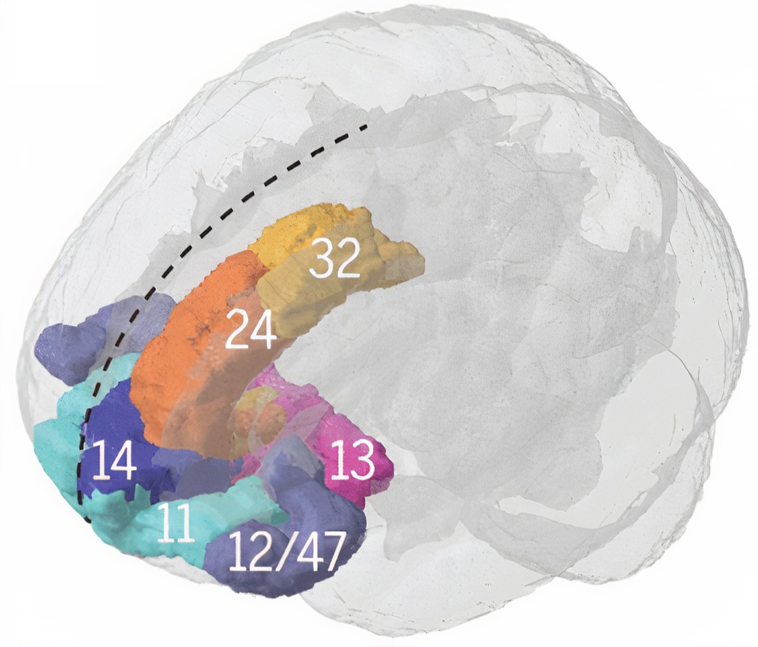
Graphics © Science and Fascija/Shutterstock.com.
.jpg)
Brodmann areas. The insular cortex is associated with Brodmann areas 13, 14, and 52.
Location. The insular cortex is located deep within the lateral sulcus, separating the frontal and parietal lobes from the temporal lobe.
Connections. The insular cortex has widespread connections with various brain regions, including the prefrontal cortex, parietal cortex, temporal cortex, and limbic structures (Augustine, 1996).
Participation in brain networks. The insular cortex is involved in multiple brain networks, including the salience network, and is responsible for detecting and responding to salient stimuli (Menon & Uddin, 2010).
Functions. The insular cortex involves various functions, including interoception, emotional processing, pain perception, and cognitive control (Craig, 2009).
Role in clinical disorders. Abnormalities in the insular cortex have been implicated in several neurological and psychiatric disorders, such as anxiety, depression, autism, and schizophrenia (Menon, 2011).
Brodmann Area Involvement in Clinical Disorders
The area profiles above run from anatomy toward behavior. This section runs the other way, from a diagnosis toward the areas implicated in it, which is the direction a referral question usually arrives in. The correlations between psychiatric and neurological disorders and specific Brodmann areas provide insight into the neural mechanisms of these conditions and give you a set of candidate regions to hold in mind while you read a record. Treat them as prior expectations to test, not as findings to confirm.
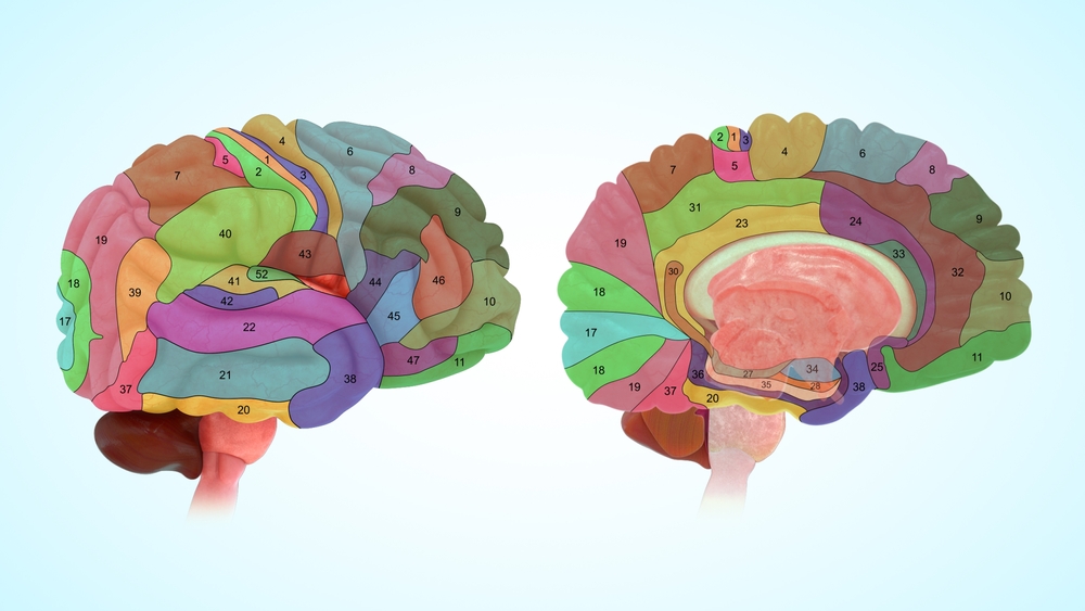
Brodmann graphic © sciencepics/Shutterstock.com.
Attention-Deficit Hyperactivity Disorder (ADHD)
ADHD is associated with abnormalities in multiple Brodmann areas, including BAs 44 and 45 (Broca's area), 8, 9, 10, 11, and 46 (frontal regions), 7, 39, and 40 (parietal regions), 4 (motor cortex), 30 (cingulate gyrus), 21 and 38 (temporal regions), and 6 (premotor cortex). These abnormalities contribute to the diverse cognitive, attentional, and motor deficits observed in individuals with ADHD.
In individuals with ADHD, Broca's area (BA 44 and 45) exhibits functional disturbances, particularly a lower fractional amplitude of low-frequency fluctuations (fALFF). This dysfunction is linked to language deficits, with different ADHD subtypes affecting distinct parts of Broca's area: Area 44 in the inattentive subtype and Area 45 in the combined subtype (Pikusa & Jończyk, 2015; Silk et al., 2005).
In the frontal regions, children with ADHD display grey matter deficits in the right superior frontal gyrus (BA 8 and 9), suggesting disruptions in attentional networks (Overmeyer et al., 2001). Adolescents with ADHD show decreased activation in the middle frontal gyrus (Area 10) and the dorsolateral prefrontal cortex (Area 46), indicating widespread frontal lobe dysfunction (Silk et al., 2005). Abnormalities in the ventromedial orbitofrontal cortex (Area 11) are linked to motivational deficits in adults with ADHD (Farré-Colomés et al., 2021).
The parietal regions, including the superior parietal lobe (Area 7) and the inferior parietal lobule (Areas 39 and 40), also show decreased activation in ADHD, implicating these areas in attentional and action-attentional systems (Silk et al., 2005). Children with ADHD exhibit decreased activation in the primary motor cortex (Area 4) during motor tasks, suggesting anomalies in motor development (Gaddis et al., 2015; Gilbert et al., 2011, 2019).
The right posterior cingulate gyrus (Area 30) shows grey matter deficits in children with ADHD, which may contribute to difficulties in attentional control (Overmeyer et al., 2001). Structural alterations are also observed in the temporal regions, with lower cortical thickness in the fusiform gyrus and temporal pole (Areas 21 and 38) in children with ADHD (Hoogman et al., 2019; Karalok et al., 2019; Lake et al., 2019; McLaughlin et al., 2014). Increased connectivity in the premotor cortex (Area 6) may be associated with challenges in motor planning and execution (Hoshi & Tanji, 2007; Oldehinkel et al., 2016; Sörös et al., 2019; Suskauer et al., 2008).
Autism Spectrum Disorder (ASD)
Research indicates that abnormalities in Brodmann areas 24, 44, 45, and 10 are associated with autism spectrum disorder. These abnormalities include changes in neuron size and density, gray matter volume, and functional connectivity, which are linked to the social and communication deficits characteristic of ASD. In the anterior cingulate cortex (BA 24), there is a notable reduction in cell size and cell packing density, specifically in areas 24b and 24c, along with elevated levels of glial fibrillary acidic protein (GFAP) in the white matter, indicating increased astrocyte activation. During verbal memory tasks, decreased glucose metabolism is observed in BA 24, and dysregulated DNA methylation patterns in this area affect genes related to immune functions and synaptic membranes.
In the inferior frontal cortex (BA 44 and 45), there is a decrease in the size of pyramidal neurons, which play crucial roles in language processing and social behaviors (Jacot-Descombes et al., 2012). Reduced gray matter volume in the pars opercularis (BA 44) and pars triangularis (BA 45) is linked to difficulties in social communication (Yamasaki et al., 2010).
The anterior prefrontal cortex (BA 10) exhibits abnormal functional connectivity between the right and left hemispheres, correlating with the severity of social deficits (Kikuchi et al., 2013). Similar to BA 24, BA 10 also shows dysregulated DNA methylation affecting genes involved in immune response and synaptic function (Nardone et al., 2014).
Other areas affected include the medial and cingulate regions (BAs 32, 24, and 25), where lower glucose metabolism is observed during verbal memory tasks (Deery et al., 2022; Hazlett et al., 2004, 2010). There is also a reduction in white matter volumes in the posterior frontal lobe and along the cingulate arch (Aalst et al., 2021; Gardener et al., 2016).
Bipolar Disorder (BD)
Research indicates that abnormalities in several Brodmann areas, including BAs 9, 10, 24, 38, 41, 42, and 46, are associated with bipolar disorder. These abnormalities span structural changes such as reduced glial numbers and cortical volume, as well as functional and molecular disruptions including altered gene expression and DNA methylation. In BA 9, part of the dorsolateral prefrontal cortex, abnormalities in DNA methylation and gene expression indicate a role in the pathophysiology of BD, and this area shows decreased numbers and density of glial cells, suggesting disruptions in neurochemical regulation.
The subgenual prefrontal cortex (BA 24) exhibits reduced volume and fewer glial cells, associated with familial forms of BD. This area is significant for mood regulation and reflects neurobiological changes linked to the disorder (Öngür et al., 1998; Scarr et al., 2019).
In the temporal pole (BA 38), notable differences in DNA methylation are associated with BD, indicating involvement in the molecular mechanisms underlying the disorder (Ho et al., 2019). In the primary auditory cortex (BA 41 and 42), decreased BOLD signals in response to auditory stimuli have been identified as potential biomarkers for BD (Okamoto et al., 2022).
BA 46, another region of the dorsolateral prefrontal cortex, shows altered gene expression, with specific genes exhibiting differential expression in BD, suggesting disruptions in neuronal network functions (Nakatani et al., 2006; Vizueta et al., 2012). The prefrontal cortex (BA 10) also presents changes in gene expression in BD, highlighting complex region-specific changes in cortical gene expression (Scarr et al., 2019; Vizueta et al., 2012).
Major Depressive Disorder (MDD)
Research consistently implicates several Brodmann areas in the pathology of major depressive disorder, including the anterior cingulate cortex (BA 24 and 32), dorsolateral prefrontal cortex (BA 9 and 46), ventromedial prefrontal cortex (BA 10), orbitofrontal cortex (BA 13 and 47/12), and subgenual cingulate cortex (BA 25). In the anterior cingulate cortex, structural abnormalities and reduced volume in the subgenual part of BA 24 are linked to familial forms of MDD, and elevated levels of tumor necrosis factor (TNF) have been observed in BA 24 among MDD patients, suggesting the involvement of pro-inflammatory pathways. Abnormalities in BA 32 have been associated with antidepressant treatment.
In the dorsolateral prefrontal cortex (BA 9 and 46), both structural and functional abnormalities have been connected to MDD. In BA 9, changes in connectivity and increased fractional amplitude of low-frequency fluctuation (fALFF) are noted (Gao et al., 2021; Lai & Wu, 2015; Vasic et al., 2008; Ye et al., 2012; Zhukovsky et al., 2020). Elevated TNF levels in BA 46 indicate a role in the disorder's pathophysiology (Dean et al., 2010). These are the same dorsolateral sites that underlie F3 and F4, which is why the frontal alpha asymmetry finding discussed earlier belongs to a much larger picture.
The ventromedial prefrontal cortex (BA 10) shows abnormal functional connectivity linked to emotional regulation deficits in MDD patients (Almeida et al., 2011; Johnstone et al., 2007; Wackerhagen et al., 2017; Young et al., 2016). Epigenetic modifications, such as methylation changes, have been identified in BA 9 and BA 38 (Ho et al., 2019).
In the orbitofrontal cortex (BA 13 and 47/12), reduced functional connectivity in BA 13 is associated with depressive symptoms, while increased functional connectivity in BA 47/12 correlates with negative self-perception in MDD (Cheng et al., 2016). The subgenual cingulate cortex (BA 25) exhibits reduced functional connectivity with regions involved in emotional regulation (Peng et al., 2020), and methylation changes in BA 25 have been consistently found in independent brain samples (Åberg et al., 2018).
Panic Disorder
Research indicates that abnormalities in Brodmann areas 11, 15, 25, and 32, as well as regions within the prefrontal cortex and parahippocampal gyrus, are associated with panic disorder. During panic attacks, there is a noticeable decrease in regional cerebral blood flow (rCBF) in the right orbitofrontal cortex (BA 11) and the prelimbic cortex (BA 25). This reduction is also observed in the anterior cingulate cortex (BA 32), which exhibits mixed patterns of hyperactivation and hypoactivation during emotional processing.
The parahippocampal gyrus in panic disorder patients shows significant abnormal asymmetry in cerebral blood flow. The anterior temporal cortex (BA 15) experiences decreased rCBF during panic attacks (Hasler et al., 2007). Key regions within the prefronto-limbic network, particularly areas within the PFC, show selective deficits in emotional processing among panic disorder patients (Hasler et al., 2007; Shang et al., 2014). Dysfunctional communication within frontotemporal structures is indicated by decreased coherence imaging values (Shang et al., 2014; Speer et al., 2003).
Increased resting-state functional connectivity between the thalamus and insula suggests excessive sensitivity to external information in panic disorder patients, highlighting a potential neural mechanism underlying their heightened responsiveness to stimuli (Feldker et al., 2016, 2019; Zhou et al., 2022).
Post-Traumatic Stress Disorder (PTSD)
PTSD is associated with abnormalities in the anterior cingulate cortex (BA 24 and 32), medial prefrontal cortex (BA 10 and 11), dorsolateral prefrontal cortex (BA 46), insula (BA 13), orbitofrontal cortex (BA 25), and sensorimotor areas (BA 4 and 6). These regions are involved in emotional regulation, memory processing, and response to trauma-related stimuli. In individuals with PTSD, the DLPFC (BA 46) shows decreased activity and signs of mitochondrial dysfunction, and the anterior cingulate gyrus (BA 32) exhibits less activation compared to those without PTSD.
The medial prefrontal cortex (BA 10 and 11) shows reduced activation in PTSD subjects (Etkin & Wager, 2007; Herringa et al., 2012; Manthey et al., 2021; Sartory et al., 2013). The activation levels in BA 10 correlate with PTSD symptoms during threat processing. The insula (BA 13) demonstrates increased delta slow waves linked to worsening PTSD symptoms over time (Harricharan et al., 2019; Herringa et al., 2012; Rabinak et al., 2011), and differences in functional connectivity in the insula are observed between PTSD and non-PTSD individuals (Rabinak et al., 2011; Sripada et al., 2012; Zhang et al., 2016).
During symptom provocation in PTSD patients, there is increased regional cerebral blood flow in the right sensorimotor areas (BA 4 and 6), while lowered rCBF is observed in the right retrosplenial cortex (BA 26, 29, and 30) (Pissiota et al., 2000). Structural and functional changes in the hippocampus and amygdala are consistently observed in PTSD, highlighting their crucial roles in memory and emotional processing (Chen et al., 2018; Hull, 2002).
Schizophrenia
Abnormalities in frontal (BAs 4, 6, 8, 9, 10, 44, 46, and 47), temporal (BAs 20, 21, 22, 37, 39, and 42), and cingulate (BAs 24, 25, 29, 30, and 31) cortices are associated with schizophrenia. These include disrupted intercorrelations, altered receptor binding, gene expression changes, and structural deficits. Significant reductions in gray matter volume have been observed in the left anterior hippocampus and amygdala, left parahippocampal gyrus, and left superior temporal gyrus, with correlations between the volume of the left posterior superior temporal gyrus and the severity of thought disorders.
Disruptions in D2 dopamine receptor patterns have been identified in the perirhinal, superior, and inferior temporal cortices (BA 20, 22, 37, 39, and 42). These disruptions are distinct to the temporal lobe and are not attributable to long-term antipsychotic treatment, suggesting a specific role in auditory hallucinations and other positive symptoms (Goldsmith et al., 1997; Joyce et al., 1997).
In schizotypal personality disorder, lower fractional anisotropy in the left temporal lobe (BA 20, 21, and 22) indicates alterations in white matter microstructure (Chan et al., 2018; Ellison-Wright & Bullmore, 2009; Hazlett et al., 2011; Lee et al., 2016; Szeszko et al., 2008). Schizophrenia is also associated with a thinner cortex and reduced surface area in the temporal lobe, with the most significant cortical thickness reductions at the temporal pole (Kaur et al., 2020; McCarley et al., 1999; Shenton et al., 1992). Progressive volumetric changes over time, particularly decreased temporal white matter volume, indicate ongoing structural abnormalities (Kaur et al., 2020; Mathew et al., 2014; Olabi et al., 2011).
Enhanced functional connectivity between the left DLPFC and the left mid-posterior temporal lobe has been identified in patients experiencing their first episode of schizophrenia (Zhou et al., 2007), a connectivity pattern that may contribute to the cognitive and perceptual disturbances characteristic of the disorder.
Substance Use Disorder (SUD)
Abnormalities in specific Brodmann areas, particularly BAs 10, 18, 19, 21, 22, 24, and 30, are associated with substance use disorder. These areas serve critical functions such as self-reflection, emotional regulation, memory, and executive function, which are often impaired in individuals with SUD.
In adolescents with SUD, there is reduced activity in BA 10 (superior, medial, and middle frontal gyrus), suggesting difficulties in self-referential evaluation and future planning (Dalwani et al., 2014). The anterior cingulate cortex (BA 24) shows abnormal activity, affecting emotional behavior and executive function in individuals with SUD.
Reading These Correlations Responsibly
The examination of specific Brodmann areas reveals correlations with various psychiatric and neurological disorders. ADHD features disruptions in regions related to attention and executive function. Autism spectrum disorder is characterized by changes in areas affecting social and communication skills. Bipolar disorder involves alterations in areas associated with mood regulation. Panic disorder and PTSD both involve the prefronto-limbic network, affecting emotional and stress responses. Schizophrenia is associated with widespread disruptions affecting cognition and perception, and substance use disorder involves regions critical for executive function and emotional regulation.
Two cautions belong with this material. First, most of these findings come from fMRI, PET, receptor binding, and postmortem tissue rather than from the EEG, so they tell you where to look and not what you will see. Second, the correlations are group differences, not diagnostic signs, and no combination of them identifies a disorder in an individual client. Used well, they give a qEEG report a neuroanatomical frame for the patterns you actually measured.
Brodmann's cytoarchitectural map remains the common vocabulary for describing cortical location, even though modern parcellations reveal finer divisions than his numbered areas captured. Most cortical functions recruit several Brodmann areas working as a network, so an area profile is a starting point rather than a destination. Reading each profile the same way, location, connections, network membership, functions, and clinical associations, lets you move from a client's presenting problem to a defensible set of candidate sites. Reading a diagnosis back toward its implicated areas gives you candidate regions to test, but those correlations come from imaging and tissue studies rather than the EEG, and they describe groups rather than individuals. Use the profiles as a lookup table when planning montages, and confirm your reasoning against the network architecture rather than treating any single area as the seat of a behavior.
Check Your Understanding
- On what basis did Brodmann distinguish his cortical areas, what staining method did he use, and why are numbers such as 12 through 16 absent from most human maps?
- Why do modern neuroimaging findings and probabilistic atlases complicate a strictly Brodmann-based account of cortical function?
- Which Brodmann areas correspond to the primary motor cortex, the primary visual cortex, Broca's area, and Wernicke's area?
- Choose one area profile and explain how its network membership would shape your choice of electrode sites for a recording.
- Name two disorders from this section and one Brodmann-area abnormality associated with each, then explain why that association cannot by itself support a diagnosis.
- Why should an area profile be treated as a starting point rather than a conclusion?
Assignment
Pick a client presentation you encounter often and work backward from behavior to anatomy. Which cortical regions and Brodmann areas would you expect to be involved, what electrode sites overlie them, and which networks do those areas join? Then argue the other side: what else could produce the same presentation, and what would you need to see in the recording before you committed to your first hypothesis?
Glossary
akinetic mutism: a state of markedly reduced spontaneous movement and speech despite preserved wakefulness, in which orienting responses are absent or greatly diminished, associated with cingulate malfunction.
alpha asymmetry: a difference in alpha amplitude between homologous sites, most often F3 and F4; because it compares two sites, unequal impedance, drowsy epochs, and reference choice can all manufacture one.
amygdala: a group of medial temporal-lobe nuclei involved in salience evaluation, associative learning, affective memory, and coordination of defensive and autonomic responses.
angular gyrus: a region of the parietal lobe near the junction of the temporal and occipital lobes that corresponds to Brodmann area 39. It plays a role in language processing, attention, spatial cognition, and the integration of sensory information.
anterior cingulate cortex (ACC): medial frontal cortex involved in cognitive control, motivation, affect, pain processing, and autonomic regulation, with functionally heterogeneous subdivisions.
anterior commissure: a compact white-matter tract crossing the midline and connecting portions of the temporal lobes, including olfactory and amygdalar regions.
anterior prefrontal cortex (aPFC): the most anterior region of the prefrontal cortex, corresponding to Brodmann area 10. It supports complex cognitive processes such as planning, decision-making, working memory, prospective memory, and abstract reasoning.
association bundles: white matter tracts that link cortical regions within the same hemisphere. Short association fibers, or U-fibers, arch beneath a sulcus to join adjacent gyri, while long fasciculi such as the arcuate, uncinate, and superior longitudinal fasciculi connect widely separated regions.
auditory cortex: the region of the temporal lobe that processes auditory information within the dorsal and ventral streams, supporting sound localization, pitch discrimination, and speech comprehension.
basal ganglia: interconnected subcortical nuclei, including striatum, globus pallidus, subthalamic nucleus, and substantia nigra, that regulate action selection, movement, learning, and motivation.
Broca's aphasia: an inability to produce fluent speech despite relatively intact comprehension, following lesions to Broca's area (BA 44 and 45).
Broca's area: the area located in the inferior frontal gyrus (BA 44 and 45) of the dominant hemisphere (F7-T3 in the left hemisphere) concerned with speech production, grammar, language comprehension, and sequencing.
Brodmann area (BA): a numbered cytoarchitectural zone of the cerebral cortex mapped by Brodmann from Nissl staining; the numbering runs to 52, but areas 12-16 and 48-51 were defined in non-human species.
central nucleus of the amygdala: an amygdalar output nucleus coordinating autonomic, endocrine, attentional, and defensive responses through projections to hypothalamic and brainstem targets.
cingulate cortex: a limbic region on the medial surface of the frontal and parietal lobes, comprising anterior (Brodmann areas 24, 32, 33) and posterior (areas 23, 29, 30, 31) divisions serving emotion, memory, attention, and cognitive control.
commissure: a tract crossing the midline and connecting corresponding or related regions on the two sides of the central nervous system.
corpus callosum: the largest cerebral commissure, connecting homologous and heterologous cortical regions across the two hemispheres.
dorsal entorhinal cortex (DEC): a medial temporal lobe region often associated with Brodmann areas 28 and 34, although the entorhinal cortex is allocortex and does not map neatly onto Brodmann's cytoarchitectonic divisions. It is involved in spatial memory and navigation.
dorsal posterior cingulate cortex (dPCC): the dorsal part of the posterior cingulate cortex, composed primarily of Brodmann areas 23 and 31. It is involved in self-referential thought, memory, and spatial awareness.
dorsolateral prefrontal cortex: lateral prefrontal cortex involved in working memory, cognitive control, planning, rule use, and goal-directed behavior; simple left-positive and right-negative mappings are inadequate.
ectosplenial region: a region of the retrosplenial cortex (BA 26) that is not widely recognized as a well-established human subdivision, adjacent to a cortex involved in spatial memory and navigation.
frontal eye field (FEF): a premotor region in the caudal middle frontal gyrus at its junction with the precentral gyrus, corresponding to Brodmann area 8, that controls voluntary eye movements and directs gaze toward attended targets.
frontal lobe: either cerebral lobe anterior to the central sulcus, involved in motor control, executive functions, language, motivation, and social behavior.
fusiform gyrus: a region on the ventral surface of the temporal and occipital lobes associated with Brodmann areas 37 and 19. It is involved in face recognition, object recognition, and the processing of color and visual form.
higher-order extrastriate cortex: the visual cortex beyond primary area V1 responsible for advanced processing such as object recognition, face perception, and the analysis of complex visual scenes.
hippocampus: a medial temporal-lobe structure essential to episodic-memory formation, spatial representation, and contextual regulation of stress and emotion.
inferior temporal gyrus (ITG): a region of the inferior temporal lobe, primarily Brodmann areas 20 and 37, that supports visual object recognition and semantic memory.
insula: cortex buried within the lateral sulcus that integrates interoceptive, affective, sensory, cognitive, and autonomic information.
insular cortex (insula): a cortical region buried within the lateral sulcus, comprising Brodmann areas 13, 14, 15, 16, and parts of area 52, that supports interoception, self-awareness, emotion, pain perception, and taste and serves as an integrative hub for the salience network.
lateral nucleus: the amygdalar nucleus that processes sensory information and distributes it throughout the amygdala.
left DLPFC: the left dorsolateral prefrontal cortex, concerned with approach behavior and positive affect, which helps select positive goals and organize behavior to achieve them.
limbic system: a widespread network of nuclei involved in emotion, motivation, learning, memory, and navigation, including the hippocampus, amygdala, and septal nuclei along with the hypothalamus, anterior thalamus, and cingulate gyrus.
middle temporal gyrus (MTG): a temporal lobe convolution between the superior and inferior temporal gyri, corresponding chiefly to Brodmann area 21 and extending posteriorly into area 37, that supports language processing, semantic memory, and visual motion processing.
motor association cortex (premotor cortex): the frontal lobe region rostral to the primary motor cortex, corresponding to Brodmann area 6, that helps program and execute movements.
occipital lobe: the posterior cerebral lobe containing primary and association visual cortices.
orbitofrontal cortex: prefrontal cortex above the orbits involved in valuation, outcome updating, emotion, sensory integration, and context-appropriate behavior.
parahippocampal gyri: structures located within the medial temporal lobe that form spatial and nonspatial contextual associations, which serve as building blocks for contextual processing, episodic memory, navigation, and scene processing.
parainsular area: a region at the junction of the temporal lobe and insula that is associated with Brodmann areas 13, 14, and 52. It participates in auditory and somatosensory integration and in processing pain and temperature sensations.
parietal lobe: a cerebral lobe posterior to the central sulcus involved in somatosensation, spatial representation, attention, body schema, and sensorimotor integration.
pars opercularis: the opercular part of the inferior frontal gyrus, corresponding to Brodmann area 44. It plays a role in language production and forms part of Broca's area.
pars orbitalis: the orbital part of the inferior frontal gyrus, corresponding to Brodmann area 47. It is involved in language processing, social cognition, and emotional regulation.
pars triangularis: the triangular part of the inferior frontal gyrus, corresponding to Brodmann area 45. It is involved in language processing and forms part of Broca's area.
perirhinal cortex (PRC): a medial temporal lobe region comprising Brodmann areas 35 and 36 that supports object recognition, associative memory, and contextual processing.
posterior commissure: a small midline fiber tract in the dorsal midbrain connecting bilateral pretectal and related nuclei, including pathways involved in the pupillary light reflex.
prefrontal cortex (PFC): the anterior association cortex of the frontal lobes involved in executive control, planning, working memory, valuation, social cognition, and emotion regulation.
prestriate cortex: the secondary visual cortex (V2), bordering the striate cortex, involved in the further processing and integration of visual information received from the primary visual cortex.
presubiculum: a part of the hippocampal formation involved in spatial navigation and memory.
primary gustatory cortex (PGC): a region associated with Brodmann area 43, which lies at the opercular part of the inferior frontal gyrus and extends into the insular cortex (Brodmann area 13), responsible for processing taste information.
primary motor cortex: cortex in the precentral gyrus that contributes substantially to execution and control of voluntary movement.
primary olfactory cortex: the pyriform cortex region (BA 27) that processes olfactory information.
primary somatosensory cortex (S1): cortex in the postcentral gyrus that receives organized thalamic somatosensory input and represents touch, proprioception, and nociceptive features.
primary visual cortex (V1): the occipital lobe region located in the calcarine sulcus (BA 17, also called striate cortex) that receives most visual information from the lateral geniculate nucleus of the thalamus and performs the initial processing of visual information received from the retinas.
projection fibers: white matter tracts that connect the cortex with structures deep in the brain, the brainstem, and the spinal cord, including the frontostriatal, thalamocortical, optic, and pyramidal tracts.
pyriform cortex: the ventral temporal lobe region that processes olfactory input, also called primary olfactory cortex and conventionally placed at Brodmann area 27 although it is allocortex.
retrosplenial cingulate cortex: a region in the posterior part of the cingulate cortex comprising Brodmann areas 29 and 30, involved in spatial memory, navigation, and contextual processing.
retrosubicular area: a small medial temporal lobe region of the hippocampal formation designated Brodmann area 48, involved in spatial navigation and memory.
right DLPFC: the right dorsolateral prefrontal cortex, which organizes withdrawal-related behavior and negative affect, mediates threat-related vigilance, and supports working memory for object location.
secondary somatosensory cortex (S2): parietal-opercular cortex receiving bilateral somatosensory input and contributing to integration, learning, and higher-order perception of bodily stimuli.
secondary visual cortex (V2): a region of the occipital lobe adjacent to the primary visual cortex, corresponding to Brodmann area 18, that processes shape, color, and spatial orientation.
septal nuclei: basal forebrain nuclei with reciprocal hippocampal and hypothalamic connections involved in theta modulation, memory, motivation, and autonomic function.
somatosensory association cortex (SAC): a posterior parietal region encompassing Brodmann areas 5 and 7 that integrates and interprets somatosensory information such as touch, pain, temperature, and proprioception.
striate cortex: the primary visual cortex (BA 17), responsible for processing basic visual information such as orientation, spatial frequency, and color.
Stroop task: a cognitive-interference task requiring a response to one stimulus dimension while inhibiting a conflicting, more automatic dimension.
subgenual region of the ventromedial prefrontal cortex (vmPFC): the ventromedial prefrontal region beneath the genu of the corpus callosum (BA 25 and 24b) involved in cognitive and emotional processes.
superior temporal gyrus (STG): the uppermost gyrus of the temporal lobe, spanning Brodmann areas 22, 41, and 42, which supports auditory processing, language comprehension, and social cognition; its posterior part contains Wernicke's area.
supplementary motor cortex (SMA) and premotor cortex (PMC): BA 6 regions critical for planning and executing voluntary movements.
supramarginal gyrus: a part of the inferior parietal lobule corresponding to Brodmann area 40, involved in phonological processing, language perception, attention, spatial cognition, and the integration of sensory information.
temporal lobe: a cerebral lobe inferior to the lateral sulcus involved in auditory processing, language, memory, and recognition of objects and faces.
temporal pole: the temporopolar area (BA 38) involved in a range of cognitive and emotional processes.
temporopolar area: the most anterior part of the temporal lobe, associated with Brodmann area 38. It is involved in olfactory processing, social cognition, and semantic memory.
thalamus: a paired diencephalic structure that relays and modulates most sensory and motor information reaching cortex and contributes to arousal and cortical rhythms.
ventral anterior cingulate cortex (vACC): the ventral subdivision of the anterior cingulate cortex, comprising Brodmann areas 24 and 25 and, in some parcellations, area 33. It is involved in emotional regulation, attention, and pain processing.
ventral entorhinal cortex (vEC): a medial temporal lobe region associated with Brodmann area 28, although the entorhinal cortex is allocortex and is not readily defined by Brodmann areas. It is involved in object recognition, memory, and contextual processing.
ventral posterior cingulate cortex (vPCC): the ventral part of the posterior cingulate cortex, comprising primarily Brodmann area 23. It supports self-referential thought, episodic memory retrieval, and emotional processing.
ventromedial prefrontal cortex: a medial and ventral prefrontal region involved in valuation, emotion regulation, extinction, decision-making, and integration of affective and autonomic information.
visual association cortex: a set of occipital and temporal lobe regions comprising Brodmann areas 18, 19, 21, 22, and 37 that includes areas V3, V4, and V5. It is responsible for higher-level visual processing, including object recognition, motion perception, and spatial awareness.
Wernicke's aphasia: a language disorder following damage to Wernicke's area, characterized by fluent but nonsensical speech and impaired comprehension of language.
Wernicke's area: the area of the temporoparietal cortex (BA 22) of the dominant hemisphere specialized for speech comprehension. Damage can result in an inability to understand the meaning of speech and construct intelligible sentences.
Test Yourself on ClassMarker
Click the button below to take a 10-question exam over this entire unit. There is no password.
Review Flash Cards on Quizlet
Click the button below to review our chapter flash cards.
References
Aalst, J., Devrome, M., Weehaeghe, D., Rezaei, A., Radwan, A., Schramm, G., Ceccarini, J., Sunaert, S., Koole, M., & Laere, K. (2021). Regional glucose metabolic decreases with ageing are associated with microstructural white matter changes: A simultaneous PET/MR study. European Journal of Nuclear Medicine and Molecular Imaging. https://doi.org/10.1007/s00259-021-05518-6
Aberg, K., Dean, B., Shabalin, A., Chan, R., Han, L., Zhao, M., Grootheest, G., Xie, L., Milaneschi, Y., Clark, S., Turecki, G., Penninx, B., & Oord, E. (2018). Methylome-wide association findings for major depressive disorder overlap in blood and brain and replicate in independent brain samples. Molecular Psychiatry, 25, 1344-1354. https://doi.org/10.1038/s41380-018-0247-6
Aggleton, J. P., & Brown, M. W. (1999). Episodic memory, amnesia, and the hippocampal-anterior thalamic axis. The Behavioral and Brain Sciences, 22(3), 425–489.
Aggleton, J. P., O'Mara, S. M., Vann, S. D., Wright, N. F., Tsanov, M., & Erichsen, J. T. (2010). Hippocampal-anterior thalamic pathways for memory: Uncovering a network of direct and indirect actions. Eur J Neurosci, 31(12), 2292-2307. https://doi.org/10.1111/j.1460-9568.2010.07251.x
Ahuja, A., & Yusif Rodriguez, N. (2022). Is the dorsolateral prefrontal cortex actually several different brain areas? The Journal of Neuroscience: The Official Journal of the Society for Neuroscience, 42(33), 6310–6312. https://doi.org/10.1523/JNEUROSCI.0848-22.2022
Almeida, J., Kronhaus, D., Sibille, E., Langenecker, S., Versace, A., LaBarbara, E., & Phillips, M. (2011). Abnormal left-sided orbitomedial prefrontal cortical–amygdala connectivity during happy and fear face processing: A potential neural mechanism of female MDD. Frontiers in Psychiatry, 2. https://doi.org/10.3389/fpsyt.2011.00069
Amaral, D. G., & Witter, M. P. (1995). Hippocampal formation. In G. Paxinos (Ed.), The rat nervous system (pp. 443-493). Academic Press.
Aminoff, E. M., Kveraga, K., & Bar, M. (2013). The role of the parahippocampal cortex in cognition. Trends Cogn Sci, 17(8), 379-390. https://doi.org/10.1016/j.tics.2013.06.009
Amunts, K., Schleicher, A., Bürgel, U., Mohlberg, H., Uylings, H. B., & Zilles, K. (1999). Broca's region revisited: Cytoarchitecture and intersubject variability. The Journal of Comparative Neurology, 412(2), 319–341. https://doi.org/10.1002/(sici)1096-9861(19990920)412:2<319::aid-cne10>3.0.co;2-7
Amunts, K., & Zilles, K. (2015). Architectonic mapping of the human brain beyond Brodmann. Neuron, 88, 1086-1107. https://doi.org/10.1016/j.neuron.2015.12.001
Amzica, F., & Lopes da Silva, F. H. (2018). Cellular substrates of brain rhythms. In Schomer, D. L. & F. H. Lopes da Silva (Eds.). Niedermeyer's electroencephalography: Basic principles, clinical applications, and related fields (7th ed.). Oxford University Press.
Andrews-Hanna, J. R., Reidler, J. S., Sepulcre, J., Poulin, R., & Buckner, R. L. (2010). Functional-anatomic fractionation of the brain's default network. Neuron, 65(4), 550–562. https://doi.org/10.1016/j.neuron.2010.02.005
Augustine J. R. (1996). Circuitry and functional aspects of the insular lobe in primates including humans. Brain Research. Brain Research Reviews, 22(3), 229–244. https://doi.org/10.1016/s0165-0173(96)00011-2
Avidan, G., & Behrmann, M. (2009). Functional MRI reveals compromised neural integrity of the face processing network in congenital prosopagnosia. Current Biology: CB, 19(13), 1146–1150. https://doi.org/10.1016/j.cub.2009.04.060
Bear, M. F., Connors, B. W., & Paradiso, M. A. (2020). Neuroscience: Exploring the brain (Enhanced 4th ed.). Jones & Bartlett Learning.
Beauregard, M., & Levesque, J. (2006). Functional magnetic resonance imaging investigation of the effects of neurofeedback training on the neural bases of selective attention and response inhibition in children with Attention-Deficit/Hyperactivity Disorder. Applied Psychophysiology and Biofeedback, 31(1), 3-20. https://doi.org/10.1007/s10484-006-9001-y
Bizley, J. K., & Cohen, Y. E. (2013). The what, where and how of auditory-object perception. Nature Reviews. Neuroscience, 14(10), 693–707. https://doi.org/10.1038/nrn3565
Braak, H., & Braak, E. (1991). Neuropathological stageing of Alzheimer-related changes. Acta Neuropathologica, 82(4), 239–259. https://doi.org/10.1007/BF00308809
Breedlove, S. M., & Watson, N. V. (2023). Behavioral neuroscience (10th ed.). Sinauer Associates, Inc.
Bressler, S. L., & Menon, V. (2010). Large-scale brain networks in cognition: Emerging methods and principles. Trends in Cognitive Sciences, 14(6), 277–290. https://doi.org/10.1016/j.tics.2010.04.004
Brodmann, K. (1909). Vergleichende Lokalisationslehre der Grosshirnrinde in ihren Prinzipien dargestellt auf Grund des Zellenbaues. Barth.
Broyd, S. J., Demanuele, C., Debener, S., Helps, S. K., James, C. J., & Sonuga-Barke, E. J. (2009). Default-mode brain dysfunction in mental disorders: A systematic review. Neuroscience and Biobehavioral Reviews, 33(3), 279–296. https://doi.org/10.1016/j.neubiorev.2008.09.002
Buckner, R. L., Andrews-Hanna, J. R., & Schacter, D. L. (2008). The brain's default network: Anatomy, function, and relevance to disease. Annals of the New York Academy of Sciences, 1124, 1-38. https://doi.org/10.1196/annals.1440.011
Buckner, R. L., Snyder, A. Z., Shannon, B. J., LaRossa, G., Sachs, R., Fotenos, A. F., Sheline, Y. I., Klunk, W. E., Mathis, C. A., Morris, J. C., & Mintun, M. A. (2005). Molecular, structural, and functional characterization of Alzheimer's disease: evidence for a relationship between default activity, amyloid, and memory. The Journal of Neuroscience: The Official Journal of the Society for Neuroscience, 25(34), 7709–7717. https://doi.org/10.1523/JNEUROSCI.2177-05.2005
Bush, G., Luu, P., & Posner, M. I. (2000). Cognitive and emotional influences in anterior cingulate cortex. Trends in Cognitive Sciences, 4(6), 215–222. https://doi.org/10.1016/s1364-6613(00)01483-2
Cao, Z., Ottino-Gonzalez, J., Cupertino, R. B., Schwab, N., Hoke, C., Catherine, O., Cousijn, J., Dagher, A., Foxe, J. J., Goudriaan, A. E., Hester, R., Hutchison, K., Li, C. R., London, E. D., Lorenzetti, V., Luijten, M., Martin-Santos, R., Momenan, R., Paulus, M. P., Schmaal, L., … Garavan, H. (2021). Mapping cortical and subcortical asymmetries in substance dependence: Findings from the ENIGMA Addiction Working Group. Addiction Biology, 26(5), e13010. https://doi.org/10.1111/adb.13010
Caplan, D. (2006). Why is Broca’s area involved in syntax? Cortex, 42, 469–471. https://doi.org/10.1016/S0010-9452(08)70379-4
Carlson, N. R., & Birkett, M. A. (2017). Physiology of behavior (12th ed.). Pearson Education, Inc.
Caspers, S., Geyer, S., Schleicher, A., Mohlberg, H., Amunts, K., & Zilles, K. (2006). The human inferior parietal cortex: Cytoarchitectonic parcellation and interindividual variability. NeuroImage, 33(2), 430–448. https://doi.org/10.1016/j.neuroimage.2006.06.054
Catani, M., Jones, D. K., Donato, R., & Ffytche, D. H. (2003). Occipito-temporal connections in the human brain. Brain: A Journal of Neurology, 126(Pt 9), 2093–2107. https://doi.org/10.1093/brain/awg203
Craig A. D. (2009). How do you feel--now? The anterior insula and human awareness. Nature Reviews. Neuroscience, 10(1), 59–70. https://doi.org/10.1038/nrn2555
Dean, B., Tawadros, N., Scarr, E., & Gibbons, A. (2010). Regionally-specific changes in levels of tumour necrosis factor in the dorsolateral prefrontal cortex obtained postmortem from subjects with major depressive disorder. Journal of Affective Disorders, 120(1-3), 245-248. https://doi.org/10.1016/j.jad.2009.04.027
deCharms, R. C., Fumiko, M., Glover, G. H., Ludlow, D., Pauly, J. M., Soneji, D., Gabrieli, J. D. E., & Mackey, S. C. (2005). Control over brain activation and pain learned by using real-time functional MRI. Proceedings of the National Academy of Sciences, 102(51), 18626-18631.
Demos, J. N. (2019). Getting started with neurofeedback. (2nd ed.). W. W. Norton & Company.
Devinsky, O., Morrell, M. J., & Vogt, B. A. (1995). Contributions of anterior cingulate cortex to behaviour. Brain: A Journal of Neurology, 118 ( Pt 1), 279–306. https://doi.org/10.1093/brain/118.1.279
Diamond, A. (2013). Executive functions. Annu Rev Psychol, 64, 135-168. https://doi.org/10.1146/annurev-psych-113011-143750
Dosenbach, N. U., Fair, D. A., Miezin, F. M., Cohen, A. L., Wenger, K. K., Dosenbach, R. A., Fox, M. D., Snyder, A. Z., Vincent, J. L., Raichle, M. E., Schlaggar, B. L., & Petersen, S. E. (2007). Distinct brain networks for adaptive and stable task control in humans. Proceedings of the National Academy of Sciences of the United States of America, 104(26), 11073–11078. https://doi.org/10.1073/pnas.0704320104
Do Tran, A., Kelway, C., Sellier, A., Dagain, A., & Beucler, N. (2025). Diffuse axonal injury unveiling white matter tracts anatomy. Neurochirurgie, 71(4), Article 101690. https://doi.org/10.1016/j.neuchi.2025.101690
Doty R. L. (2008). The olfactory vector hypothesis of neurodegenerative disease: Is it viable? Annals of Neurology, 63(1), 7–15. https://doi.org/10.1002/ana.21327
Drevets, W. C., Price, J. L., & Furey, M. L. (2008). Brain structural and functional abnormalities in mood disorders: Implications for neurocircuitry models of depression. Brain Structure & Function, 213(1-2), 93–118. https://doi.org/10.1007/s00429-008-0189-x
Du, F., Whetsell, W. O., Jr, Abou-Khalil, B., Blumenkopf, B., Lothman, E. W., & Schwarcz, R. (1993). Preferential neuronal loss in layer III of the entorhinal cortex in patients with temporal lobe epilepsy. Epilepsy Research, 16(3), 223–233. https://doi.org/10.1016/0920-1211(93)90083-j
Eichenbaum, H., Yonelinas, A. P., & Ranganath, C. (2007). The medial temporal lobe and recognition memory. Annual Review of Neuroscience, 30, 123–152. https://doi.org/10.1146/annurev.neuro.30.051606.094328
Eickhoff, S. B., Stephan, K. E., Mohlberg, H., Grefkes, C., Fink, G. R., Amunts, K., & Zilles, K. (2005). A new SPM toolbox for combining probabilistic cytoarchitectonic maps and functional imaging data. NeuroImage, 25(4), 1325–1335. https://doi.org/10.1016/j.neuroimage.2004.12.034
Eisenberger, N. I., Lieberman, M. D., & Williams, K. D. (2003). Does rejection hurt? An fMRI study of social exclusion. Science, 302, 290-292. https://doi.org/10.1126/science.1089134
Ellison-Wright, I., & Bullmore, E. (2009). Meta-analysis of diffusion tensor imaging studies in schizophrenia. Schizophrenia Research, 108, 3-10. https://doi.org/10.1016/j.schres.2008.11.021
Epstein R. A. (2008). Parahippocampal and retrosplenial contributions to human spatial navigation. Trends in Cognitive Sciences, 12(10), 388–396. https://doi.org/10.1016/j.tics.2008.07.004
Etkin, A., Egner, T., & Kalisch, R. (2011). Emotional processing in anterior cingulate and medial prefrontal cortex. Trends in Cognitive Sciences, 15(2), 85–93. https://doi.org/10.1016/j.tics.2010.11.004
Farré-Colomés, Á., Gerhardt, S., Luderer, M., Sobanski, E., Kiefer, F., & Vollstädt-Klein, S. (2021). Common and distinct neural connectivity in Attention Deficit/Hyperactivity Disorder and Alcohol Use Disorder: A study using resting-state functional magnetic resonance imaging. Alcoholism, Clinical and Experimental Research. https://doi.org/10.1111/acer.14593
Frank, G. K., Shott, M. E., Riederer, J., & Pryor, T. L. (2016). Altered structural and effective connectivity in anorexia and bulimia nervosa in circuits that regulate energy and reward homeostasis. Translational Psychiatry, 6(11), e932. https://doi.org/10.1038/tp.2016.199
Friederici A. D. (2011). The brain basis of language processing: From structure to function. Physiological Reviews, 91(4), 1357–1392. https://doi.org/10.1152/physrev.00006.2011
Fuster, J. (2015). The prefrontal cortex (5th ed.). Academic Press.
Fuster J. M. (2001). The prefrontal cortex--An update: Time is of the essence. Neuron, 30(2), 319–333. https://doi.org/10.1016/s0896-6273(01)00285-9
Garey, L. J. (Ed. & Trans.). (1994). Brodmann's ‘Localisation in the cerebral cortex’. Smith-Gordon.
Glasser, M. F., Coalson, T. S., Robinson, E. C., Hacker, C. D., Harwell, J., Yacoub, E., Ugurbil, K., Andersson, J., Beckmann, C. F., Jenkinson, M., Smith, S. M., & Van Essen, D. C. (2016). A multi-modal parcellation of human cerebral cortex. Nature, 536(7615), 171–178. https://doi.org/10.1038/nature18933
Gottfried J. A. (2010). Central mechanisms of odour object perception. Nature Reviews. Neuroscience, 11(9), 628–641. https://doi.org/10.1038/nrn2883
Greicius, M. D., Flores, B. H., Menon, V., Glover, G. H., Solvason, H. B., Kenna, H., Reiss, A. L., & Schatzberg, A. F. (2007). Resting-state functional connectivity in major depression: Abnormally increased contributions from subgenual cingulate cortex and thalamus. Biological Psychiatry, 62(5), 429–437. https://doi.org/10.1016/j.biopsych.2006.09.020
Griffiths, T. D., & Warren, J. D. (2002). The planum temporale as a computational hub. Trends in Neurosciences, 25(7), 348–353. https://doi.org/10.1016/s0166-2236(02)02191-4
Hafting, T., Fyhn, M., Molden, S., Moser, M. B., & Moser, E. I. (2005). Microstructure of a spatial map in the entorhinal cortex. Nature, 436(7052), 801–806. https://doi.org/10.1038/nature03721
Hale, J. B., & Fiorello, C. A. (2004). School neuropsychology: A practitioner's handbook. Guilford Press.
Heckers, S., Rauch, S. L., Goff, D., Savage, C. R., Schacter, D. L., Fischman, A. J., & Alpert, N. M. (1998). Impaired recruitment of the hippocampus during conscious recollection in schizophrenia. Nature Neuroscience, 1(4), 318–323. https://doi.org/10.1038/1137
Herbet, G., & Duffau, H. (2020). Revisiting the functional anatomy of the human brain: Toward a meta-networking theory of cerebral functions. Physiological Reviews, 100(3), 1181-1228. https://doi.org/10.1152/physrev.00033.2019
Hoeft, F., Meyler, A., Hernandez, A., Juel, C., Taylor-Hill, H., Martindale, J. L., McMillon, G., Kolchugina, G., Black, J. M., Faizi, A., Deutsch, G. K., Siok, W. T., Reiss, A. L., Whitfield-Gabrieli, S., & Gabrieli, J. D. (2007). Functional and morphometric brain dissociation between dyslexia and reading ability. Proceedings of the National Academy of Sciences of the United States of America, 104(10), 4234–4239. https://doi.org/10.1073/pnas.0609399104
Hyman, B. T., Van Hoesen, G. W., Damasio, A. R., & Barnes, C. L. (1984). Alzheimer's disease: Cell-specific pathology isolates the hippocampal formation. Science, 225(4667), 1168–1170. https://doi.org/10.1126/science.6474172
Jasper, H. H. (1958). The Ten-Twenty Electrode System of the International Federation. Electroencephalography and Clinical Neurophysiology, 10, 371-375.
Johnstone, T., Reekum, C., Urry, H., Kalin, N., & Davidson, R. (2007). Failure to regulate: Counterproductive recruitment of top-down prefrontal-subcortical circuitry in Major Depression. The Journal of Neuroscience, 27, 8877 - 8884. https://doi.org/10.1523/JNEUROSCI.2063-07.2007
Kennerley, S. W., Behrens, T. E., & Wallis, J. D. (2011). Double dissociation of value computations in orbitofrontal and anterior cingulate neurons. Nat Neurosci, 14(12), 1581-1589. doi:10.1038/nn.2961
Khan, U. A., Liu, L., Provenzano, F. A., Berman, D. E., Profaci, C. P., Sloan, R., Mayeux, R., Duff, K. E., & Small, S. A. (2014). Molecular drivers and cortical spread of lateral entorhinal cortex dysfunction in preclinical Alzheimer's disease. Nature Neuroscience, 17(2), 304–311. https://doi.org/10.1038/nn.3606
Kleen, J. K., Chung, J. E., Sellers, K. K., Zhou, J., Triplett, M., Lee, K., Tooker, A., Haque, R., & Chang, E. F. (2021). Bidirectional propagation of low frequency oscillations over the human hippocampal surface. Nature Communications, 12(2764). https://doi.org/10.1038/s41467-021-22850-5
Kringelbach M. L. (2005). The human orbitofrontal cortex: Linking reward to hedonic experience. Nature Reviews. Neuroscience, 6(9), 691–702. https://doi.org/10.1038/nrn1747
Kurth, F., Zilles, K., Fox, P. T., Laird, A. R., & Eickhoff, S. B. (2010). A link between the systems: Functional differentiation and integration within the human insula revealed by meta-analysis. Brain Structure & Function, 214(5-6), 519–534. https://doi.org/10.1007/s00429-010-0255-z
Leech, R., & Sharp, D. J. (2014). The role of the posterior cingulate cortex in cognition and disease. Brain: A Journal of Neurology, 137(Pt 1), 12–32. https://doi.org/10.1093/brain/awt162
Liston, C., Malter Cohen, M., Teslovich, T., Levenson, D., & Casey, B. J. (2011). Atypical prefrontal connectivity in attention-deficit/hyperactivity disorder: Pathway to disease or pathological end point? Biological Psychiatry, 69(12), 1168–1177. https://doi.org/10.1016/j.biopsych.2011.03.022
Manthey, A., Sierk, A., Brakemeier, E., Walter, H., & Daniels, J. (2021). Does trauma-focused psychotherapy change the brain? A systematic review of neural correlates of therapeutic gains in PTSD. European Journal of Psychotraumatology, 1. https://doi.org/10.1080/20008198.2021.1929025
Margulies, D. S., Vincent, J. L., Kelly, C., Lohmann, G., Uddin, L. Q., Biswal, B. B., Villringer, A., Castellanos, F. X., Milham, M. P., & Petrides, M. (2009). Precuneus shares intrinsic functional architecture in humans and monkeys. Proceedings of the National Academy of Sciences of the United States of America, 106(47), 20069–20074. https://doi.org/10.1073/pnas.0905314106
Mayberg H. S. (2003). Modulating dysfunctional limbic-cortical circuits in depression: Towards development of brain-based algorithms for diagnosis and optimised treatment. British Medical Bulletin, 65, 193–207. https://doi.org/10.1093/bmb/65.1.193
McCarley, R., Wible, C., Frumin, M., Hirayasu, Y., Levitt, J., Fischer, I., & Shenton, M. (1999). MRI anatomy of schizophrenia. Biological Psychiatry, 45, 1099-1119. https://doi.org/10.1016/S0006-3223(99)00018-9
Menon, V. (2011). Large-scale brain networks and psychopathology: A unifying triple network model. Trends in Cognitive Sciences, 15(10), 483-506. https://doi.org/10.1016/j.tics.2011.08.003
Menon, V., & Uddin, L. Q. (2010). Saliency, switching, attention and control: A network model of insula function. Brain Struct Funct, 214(5-6), 655-667. https://dx.doi.org/10.1007%2Fs00429-010-0262-0
Miller, B. L., & Cummings, J. L. (Eds.) (2007). The human frontal lobes: Functions and disorders (2nd ed.). Guilford Press.
Morosan, P., Rademacher, J., Schleicher, A., Amunts, K., Schormann, T., & Zilles, K. (2001). Human primary auditory cortex: Cytoarchitectonic subdivisions and mapping into a spatial reference system. NeuroImage, 13(4), 684–701. https://doi.org/10.1006/nimg.2000.0715
Neef, N. E., Anwander, A., Bütfering, C., Schmidt-Samoa, C., Friederici, A. D., Paulus, W., & Sommer, M. (2018). Structural connectivity of right frontal hyperactive areas scales with stuttering severity. Brain: A Journal of Neurology, 141(1), 191–204. https://doi.org/10.1093/brain/awx316
Neville, K. R., & Haberly, L. B. (2004). Olfactory cortex. The synaptic organization of the brain. In G. M. Shepherd (Ed.). Oxford University Press, 415–454. https://doi.org/10.1093/acprof:oso/9780195159561.003.0010
Niendam, T. A., Laird, A. R., Ray, K. L., Dean, Y. M., Glahn, D. C., & Carter, C. S. (2012). Meta-analytic evidence for a superordinate cognitive control network subserving diverse executive functions. Cognitive, Affective & Behavioral Neuroscience, 12(2), 241–268. https://doi.org/10.3758/s13415-011-0083-5
Olabi, B., Ellison-Wright, I., McIntosh, A., Wood, S., Bullmore, E., & Lawrie, S. (2011). Are there progressive brain changes in schizophrenia? A meta-analysis of structural magnetic resonance imaging studies. Biological Psychiatry, 70, 88-96. https://doi.org/10.1016/j.biopsych.2011.01.032
Oldehinkel, M., Beckmann, C., Pruim, R., Oort, E., Franke, B., Hartman, C., Hoekstra, P., Oosterlaan, J., Heslenfeld, D., Buitelaar, J., & Mennes, M. (2016). Attention-Deficit/Hyperactivity Disorder symptoms coincide with altered striatal connectivity. Biological Psychiatry: Cognitive Neuroscience and Neuroimaging, 14, 353-363. https://doi.org/10.1016/J.BPSC.2016.03.008
Olson, I. R., Plotzker, A., & Ezzyat, Y. (2007). The Enigmatic temporal pole: A review of findings on social and emotional processing. Brain: A Journal of Neurology, 130(Pt 7), 1718–1731. https://doi.org/10.1093/brain/awm052
Ongür, D., Ferry, A. T., & Price, J. L. (2003). Architectonic subdivision of the human orbital and medial prefrontal cortex. The Journal of Comparative Neurology, 460(3), 425–449. https://doi.org/10.1002/cne.10609
Öngür, D., & Price, J. L. (2000). The organization of networks within the orbital and medial prefrontal cortex of rats, monkeys and humans. Cereb Cortex, 10(3), 206-219. https://doi.org/10.1093/cercor/10.3.206
Padmanabhan, A., Lynch, C. J., Schaer, M., & Menon, V. (2017). The Default Mode Network in autism. Biological Psychiatry. Cognitive Neuroscience and Neuroimaging, 2(6), 476–486. https://doi.org/10.1016/j.bpsc.2017.04.004
Penfield, W., & Boldrey, E. (1937). Somatic motor and sensory representation in the cerebral cortex of man as studied by electrical stimulation. Brain, 60(4), 389–443. https://doi.org/10.1093/brain/60.4.389
Peng, K., Steele, S. C., Becerra, L., & Borsook, D. (2018). Brodmann area 10: Collating, integrating and high level processing of nociception and pain. Progress in Neurobiology, 161, 1–22. https://doi.org/10.1016/j.pneurobio.2017.11.004
Pikusa, M., & Jończyk, R. (2015). Functional abnormalities in Broca’s area in adolescents with ADHD: A resting-state fMRI study. Poznan Studies in Contemporary Linguistics, 51, 163 - 177. https://doi.org/10.1515/psicl-2015-0006
Pissiota, A., Frans, Ö., Fernández, M., Knorring, L., Fischer, H., & Fredrikson, M. (2000). Neurofunctional correlates of posttraumatic stress disorder: A PET symptom provocation study. European Archives of Psychiatry and Clinical Neuroscience, 252, 68-75. https://doi.org/10.1007/s004060200014
Price, J. L., & Drevets, W. C. (2010). Neurocircuitry of mood disorders. Neuropsychopharmacology: Official Publication of the American College of Neuropsychopharmacology, 35(1), 192–216. https://doi.org/10.1038/npp.2009.104
Purves, D. (2018). Neuroscience (6th ed.). Oxford University Press.
Raichle, M. E., MacLeod, A. M., Snyder, A. Z., Powers, W. J., Gusnard, D. A., & Shulman, G. L. (2001). A default mode of brain function. Proceedings of the National Academy of Sciences of the United States of America, 98(2), 676–682. https://doi.org/10.1073/pnas.98.2.676
Rajkowska, G., & Goldman-Rakic, P. S. (1995). Cytoarchitectonic definition of prefrontal areas in the normal human cortex: II. Variability in locations of areas 9 and 46 and relationship to the Talairach Coordinate System. Cerebral Cortex, 5(4), 323–337. https://doi.org/10.1093/cercor/5.4.323
Ramnani, N., & Owen, A. M. (2004). Anterior prefrontal cortex: Insights into function from anatomy and neuroimaging. Nature Reviews. Neuroscience, 5(3), 184–194. https://doi.org/10.1038/nrn1343
Rolls E. T. (2006). Brain mechanisms underlying flavour and appetite. Philosophical Transactions of the Royal Society of London. Series B, Biological Sciences, 361(1471), 1123–1136. https://doi.org/10.1098/rstb.2006.1852
Roy, A. K., Shehzad, Z., Margulies, D. S., Kelly, A. M., Uddin, L. Q., Gotimer, K., Biswal, B. B., Castellanos, F. X., & Milham, M. P. (2009). Functional connectivity of the human amygdala using resting state fMRI. NeuroImage, 45(2), 614–626. https://doi.org/10.1016/j.neuroimage.2008.11.030
Roy, M., Shohamy, D., & Wager, T. D. (2012). Ventromedial prefrontal-subcortical systems and the generation of affective meaning. Trends in Cognitive Sciences, 16(3), 147–156. https://doi.org/10.1016/j.tics.2012.01.005
Rudebeck, P. H., Putnam, P. T., Daniels, T. E., Yang, T., Mitz, A. R., Rhodes, S. E., & Murray, E. A. (2014). A role for primate subgenual cingulate cortex in sustaining autonomic arousal. Proceedings of the National Academy of Sciences of the United States of America, 111(14), 5391–5396. https://doi.org/10.1073/pnas.1317695111
Sartory, G., Cwik, J., Knuppertz, H., Schürholt, B., Lebens, M., Seitz, R., & Schulze, R. (2013). In search of the trauma memory: A meta-analysis of functional neuroimaging studies of symptom provocation in Posttraumatic Stress Disorder (PTSD). PLoS ONE, 8. https://doi.org/10.1371/journal.pone.0058150
Scarr, E., Udawela, M., & Dean, B. (2019). Changed cortical risk gene expression in major depression and shared changes in cortical gene expression between major depression and bipolar disorders. Australian & New Zealand Journal of Psychiatry, 53, 1189 - 1198. https://doi.org/10.1177/0004867419857808
Sedley, W., Parikh, J., Edden, R. A., Tait, V., Blamire, A., & Griffiths, T. D. (2015). Human auditory cortex neurochemistry reflects the presence and severity of tinnitus. The Journal of Neuroscience: The Official Journal of the Society for Neuroscience, 35(44), 14822–14828. https://doi.org/10.1523/JNEUROSCI.2695-15.2015
Seeley, W. W., Menon, V., Schatzberg, A. F., Keller, J., Glover, G. H., Kenna, H., Reiss, A. L., & Greicius, M. D. (2007). Dissociable intrinsic connectivity networks for salience processing and executive control. The Journal of Neuroscience: The Official Journal of the Society for Neuroscience, 27(9), 2349–2356. https://doi.org/10.1523/JNEUROSCI.5587-06.2007
Seghier M. L. (2013). The angular gyrus: Multiple functions and multiple subdivisions. The Neuroscientist: A Review Journal Bridging Neurobiology, Neurology and Psychiatry, 19(1), 43–61. https://doi.org/10.1177/1073858412440596
Shang, J., Fu, Y., Ren, Z., Zhang, T., Du, M., Gong, Q., Lui, S., & Zhang, W. (2014). The Common Traits of the ACC and PFC in anxiety disorders in the DSM-5: Meta-analysis of voxel-based morphometry studies. PLoS ONE, 9. https://doi.org/10.1371/journal.pone.0093432
Sheline, Y. I., Price, J. L., Yan, Z., & Mintun, M. A. (2010). Resting-state functional MRI in depression unmasks increased connectivity between networks via the dorsal nexus. Proceedings of the National Academy of Sciences of the United States of America, 107(24), 11020–11025. https://doi.org/10.1073/pnas.1000446107
Shepherd G. M. (2007). Perspectives on olfactory processing, conscious perception, and orbitofrontal cortex. Annals of the New York Academy of Sciences, 1121, 87–101. https://doi.org/10.1196/annals.1401.032
Small, D. M., Zald, D. H., Jones-Gotman, M., Zatorre, R. J., Pardo, J. V., Frey, S., & Petrides, M. (1999). Human cortical gustatory areas: A review of functional neuroimaging data. Neuroreport, 10(1), 7–14. https://doi.org/10.1097/00001756-199901180-00002
Sörös, P., Hoxhaj, E., Borel, P., Sadohara, C., Feige, B., Matthies, S., Müller, H., Bachmann, K., Schulze, M., & Philipsen, A. (2019). Hyperactivity/restlessness is associated with increased functional connectivity in adults with ADHD: A dimensional analysis of resting state fMRI. BMC Psychiatry, 19. https://doi.org/10.1186/s12888-019-2031-9
Steriade, M., & Llinás, R. R. (1988). The functional states of the thalamus and the associated neuronal interplay. Physiol Rev, 68(3), 649-742. https://doi.org/10.1152/physrev.1988.68.3.649
Suzuki, W. A., & Amaral, D. G. (1994). Perirhinal and parahippocampal cortices of the macaque monkey: Cortical afferents. The Journal of Comparative Neurology, 350(4), 497–533. https://doi.org/10.1002/cne.903500402
Thompson, M., & Thompson, L. (2015). The neurofeedback book: An introduction to basic concepts in applied psychophysiology (2nd ed.). Association for Applied Psychophysiology and Biofeedback.
Utevsky, A. V., Smith, D. V., & Huettel, S. A. (2014). Precuneus is a functional core of the default-mode network. The Journal of Neuroscience: The Official Journal of the Society for Neuroscience, 34(3), 932–940. https://doi.org/10.1523/JNEUROSCI.4227-13.2014
Van Hoesen, G., & Pandya, D. N. (1975). Some connections of the entorhinal (area 28) and perirhinal (area 35) cortices of the rhesus monkey. I. Temporal lobe afferents. Brain Research, 95(1), 1–24. https://doi.org/10.1016/0006-8993(75)90204-8
Vann, S. D., Aggleton, J. P., & Maguire, E. A. (2009). What does the retrosplenial cortex do? Nature Reviews. Neuroscience, 10(11), 792–802. https://doi.org/10.1038/nrn2733
van Strien, N. M., Cappaert, N. L., & Witter, M. P. (2009). The anatomy of memory: An interactive overview of the parahippocampal-hippocampal network. Nature Reviews. Neuroscience, 10(4), 272–282. https://doi.org/10.1038/nrn2614
Vogt B. A. (2005). Pain and emotion interactions in subregions of the cingulate gyrus. Nature Reviews. Neuroscience, 6(7), 533–544. https://doi.org/10.1038/nrn1704
Watkins, K. E., Vargha-Khadem, F., Ashburner, J., Passingham, R. E., Connelly, A., Friston, K. J., Frackowiak, R. S., Mishkin, M., & Gadian, D. G. (2002). MRI analysis of an inherited speech and language disorder: Structural brain abnormalities. Brain: A Journal of Neurology, 125(Pt 3), 465–478. https://doi.org/10.1093/brain/awf057
Wiebking, C., & Northoff, G. (2014). Interoceptive awareness and the insula - Application of neuroimaging techniques in psychotherapy. GSTF International Journal of Psychology, 1(1), 53-60. https://doi.org/10.5176/0000-0002_1.1.8
Witter, M. P., Wouterlood, F. G., Naber, P. A., & Van Haeften, T. (2000). Anatomical organization of the parahippocampal-hippocampal network. Annals of the New York Academy of Sciences, 911, 1–24. https://doi.org/10.1111/j.1749-6632.2000.tb06716.x
Ye, T., Peng, J., Nie, B., Gao, J., Liu, J., Li, Y., Wang, G., Ma, X., Li, K., & Shan, B. (2012). Altered functional connectivity of the dorsolateral prefrontal cortex in first-episode patients with major depressive disorder. European Journal of Radiology, 81(12), 4035-4040. https://doi.org/10.1016/j.ejrad.2011.04.058
Zald, D. H., & Andreotti, C. (2010). Neuropsychological assessment of the orbital and ventromedial prefrontal cortex. Neuropsychologia, 48(12), 3377-3391. https://doi.org/10.1016/j.neuropsychologia.2010.08.012
Zeki, S. (1993). A vision of the brain. Blackwell Scientific Publications.
Zhou, S., Su, S., Hong, A., Yang, C., Liu, Q., Feng, W., & Wang, Z. (2022). Abnormal functional connectivity of brain regions associated with fear network model in panic disorder. The World Journal of Biological Psychiatry, 23, 764 - 772. https://doi.org/10.1080/15622975.2022.2038389
Zhou, Y., Liang, M., Jiang, T., Tian, L., Liu, Y., Liu, Z., Liu, H., & Kuang, F. (2007). Functional dysconnectivity of the dorsolateral prefrontal cortex in first-episode schizophrenia using resting-state fMRI. Neuroscience Letters, 417, 297-302. https://doi.org/10.1016/j.neulet.2007.02.081
Zhukovsky, P., Anderson, J., Coughlan, G., Mulsant, B., Cipriani, A., & Voineskos, A. (2020). Coordinate-based network mapping of brain structure in Major Depressive Disorder in younger and older adults: A systematic review and meta-analysis. The American Journal of Psychiatry, appiajp202121010088. https://doi.org/10.2139/ssrn.3730007
Return to Top