
Cortical and Subcortical Structures Macro and Microanatomy
What You Will Learn in This Chapter
Every map you will ever read, every montage you will ever choose, and every rhythm you will ever train rests on the anatomy in this unit. When you place an electrode at Cz and watch a sensorimotor rhythm rise and fall, you are watching a conversation between the thalamus and the cortex, filtered through meninges, cerebrospinal fluid, and about 6.5 mm of bone. Knowing what sits between the generator and your electrode is what separates confident interpretation from guesswork.
This unit works from the outside in. You will start by learning to navigate the brain using anatomical planes and directional terms, then tour its protective coverings, ventricles, glymphatic clearance pathway, and blood supply. From there you will meet the thalamic and cortical generators that produce each EEG rhythm you will encounter in practice, and finish at the cellular level with neurons, glia, synaptic and extra-synaptic transmission, neuromodulation, and a set of discoveries that have overturned what many of us were taught in graduate school.
Consider a client named Marcus, a 34-year-old software engineer referred for attention complaints. His map shows excess frontal theta and a slow posterior dominant rhythm. To decide what that means, you need to know where theta is generated, why a thalamic pacemaker can produce activity recorded far from the electrode, and what medications or sleep debt might be shifting his rhythms. Every one of those questions is an anatomy question.
IQCB Blueprint Coverage: This unit addresses II. Neuroscience, A. Cortical and Subcortical Structures Macro and Microanatomy.
Learning Objectives
After completing this section, you will be able to:
Use anatomical planes, directional terms, and cortical landmarks to describe the location of a brain structure or electrode site.
Describe the protective and supporting structures of the brain, including the skull, meninges, ventricles, glymphatic system, cerebral vasculature, and blood-brain barrier, and explain how each one shapes the signal you record.
Identify the major divisions of the nervous system and the three nerve systems that connect the brain to the body.
Explain how thalamic pacemakers, cortical pyramidal neurons, and subcortical modulatory systems generate and regulate the EEG rhythms you train.
Characterize each classical frequency band by its generators, its normal amplitudes, its typical scalp distribution, and the behavioral states it accompanies.
Describe the microanatomy of neurons and glia and trace the sequence from postsynaptic potential to action potential.
Compare synaptic, extra-synaptic, and electrical transmission, and explain how neuromodulation adjusts the strength of neural signaling.
Summarize discoveries in neurogenesis, silent synapses, mirror neurons, and cortical architecture that have revised the traditional picture of the adult brain.
The simplest way to divide the cortex is into frontal and posterior regions. The frontal cortex (frontal lobe) specializes in action, encompassing cognition, emotion, autonomic control, movement, and speech. The posterior cortex (parietal, temporal, and occipital lobes) is concerned with perception and memory. Together with subcortical structures and the peripheral nervous system, these regions provide the hierarchically arranged feedback loops that allow us to interact with our environment and pursue goals successfully.
The prefrontal cortex (PFC), the cortex rostral to the motor association cortex, directs cognitive and emotional processes called perception-action cycles, which adapt and preadapt us to our environment. In essence, the PFC predicts and creates the future. Working with networked brain structures, it marshals executive functions such as planning, attention, working memory, and decision-making to develop innovative actions in pursuit of future goals (Fuster, 2015).
The nervous system uses bottom-up (feedforward) and top-down (feedback) processing to maintain homeostasis. This interconnectedness is best illustrated by the relationship between the thalamus and cortex. Ascending thalamocortical neurons distribute sensory information to appropriate cortical and subcortical regions, while descending corticothalamic neurons convey instructions back to the thalamus. The nervous system generates EEG activity ranging from DC potentials to beta-gamma rhythms, using multiple generators that operate as what neuroscientist William Calvin called a "cerebral symphony."

Graphic © adike/Shutterstock.com.
This unit covers Navigating the Brain, The Unfixed Brain, Dissecting Brains, Brain Subdivisions, Meninges, Cerebral Ventricles, the Glymphatic System, The Brain's Vascular System, General Cortical and Subcortical Divisions, Cortical Generators, Thalamic Generators, Microanatomy of Neurons and Glia, Synaptic Transmission, Extra-Synaptic Transmission, Modulation, and Discoveries Since Graduate School.
Full-Length Lecture: Macroanatomy, Part 1
Full-Length Lecture: Macroanatomy, Part 2
Full-Length Lecture: Microanatomy and Neurophysiology
Navigating the Brain
Before you can localize anything, you need a shared vocabulary for describing where it is. Neuroanatomists use three reference planes and a set of paired directional terms that behave the same way whether you are reading an MRI, describing a lesion, or explaining to a client why you placed an electrode where you did.
Orientations
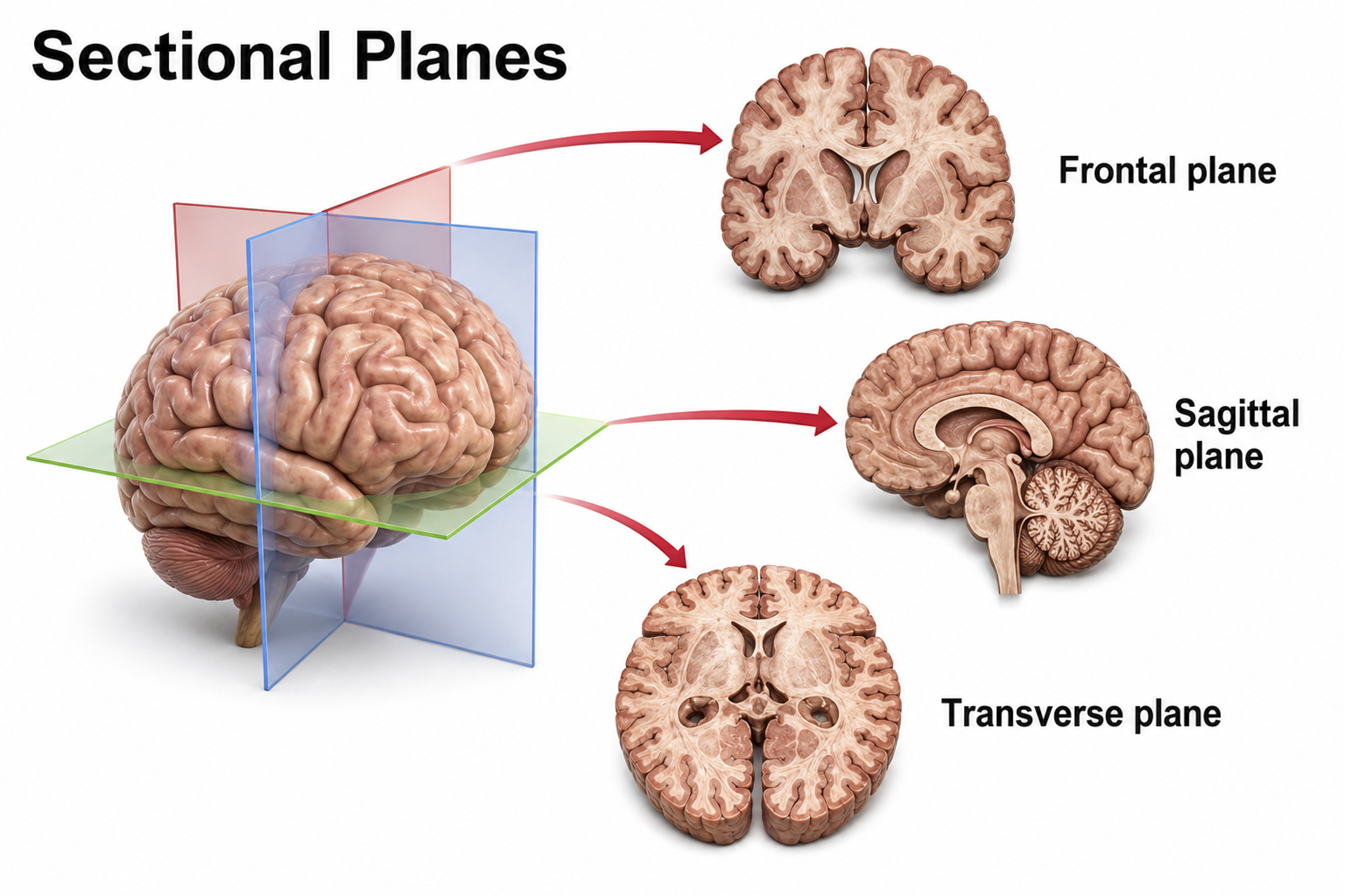
Three customary planes for viewing the body and brain are sagittal, coronal, and horizontal.
The sagittal plane divides the body into right and left halves. The coronal plane separates the body into front and back parts. The horizontal (transverse) plane divides the brain into upper and lower parts (Breedlove & Watson, 2023). These planes are used in neuroimaging and help clinicians interpret brain scans and localize areas of interest for neurofeedback training. Graphic adapted from Blausen.com staff "Blausen gallery 2014," Wikiversity Journal of Medicine.
Directional Terms
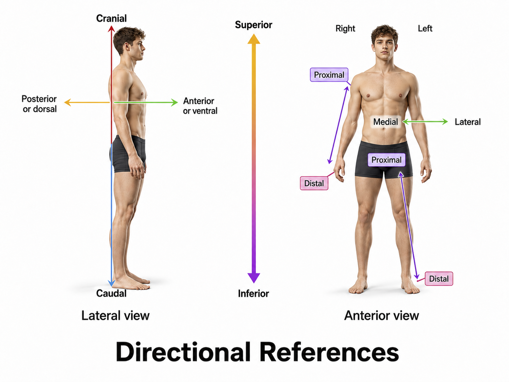
Important directional terms include medial (toward the middle) and lateral (toward the side), ipsilateral (same side) and contralateral (opposite side), superior (above) and inferior (below), anterior/rostral (toward the head) and caudal (posterior, toward or at the back), proximal (near the center) and distal (toward the periphery), and dorsal (toward or at the back) and ventral (toward the belly) (Breedlove & Watson, 2023). Graphic adapted from Blausen.com staff "Blausen gallery 2014," Wikiversity Journal of Medicine.
Cortical Features
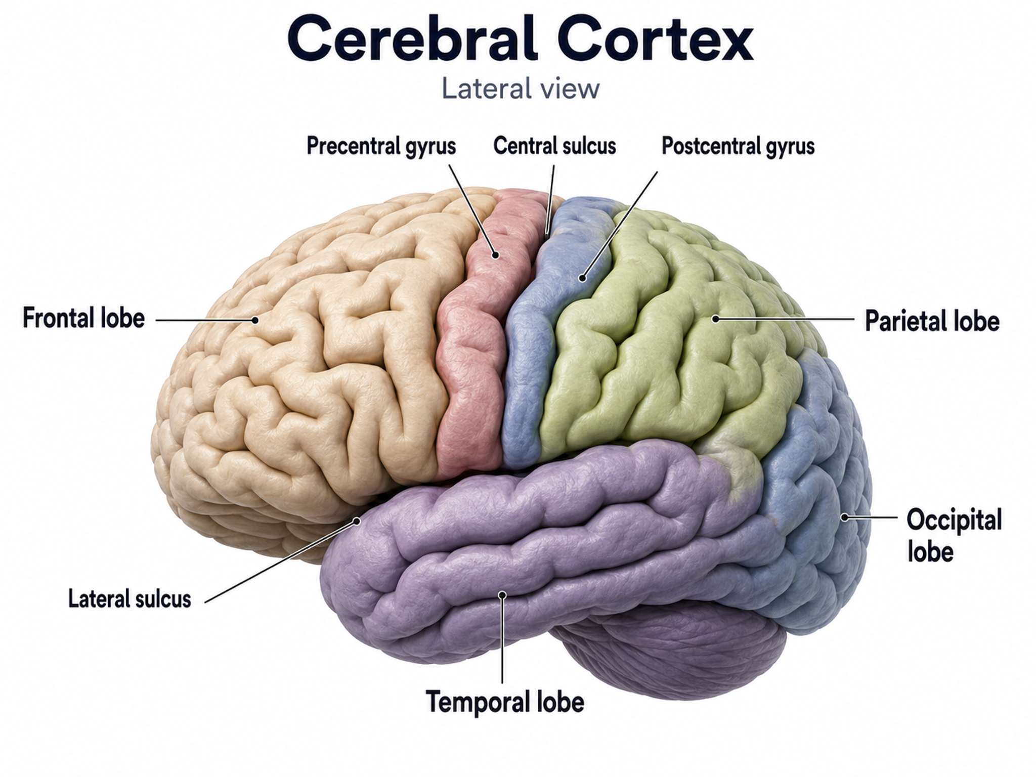
The adult human brain has a volume of roughly 1,100 to 1,300 cm3, varying with sex and body size, and requires convolutions to fit within the skull (Bear, Connors, & Paradiso, 2020). If the cortex were flattened into a sheet, its surface area would be about 2,000 to 2,500 cm2, roughly a quarter of a square meter or 2 to 2.7 square feet, with about two-thirds of the cortical surface lying within these folds (Breedlove & Watson, 2023). Anatomists distinguish three topographical features of the cerebral cortex: gyrus, sulcus, and fissure.
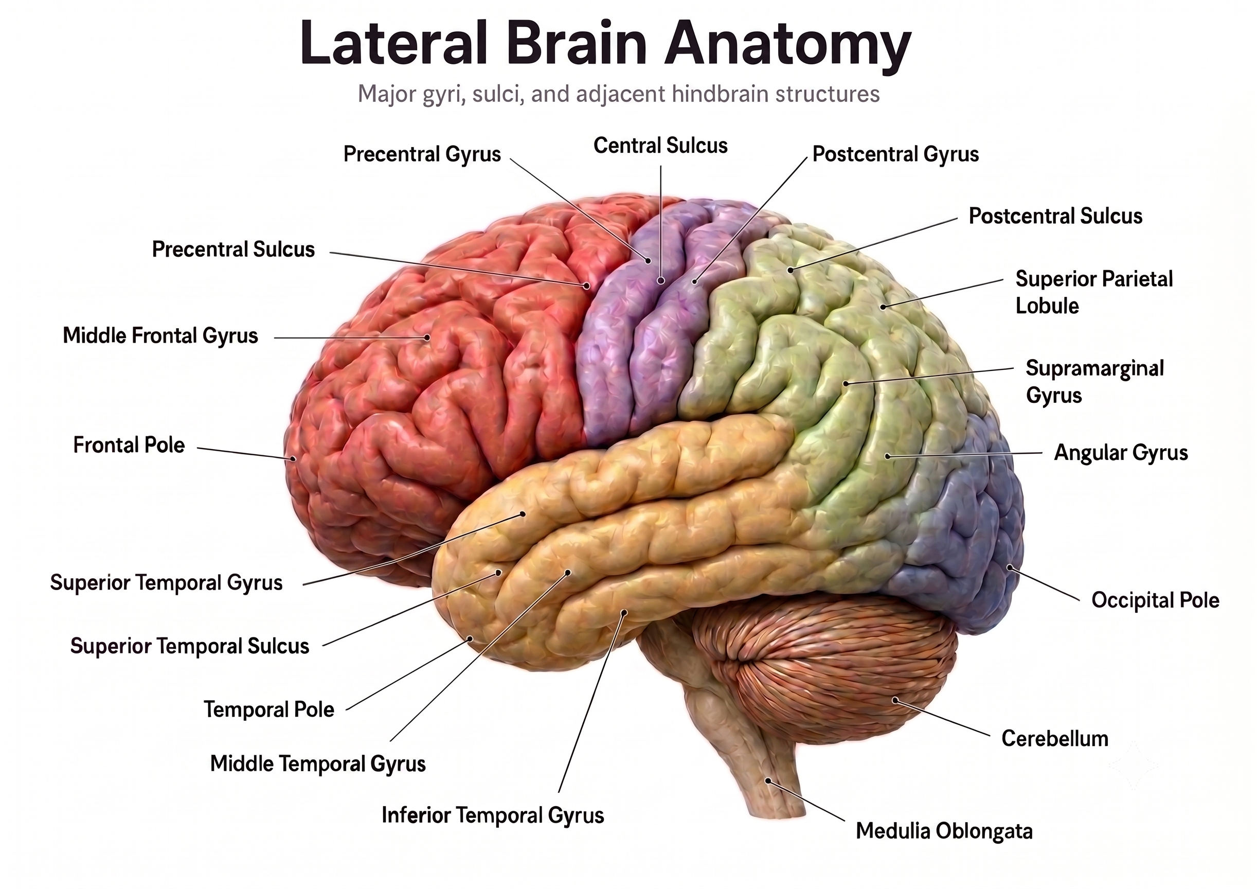
A gyrus is a ridged area of the brain. The precentral gyrus, anterior to the central sulcus, is the primary motor cortex (controlling muscles and movements). The postcentral gyrus, posterior to the central sulcus, is the primary somatosensory cortex (receiving somatosensory information).
A sulcus is a groove in the cortical surface. The central sulcus separates the primary motor cortex from the primary somatosensory cortex. A fissure is a deep groove; for example, the Sylvian fissure (also called the lateral fissure or lateral sulcus) forms the upper boundary of the temporal lobe (Breedlove & Watson, 2023). These features serve as reference landmarks for navigating brain anatomy.
The Unfixed Brain
Textbook illustrations show a firm, sculpted organ. A fresh brain is nothing like that. The video below lets you see how soft and fragile living neural tissue actually is, which explains why the skull, meninges, and cerebrospinal fluid matter so much.
This video was produced by Suzanne Stensaas, PhD, Department of Neurobiology and Anatomy, and the Spencer S. Eccles Health Sciences Library, University of Utah.
Dissecting Brains
This video is courtesy of the Wellcome Collection.
Brain Subdivisions
The brain is divided into three major subdivisions: forebrain, midbrain, and hindbrain.
Brain landmark graphic adapted from © snapgalleria/Shutterstock.com.
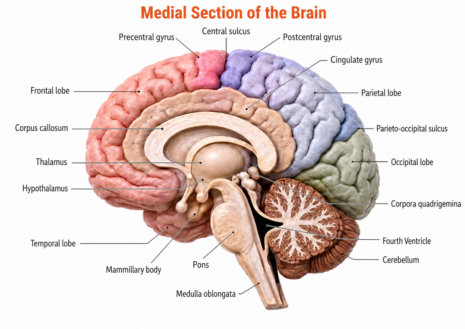
Medial brain section graphic adapted from © NatthapongSachan/Shutterstock.com.
The forebrain consists of the telencephalon (cerebral hemispheres) and the diencephalon. The telencephalon encompasses the cerebral cortex and the deeper structures of the basal ganglia and limbic system.
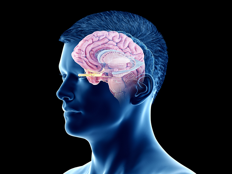
Limbic system graphic © SciePro/Shutterstock.com.
The diencephalon in the posterior forebrain contains the thalamus and hypothalamus.
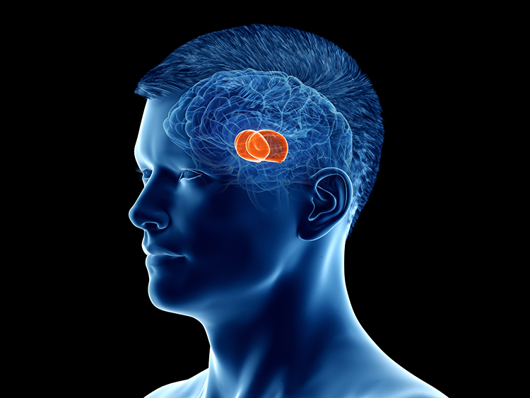
Thalamus graphic © SciePro/Shutterstock.com.
The midbrain consists of the mesencephalon, which includes the inferior colliculi, superior colliculi, and substantia nigra. The degeneration of the substantia nigra is a key step in developing Parkinson's disease.
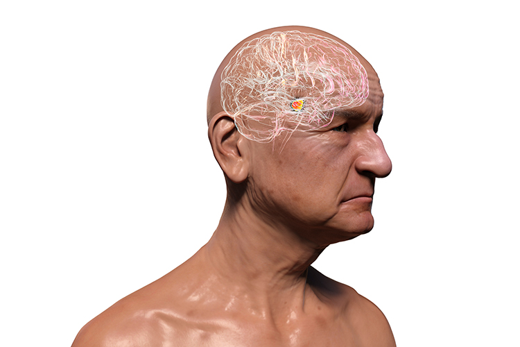
Substantia nigra graphic © Kateryna Kon/Shutterstock.com.
The hindbrain contains the metencephalon and myelencephalon. The metencephalon is comprised of the cerebellum and pons. The cerebellum plays a role in higher-level functions like emotional and cognitive regulation, influencing the speed, capacity, consistency, and appropriateness of cognitive and emotional processes. Damage to the cerebellum can impair cognitive and affective performance as well as movement.
The cerebellum provides coordination and fine-tuning of balance and movement, addressing the rate, rhythm, and force of movement as well as analogous qualities in thinking and emotional expression.
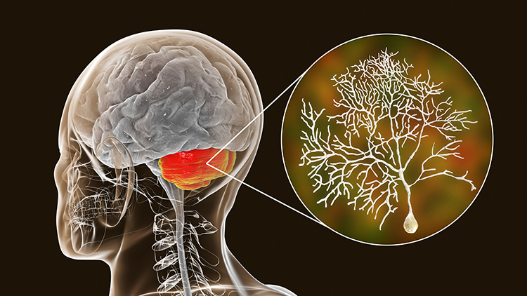
Cerebellum graphic with highlighted Purkinje neuron © Kateryna Kon/Shutterstock.com.
The myelencephalon consists of the medulla, which plays a critical role in the speeding and slowing of the heart across each breathing cycle, a phenomenon called respiratory sinus arrhythmia (RSA). This process is a key target in heart rate variability (HRV) biofeedback training. Alcohol, opioids, and sedative-hypnotics can fatally depress brainstem respiratory centers, slowing and halting breathing.
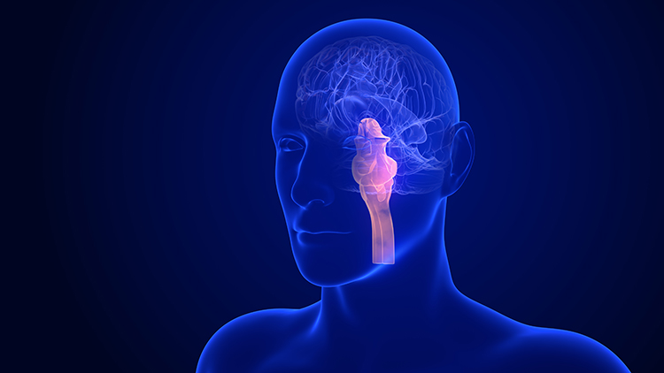
Medulla graphic © mkfilm/Shutterstock.com.
Three reference planes orient every image you will read: the sagittal plane divides the body into right and left halves, the coronal plane into front and back, and the horizontal plane into upper and lower parts. Paired directional terms, including medial and lateral, ipsilateral and contralateral, and rostral and caudal, let you describe position precisely. The cortex is heavily folded, with two-thirds of its surface hidden inside sulci and fissures, and the precentral and postcentral gyri flanking the central sulcus carry the primary motor and primary somatosensory cortices. The brain divides into forebrain, midbrain, and hindbrain, with the telencephalon and diencephalon in the forebrain, the mesencephalon in the midbrain, and the cerebellum, pons, and medulla in the hindbrain.
Check Your Understanding
- Which anatomical plane divides the body into right and left halves, and which divides it into front and back?
- Which gyrus contains the primary motor cortex, which contains the primary somatosensory cortex, and what separates them?
- Why does the cortex need convolutions, and what fraction of the cortical surface lies hidden within them?
- Name the three major subdivisions of the brain and one structure contained in each.
Meninges
Between a generator and your electrode sit several layers of tissue, each with its own electrical properties. Working outward from the cortex, a signal must cross the meninges, the cerebrospinal fluid, the skull, and the scalp before an amplifier ever sees it. Start with the bone, because it does the most damage to the signal.
The Skull
The human skull is a complex anatomical structure that plays a significant role in protecting the brain and influencing EEG signal measurement.
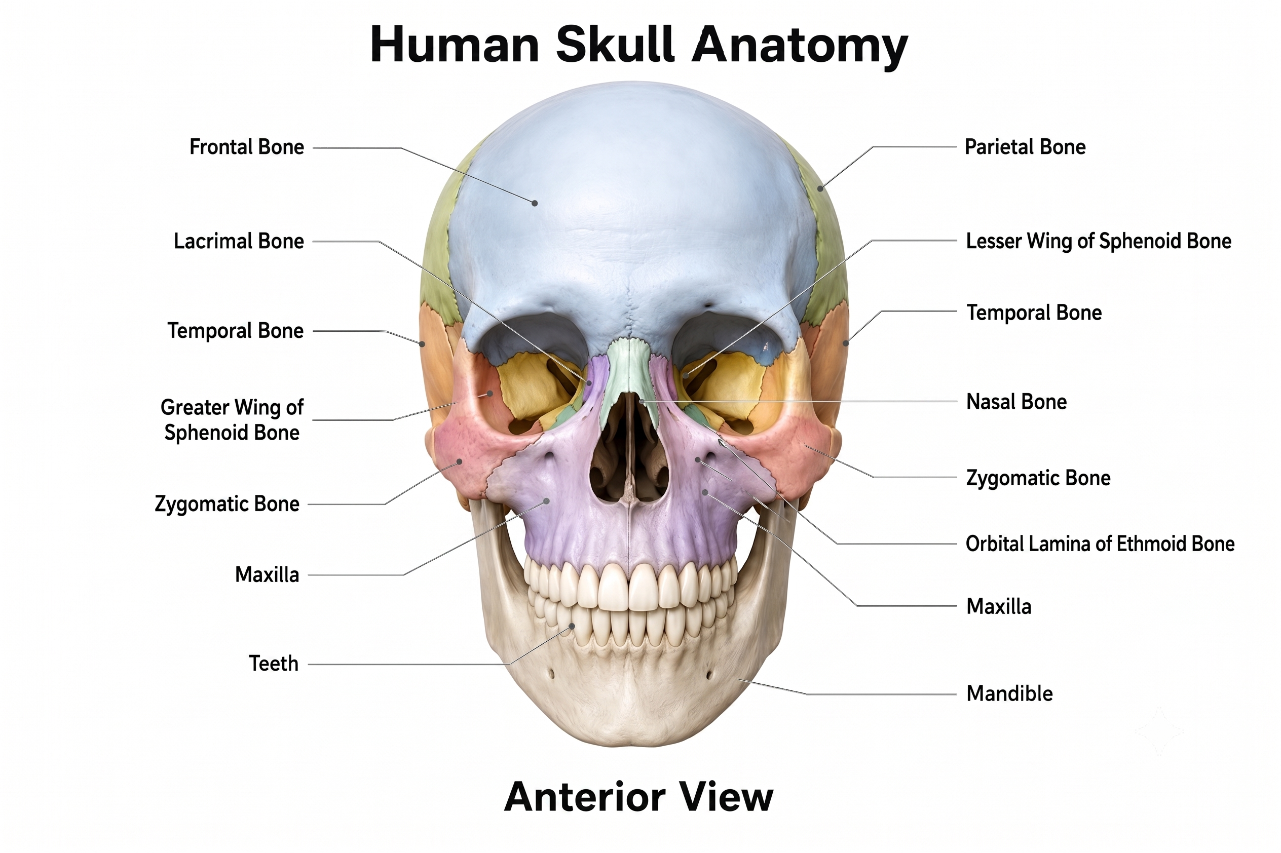
Skull anatomy graphic adapted from © Magic mine/Shutterstock.com.
Comprising 22 bones, the skull is divided into the cranium, which houses the brain, and the facial bones. The cranium is composed of several major bones, including the frontal, parietal, temporal, and occipital bones, fused together by sutures. This bony framework provides a rigid protective case for the brain while also anchoring the meninges and other protective layers (Niedermeyer & Lopes da Silva, 2004).
The skull's primary function is to protect the brain from physical damage. Its bones have an average thickness of about 6.5 mm in adults, which significantly impacts EEG signal transmission. The skull acts as a low-pass filter, attenuating high-frequency components of the brain's electrical activity while allowing lower-frequency components to pass through. The skull can reduce signal amplitude by approximately 85% (He & Li, 2010), primarily due to its low conductivity compared to brain and scalp tissues. This filtering effect is a crucial consideration in EEG signal analysis and interpretation (Sanei & Chambers, 2013).
Key Concept
The skull behaves as a low-pass filter, attenuating the high-frequency components of the brain's electrical activity while allowing lower frequencies to pass. Its low conductivity can reduce EEG signal amplitude by roughly 85 percent, and its mix of dense and porous bone produces anisotropic attenuation that varies with the direction of current flow. These properties mean that much of the neural signal is dissipated and distorted before it reaches a scalp electrode. Accounting for skull filtering is essential when interpreting amplitudes and when using source localization or connectivity techniques.
For example, if a skull defect leaves an opening, then fast-frequency EEG will be seen over the defect (i.e., breach rhythm).
The skull's high impedance means that much of the electrical activity generated by neurons is dissipated before reaching scalp electrodes, making it more challenging to detect subtle neural oscillations. Additionally, the varying thickness and composition of skull bones can introduce spatial distortions in EEG signals, complicating the localization of neural activity sources (Lopes da Silva, 2010).
The skull's heterogeneous structure, including both dense cortical bone (dense bone that forms a cortex or “bark” around the bone) and less dense cancellous bone (porous bone in the marrow-filled medullary cavity of bones), causes anisotropic attenuation, meaning that the degree of signal reduction varies depending on the direction of electrical currents (i.e., a reduction in EEG amplitude caused by the loss of uniform dipole orientation, so the underlying fields partially cancel rather than summate). This anisotropy must be accounted for in advanced EEG analysis techniques such as source localization and brain connectivity studies (Nunez & Srinivasan, 2006).
In addition to the bones, the skull's sutures and foramina (openings) influence EEG signal propagation. Sutures can act as additional barriers or channels for electrical signals, while foramina may create localized points of less attenuation. Individual variability in skull anatomy also significantly influences EEG signals: children's skulls are generally thinner and less dense than those of adults, potentially resulting in less signal attenuation (Schoffelen & Gross, 2009). The scalp and meninges add further layers of impedance that electrical signals must traverse, each with distinct electrical properties. Understanding the combined effect of these tissues is essential for accurate EEG interpretation and for the development of advanced EEG techniques (Niedermeyer & Lopes da Silva, 2004).
The Three Meninges
Three meninges protect the brain and spinal cord, which are housed within the skull and vertebrae. The choroid plexus of the ventricles produces cerebrospinal fluid, which then circulates through the ventricles and the subarachnoid space of the meninges. These membranes include the dura mater, pia mater, and middle arachnoid (Breedlove & Watson, 2023).
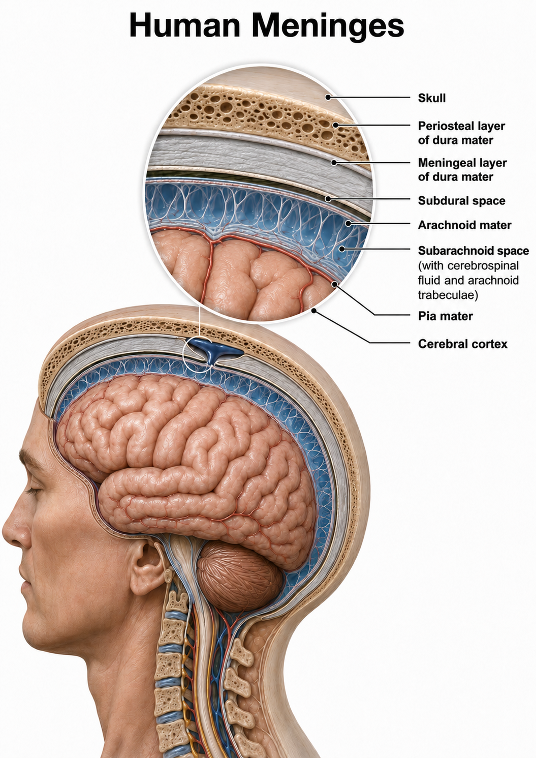
Graphic adapted from © Alilia Medical Media/Shutterstock.com.
Cerebral Ventricles
The cerebral ventricles are a network of fluid-filled chambers that protect the brain from trauma due to abrupt head movements and facilitate the exchange of nutrients and wastes between blood vessels and the brain. These cavities, found within all four lobes of each hemisphere, include the lateral, third, and fourth ventricles.
The brain contains four interconnected ventricles. A pair of lateral ventricles occupies the two cerebral hemispheres, the third ventricle sits in the midline between the left and right halves of the thalamus and hypothalamus, and the fourth ventricle lies between the pons and upper medulla in front and the cerebellum behind. Narrow channels connect these cavities: the paired interventricular foramina (foramen is the singular, meaning opening) join each lateral ventricle to the third ventricle, and the cerebral aqueduct joins the third ventricle to the fourth (Breedlove & Watson, 2023).
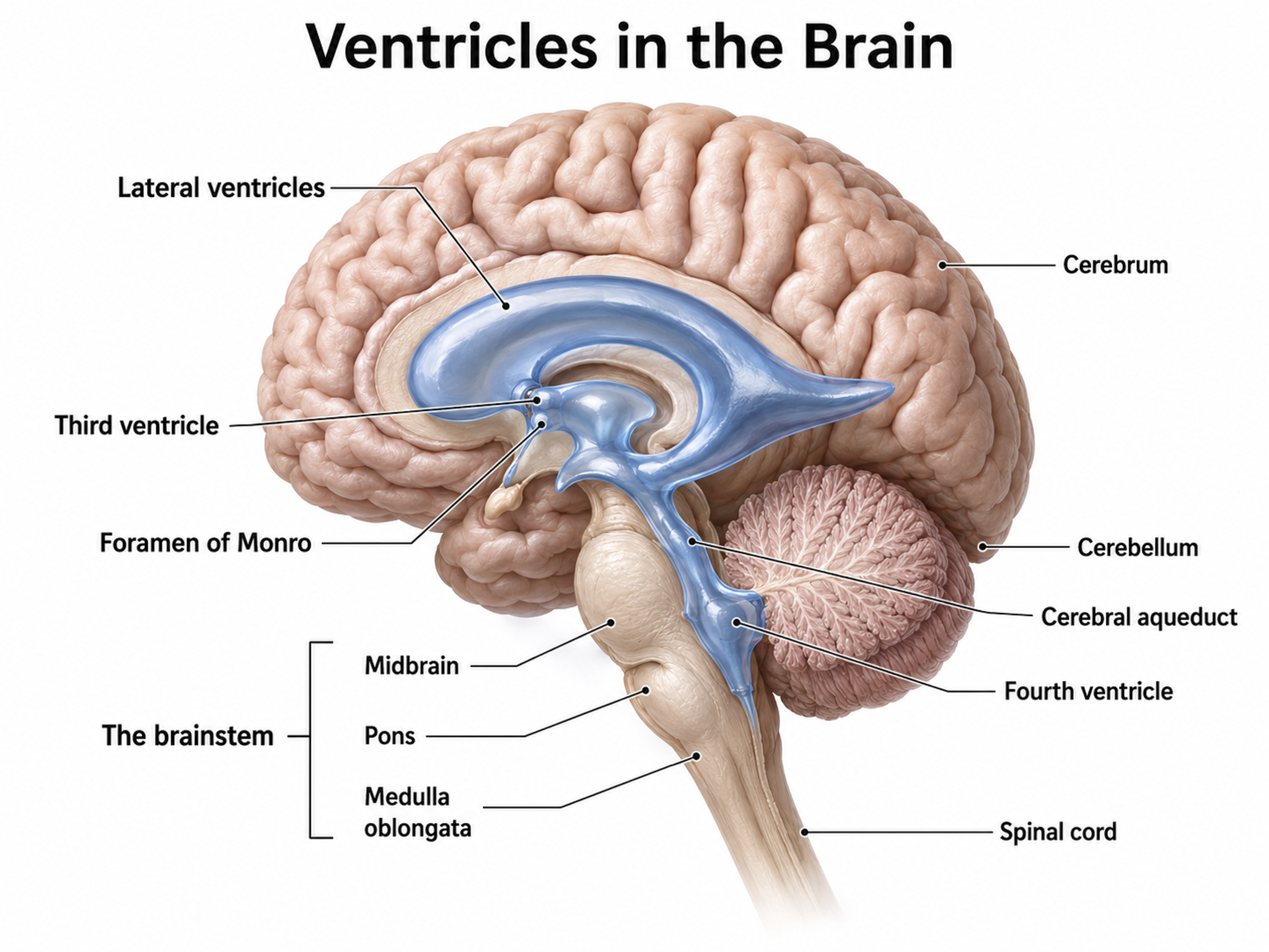
Graphic adapted from © joshya/Shutterstock.com.
Glymphatic System
For most of the history of neuroscience, the brain was thought to have no lymphatic drainage at all, which is why it was long described as immune privileged. Natale and colleagues (2021) summarized the older view this way:
The belief of an absence of conventional lymphatic vessels in the CNS contributed to the concept that the brain, in spite of its high metabolic rate, represents an immune privileged region. This idea left questioned how cerebral interstitial fluid is cleared from waste products. It was generally thought that clearance depended on cerebrospinal fluid (CSF), acting as a pseudo-lymphatic system (Natale et al., 2021).
The glymphatic system is a recently discovered lymphatic-like system in the brain. It provides a flow of CSF through the brain's interior that helps clear cellular debris, proteins, and other wastes. The pathway is astrocyte-controlled: aquaporin-4 channels concentrated in astrocyte endfeet that wrap the brain's blood vessels drive the flow of fluid through the tissue (Xie et al., 2013). This is the same cell you will meet again in the microanatomy section, doing a job that has nothing to do with signaling.
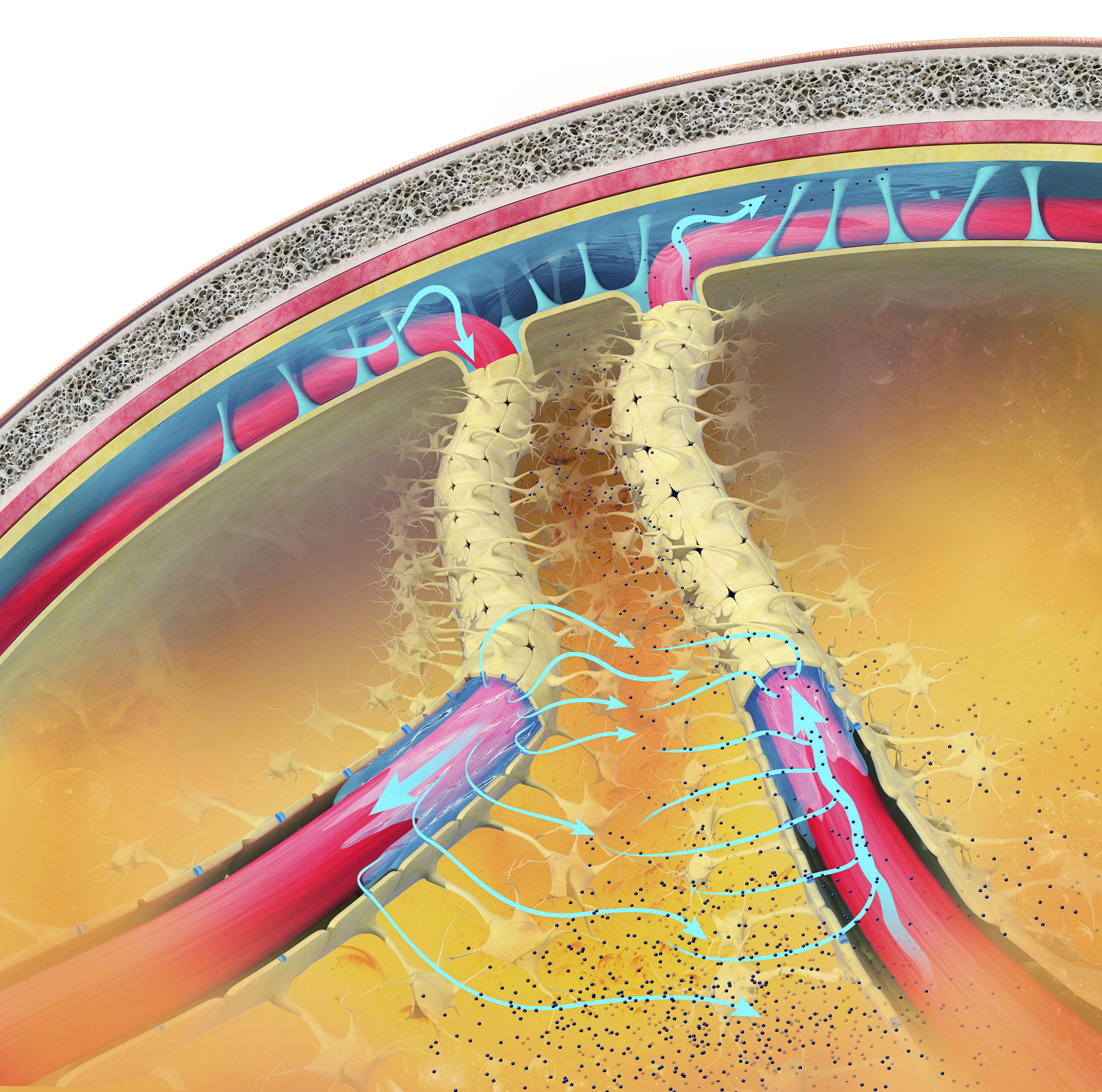
Glymphatic system graphic © Claus Lunau/Science Photo Library.
Also known as the glymphatic clearance pathway or the paravascular system, this system clears waste and fluid from the vertebrate central nervous system (CNS). Interstitial fluid is removed via the cerebrospinal fluid (CSF). It is similar to the lymphatic system, but functions to remove waste products from the brain and spinal cord. This view shows the subarachnoid space (across top) between the brain and its membranes. The blue arrows show the movement of interstitial fluid and solutes.
By removing harmful substances such as the amyloid-beta and tau proteins implicated in Alzheimer's disease and the alpha-synuclein implicated in Parkinson's disease, the glymphatic flow may protect us from various neurological disorders (Breedlove & Watson, 2023). The glymphatic system removes most of its waste during stage-3 sleep (slow-wave sleep), which underscores the clinical importance of healthy sleep, a factor biofeedback practitioners should assess in every client.
The Brain's Vascular System
The resting brain consumes over 20% of the body's energy. The internal carotid artery's anterior and middle cerebral arterial branches deliver blood to about two-thirds of the cerebral hemispheres, while the paired vertebral arteries merge to form the basilar artery, whose left and right posterior cerebral arterial branches supply blood to the posterior cerebral hemispheres, cerebellum, and brainstem (Breedlove & Watson, 2023).
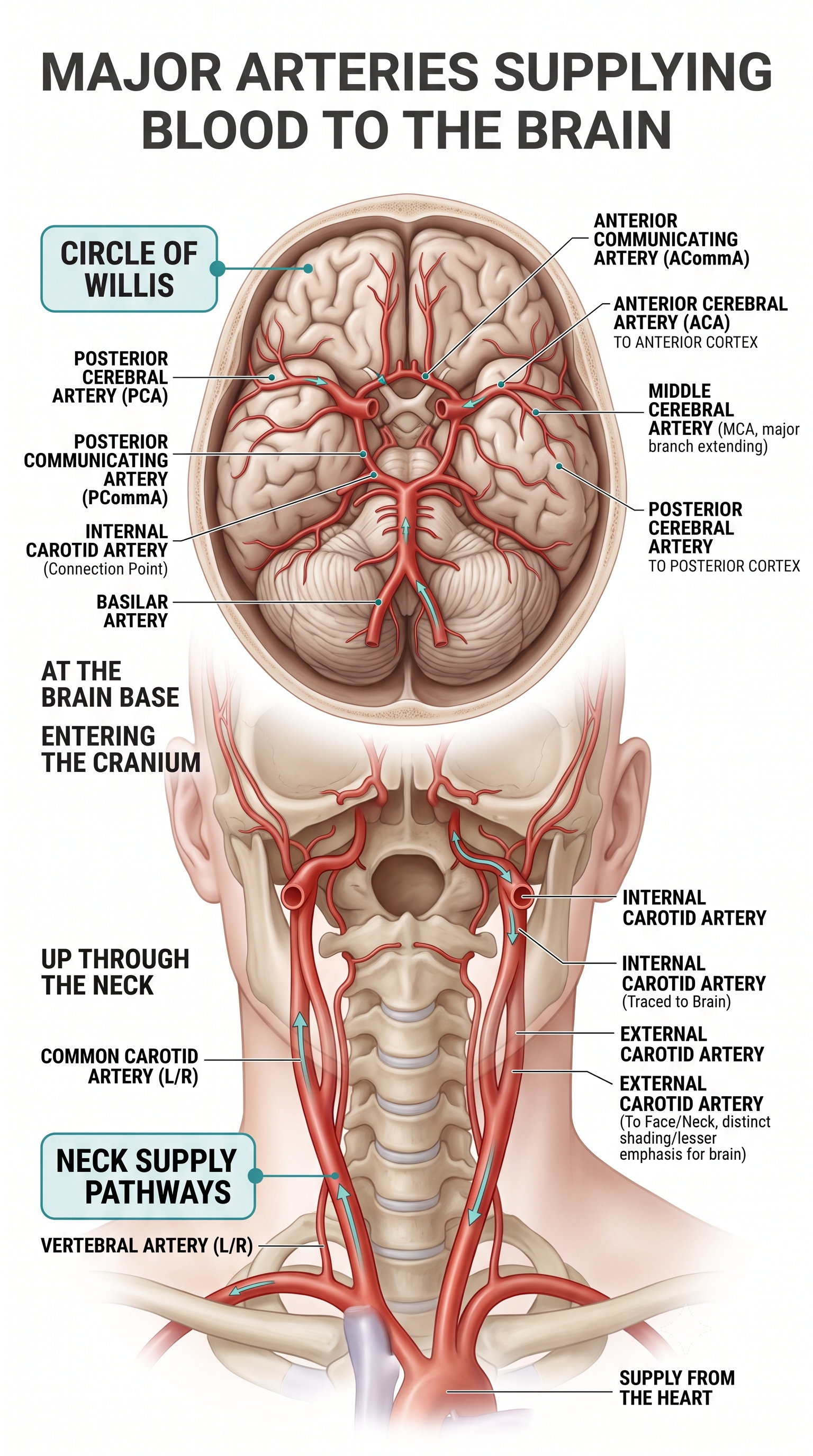
The effects of a stroke due to blood vessel blockage or rupture may be limited because paired arteries supply each brain hemisphere. The circle of Willis, a vascular network at the base of the brain comprised of the carotid and basilar arteries, may provide an alternate route for delivering blood when a major artery is compromised by disease or traumatic injury. The veins and sinuses drain deoxygenated blood, completing the circulatory loop to the heart via the jugular vein.
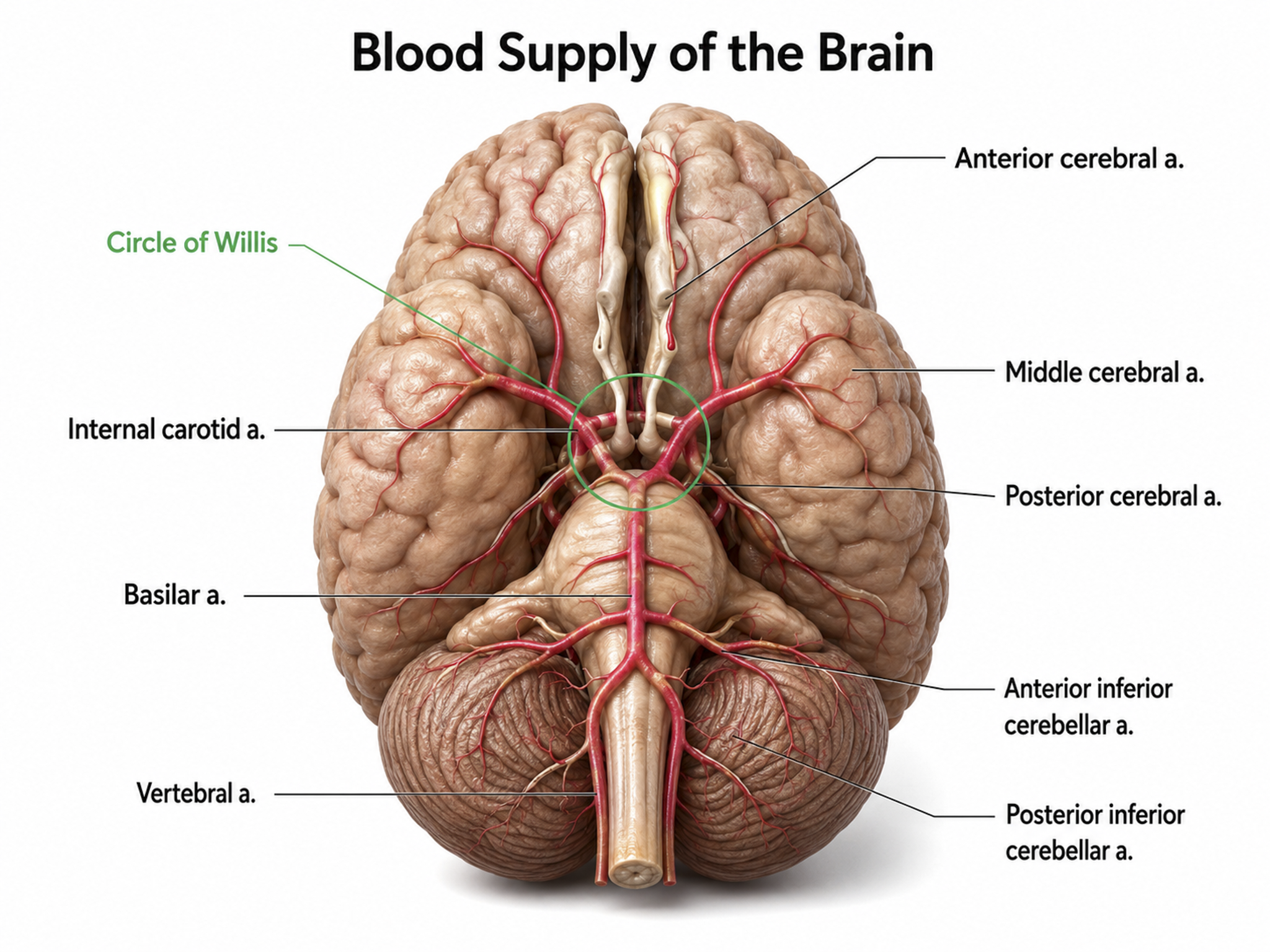
Graphic adapted from © Alilia Medical Media/Shutterstock.com.
The figure below shows areas of the cortex that may be affected by strokes affecting the three major cerebral arteries.
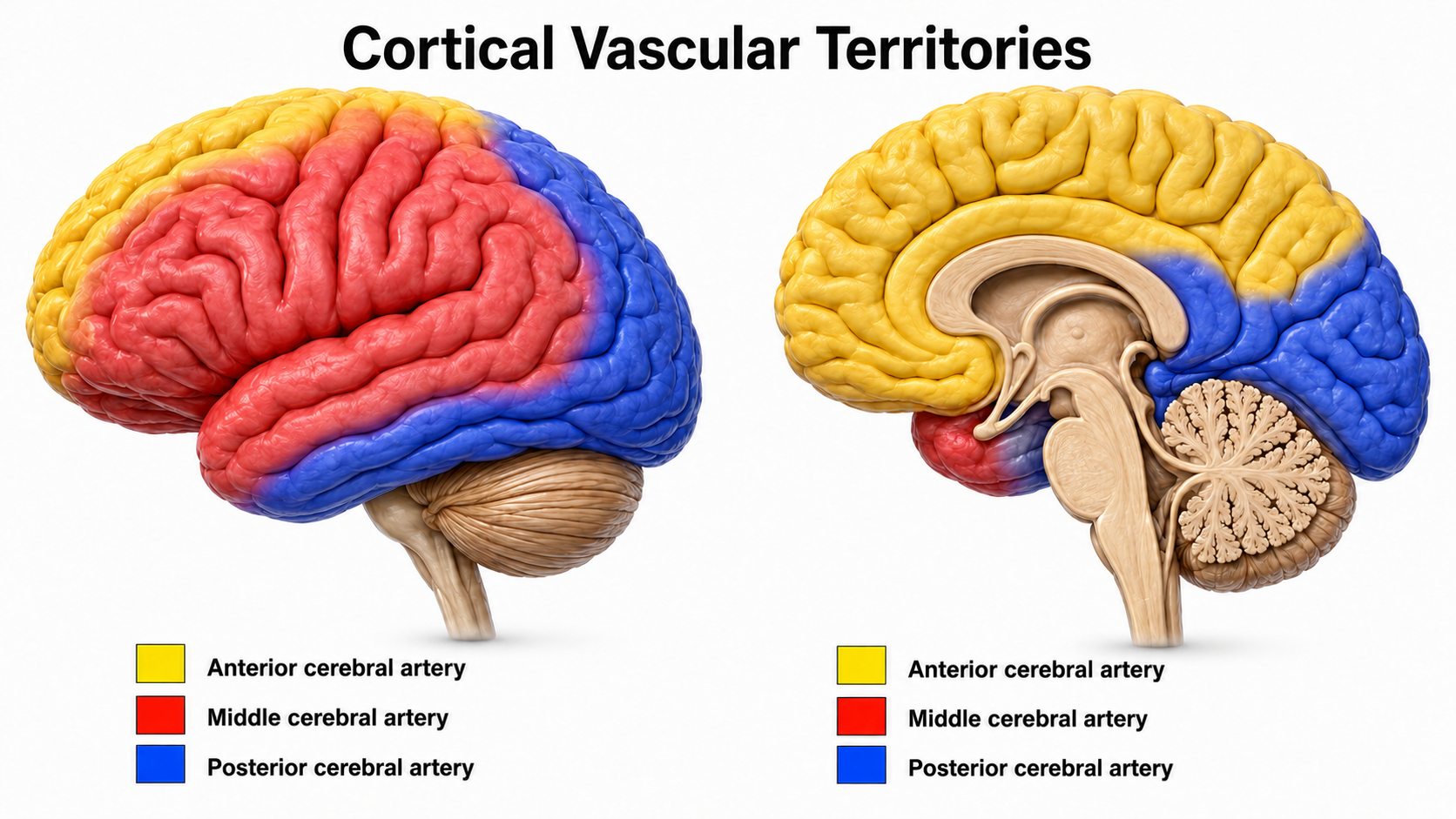
Graphic adapted from Cruces et al. (2022).
After perfusing the brain, blood depleted of oxygen, glucose, and other nutrients drains into a network of cerebral veins that differs strikingly from the venous anatomy found elsewhere in the body. Unlike most veins, cerebral veins are thin-walled, lack valves, and do not travel alongside their companion arteries, so drainage depends on pressure gradients and posture rather than on one-way valves. Superficial cortical veins collect blood from the outer surface of the cerebrum and empty into the dural venous sinuses, most notably the superior sagittal sinus, while deep veins gather blood from the basal ganglia, thalamus, and deep white matter and channel it through the great cerebral vein of Galen into the straight sinus. These sinuses converge at the confluence of the sinuses, then course through the transverse and sigmoid sinuses before exiting the skull as the internal jugular veins, which carry the blood toward the superior vena cava and the right atrium.
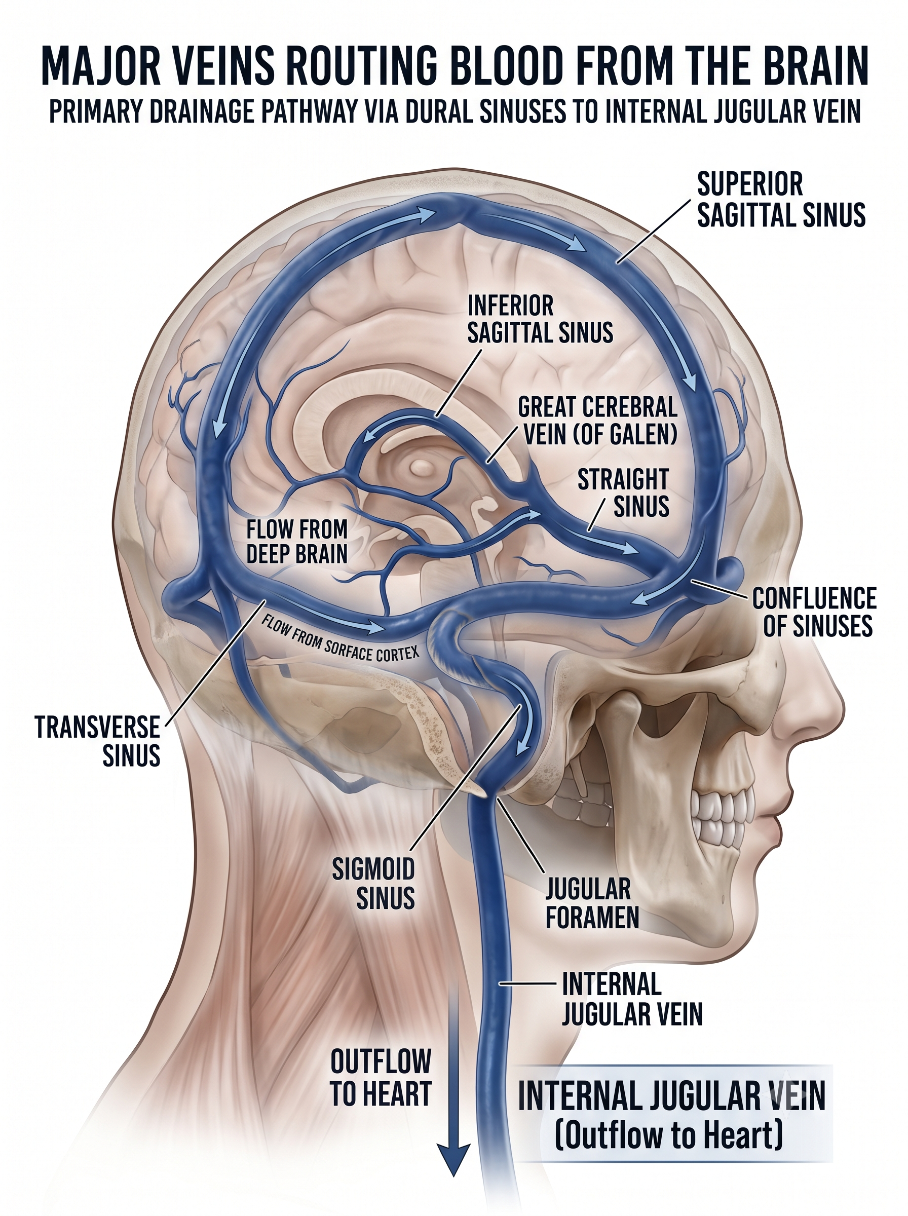
The same dural sinuses also house meningeal lymphatic vessels, a discovery that overturned the long-held belief that the central nervous system lacked lymphatic drainage and now links cerebral venous anatomy to waste clearance and immune surveillance in the brain (Aspelund et al., 2015; Louveau et al., 2015).
Blood-Brain Barrier
The blood-brain barrier (BBB) is a highly selective, semipermeable interface formed by the endothelial cells that line the brain's microvasculature, where continuous tight junctions seal the spaces between adjacent cells and force most substances to move through the cells rather than between them. This barrier is not the work of endothelial cells alone but of a coordinated neurovascular unit that also includes pericytes embedded in the capillary wall, the endfeet of astrocytes that ensheath the vessels, microglia, and the surrounding basement membrane.
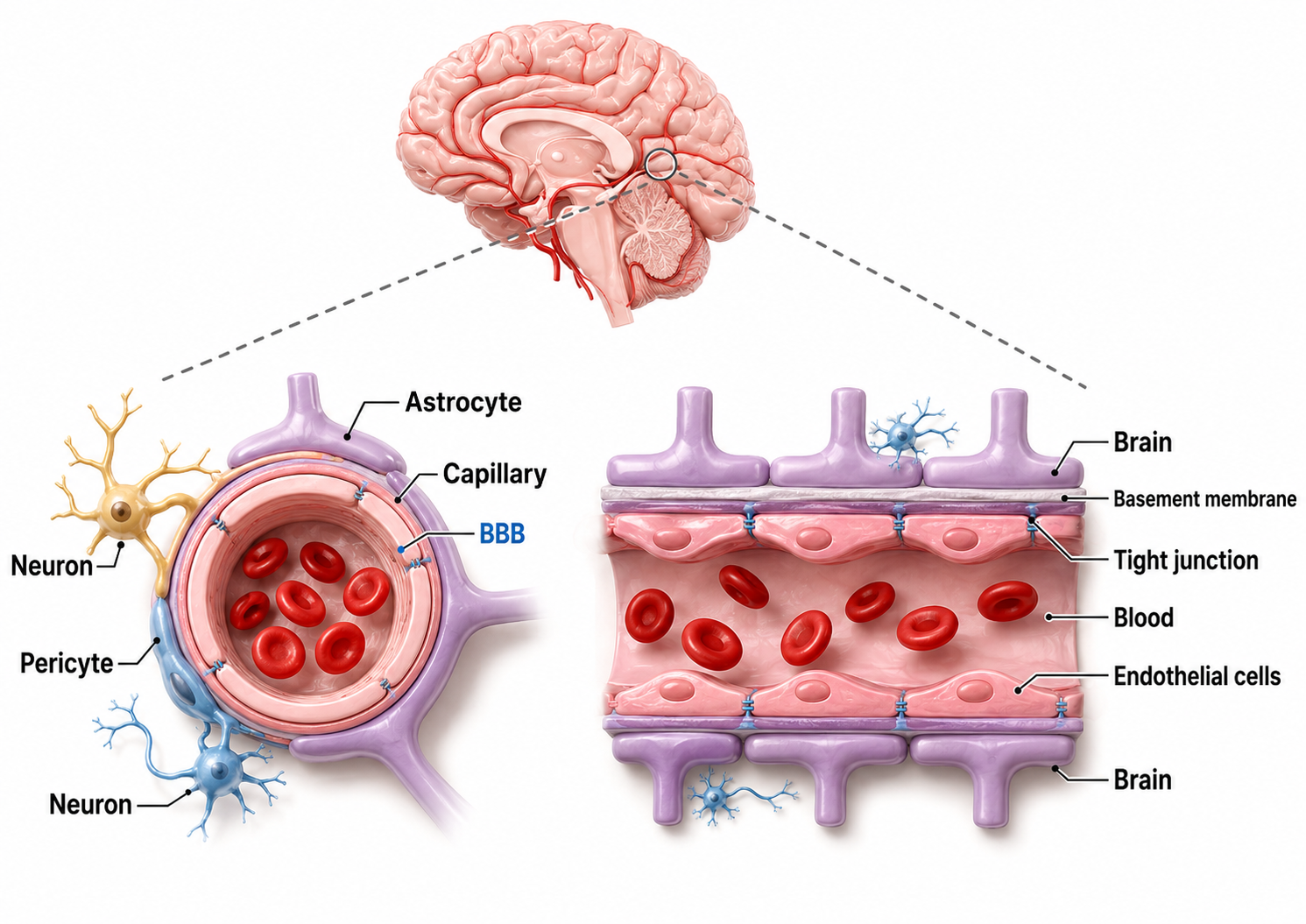
Image adapted from Ding et al. (2025).
Together these components induce and maintain the barrier's low permeability, minimal pinocytosis, and dense expression of transport proteins that set brain capillaries apart from those elsewhere in the body (Abbott et al., 2010). The result is a living border that behaves less like a passive wall and more like a tightly regulated gatekeeper for the central nervous system (Daneman & Prat, 2015).
Functionally, the BBB shields neural tissue from circulating pathogens, toxins, and swings in blood composition while precisely controlling the traffic of ions, nutrients, and signaling molecules that keep the neuronal microenvironment stable. Small lipid-soluble gases such as oxygen and carbon dioxide diffuse across freely, whereas glucose, amino acids, and other essential polar molecules depend on specific carrier and receptor-mediated systems, and efflux pumps actively return many foreign compounds to the blood (Abbott et al., 2010).
This same protective selectivity is a double-edged property, because it excludes the vast majority of small-molecule drugs and nearly all large-molecule therapeutics, making the BBB one of the central obstacles in treating neurological and psychiatric disorders. When the barrier breaks down, as it does in conditions ranging from stroke and multiple sclerosis to Alzheimer's disease, the loss of this regulation contributes directly to disease progression, which is why BBB integrity remains a priority in both neuroscience and drug development (Daneman & Prat, 2015).
A client arrives for a first session reporting that his memory has slipped noticeably over the past year, and his sleep history reveals loud snoring, witnessed apneas, and almost no time in deep sleep. Because the glymphatic system clears amyloid and tau primarily during slow-wave sleep, chronically fragmented deep sleep plausibly compromises the brain's overnight housekeeping. Screening sleep quality before you interpret a map is not a detour, it is part of the assessment, and referring for a sleep study may do more for this client than any training protocol you could design.
The skull acts as a low-pass filter that can reduce EEG amplitude by roughly 85 percent, and its mix of dense and porous bone produces anisotropic attenuation that varies with current direction. Three meninges and the cerebrospinal fluid produced by the choroid plexus cushion the brain, and four interconnected ventricles circulate that fluid. The glymphatic system clears wastes such as amyloid and tau mainly during slow-wave sleep. Paired cerebral arteries and the circle of Willis supply the brain and can limit stroke damage by providing alternate routes, while dural venous sinuses drain it and house the recently discovered meningeal lymphatics. The blood-brain barrier, now understood as a coordinated neurovascular unit, tightly regulates what reaches neural tissue and excludes most therapeutic drugs.
Check Your Understanding
- Why is the skull described as a low-pass filter, and approximately how much does it reduce EEG signal amplitude?
- What is a breach rhythm, and what skull condition produces it?
- Name the three meninges and identify the structure that produces cerebrospinal fluid.
- When is the glymphatic system most active, and why does that matter clinically?
- What is the neurovascular unit, and why does the blood-brain barrier complicate drug treatment of neurological disorders?
General Cortical and Subcortical Divisions
With the coverings and circulation in place, step back and look at how the nervous system as a whole is organized. The divisions below give you the framework into which every structure in this unit, and in the units that follow, will fit.
Major Divisions
The human nervous system consists of the central nervous system and peripheral nervous system. The central nervous system (CNS) consists of the brain, spinal cord, and retina.
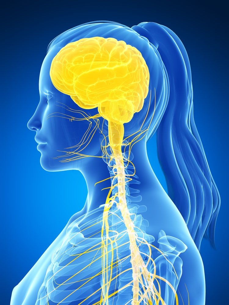
Central nervous system graphic © SciePro/Shutterstock.
The 3-pound brain consists of approximately 86 billion neurons (Azevedo et al., 2009; Voytek, 2013).
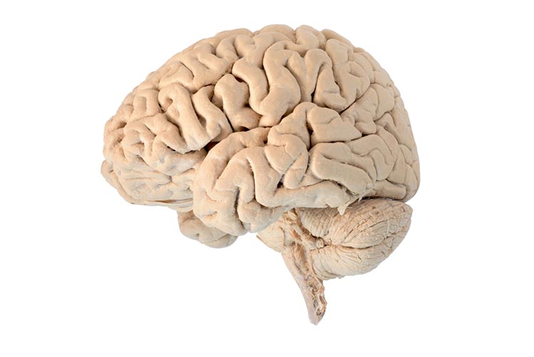
Graphic © Jasada Sabai/Shutterstock.com.
The cylindrical spinal cord extends from the medulla (brainstem) to the lumbar (lower back) segment of the vertebral column. It distributes sensory information from the body to the brain and CNS commands from the brain to the body, and also contains networks that control reflexes and central pattern generators.
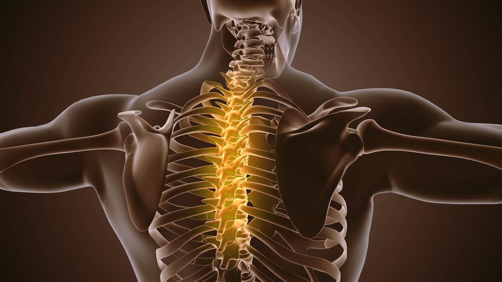
Graphic © Silver Place/Shutterstock.com.
The peripheral nervous system (PNS) consists of neurons and nerves outside of the brain and spinal cord and is comprised of the autonomic nervous system and somatic nervous system.
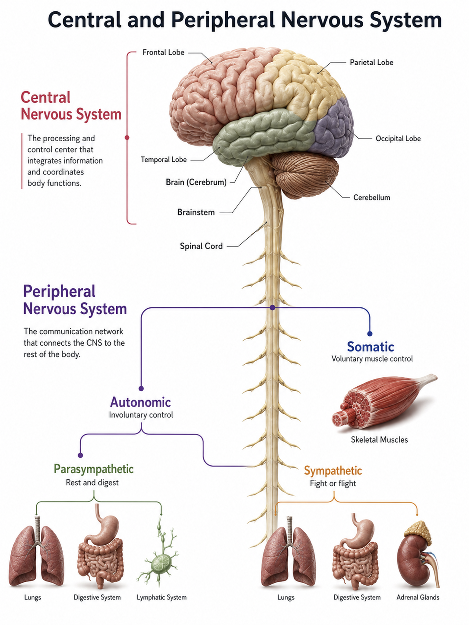
Peripheral nervous system graphic adapted from © Elena Ladanovskaya/Shutterstock.com.
Nerves
Nerves are bundles of axons that lie outside of the central nervous system. Motor nerves distribute instructions from the CNS to the rest of the body, while sensory nerves transmit information from sensory receptors to the CNS.
There are three major systems of nerves: cranial nerves, spinal nerves, and the autonomic nervous system.
The 12 pairs of cranial nerves distribute sensory and motor information. Three are exclusively sensory pathways to the brain: olfactory (I), optic (II), and vestibulocochlear (VIII). Five are exclusively motor pathways from the brain: oculomotor (III), trochlear (IV), abducens (VI), spinal accessory (XI), and hypoglossal (XII). Four cranial nerves carry both sensory and motor information: trigeminal (V), facial (VII), glossopharyngeal (IX), and vagus (X).
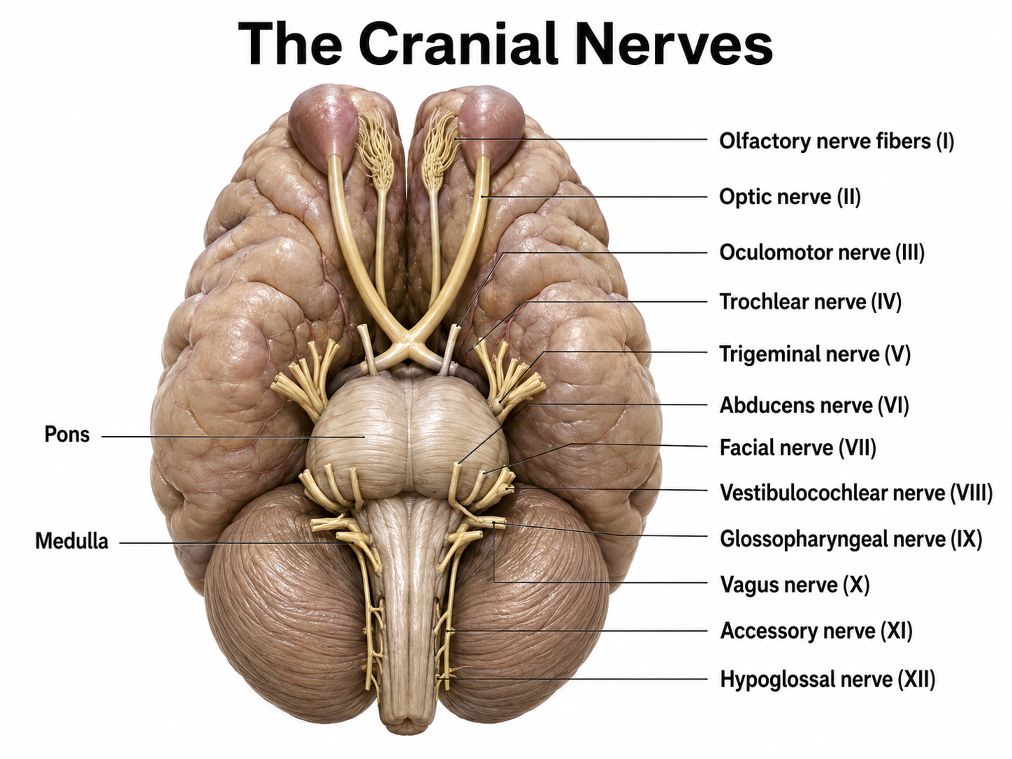
Graphic adapted from © Alila Medical Media/Shutterstock.com.
Thirty-one pairs of spinal nerves, each member serving one side of the body, leave the spinal cord through openings in the backbone.
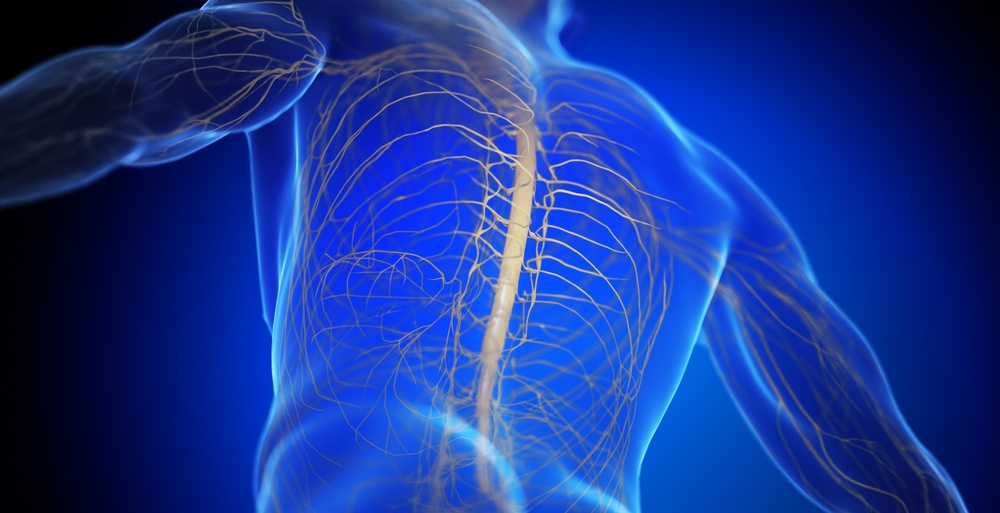
Spinal nerve graphic © SciePro/Shutterstock.com.
Each spinal nerve carries sensory projections from the body (dorsal root) and motor commands from the spinal cord to skeletal muscles (ventral root).
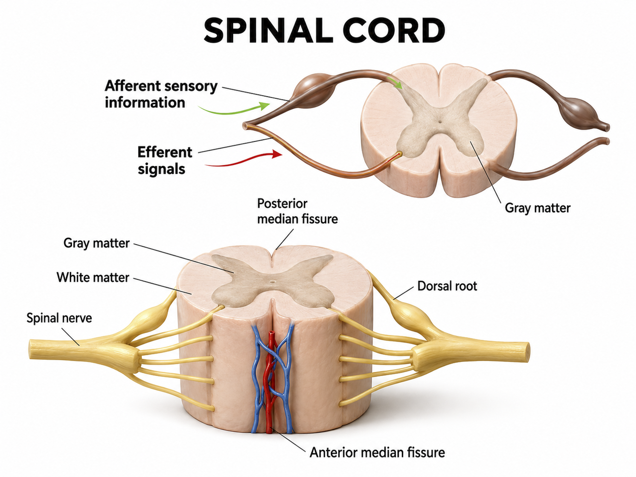
Graphic adapted from © Designua/Shutterstock.com.
The central nervous system comprises the brain, spinal cord, and retina and contains roughly 86 billion neurons, while the peripheral nervous system divides into the autonomic and somatic systems. Nerves are bundles of axons outside the central nervous system, with motor nerves carrying instructions outward and sensory nerves carrying information inward. Twelve pairs of cranial nerves route sensory and motor information, three exclusively sensory, five exclusively motor, and four mixed. Thirty-one pairs of spinal nerves leave the cord through openings in the backbone, each carrying sensory projections through its dorsal root and motor commands through its ventral root.
Check Your Understanding
- Name the three structures that make up the central nervous system.
- How do motor nerves and sensory nerves differ in the direction they carry information?
- How many cranial nerve pairs are exclusively sensory, exclusively motor, and both, and give one example of each?
- What is the functional difference between the dorsal root and the ventral root of a spinal nerve?
Cortical Generators
You now know what lies between the cortex and your electrode. The next question is what actually produces the signal. Two answers matter most: the cortex generates it, and the thalamus paces much of it.
The cerebral cortex (gray matter) consists of neuronal cell bodies, glial cells, and blood vessels, while white matter lies beneath it, composed of myelinated nerves, nonmyelinated fibers, and glial cells. The EEG mainly originates from pyramidal neurons in layers III, V, and VI of the gray matter, the six-layered outer portion of the cerebrum that contains neuronal cell bodies. Cortical thickness ranges from about 1 to 4.5 mm and averages roughly 2.5 mm.
Why do pyramidal neurons dominate the scalp EEG? Their long apical dendrites run parallel to one another and perpendicular to the cortical surface, so that synchronous synaptic currents along these dendrites create aligned electrical dipoles whose fields add together rather than cancel, a configuration known as an open field (Kirschstein & Köhling, 2009). Neurons whose dendrites radiate symmetrically in all directions, such as many stellate and thalamocortical cells, instead form a closed field in which opposing currents largely cancel, so they contribute little to the scalp signal (Buzsáki, Anastassiou, & Koch, 2012). Because each dipole is tiny, thousands of neighboring pyramidal neurons must depolarize or hyperpolarize together for their summed field to survive volume conduction through the meninges, skull, and scalp and reach a recording electrode (Buzsáki et al., 2012).
Key Concept
The scalp EEG is dominated by cortical pyramidal neurons because their long apical dendrites run parallel to one another and perpendicular to the cortical surface. When these neurons are active together, their aligned dipoles form an open field whose electrical fields summate rather than cancel. Neurons whose dendrites radiate symmetrically instead form a closed field in which opposing currents largely cancel, so they contribute little to the scalp signal. Because each dipole is tiny, thousands of neighboring pyramidal neurons must fire synchronously, and a reliable scalp EEG requires at least six square centimeters of synchronized cortex.
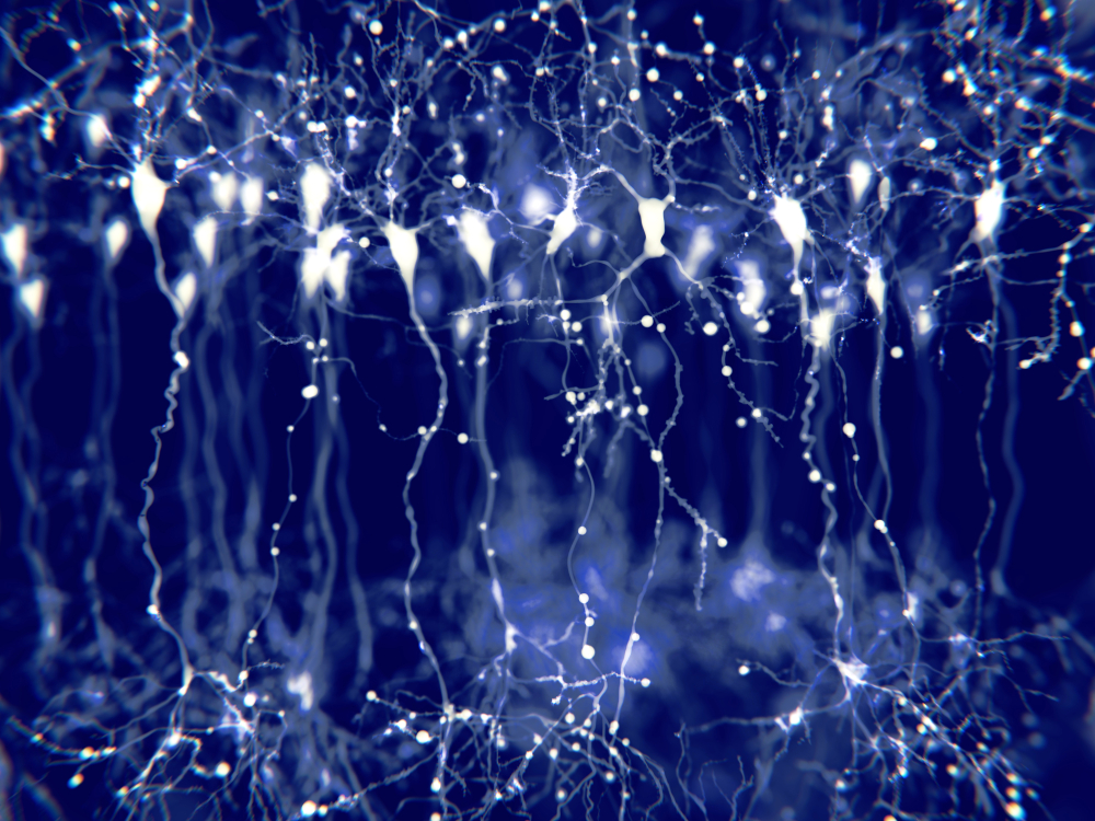
Pyramidal neuron graphic © Juan Gaertner/Shutterstock.com.
Vertical cortical macrocolumns contain hundreds of pyramidal neurons and supporting stellate and basket cells (Thompson & Thompson, 2015b). Each pyramidal neuron may receive more than 100,000 synapses. These macrocolumns are positioned side by side and perpendicular to the cortical surface, and since neighboring macrocolumns often receive the same afferent messages, they tend to fire together and generate a potential detectable from the scalp. A reliable scalp EEG requires a minimum of 6 cm² of synchronized cortex (Dyro, 1989).
Although thalamic pacemakers generate EEG rhythms, resonant loops between cortical macrocolumns represent another source (Traub et al., 1989). Over 97 percent of the brain's conversations are cortical-to-cortical, primarily within the same hemisphere (Thompson & Thompson, 2015b). Only about 3 percent of these linkages are thalamocortical, yet they exert an outsized influence on the EEG, because they connect distant cortical regions subcortically and produce most of the synchronous activity you record (Steriade, 1990). A resonant loop develops when macrocolumns that share afferent input fire synchronously to generate an electrical potential. The distance between participating macrocolumns is one determinant of EEG frequency: the closer the macrocolumns, the higher the frequency they can generate (Lubar, 1997).
There are three types of resonant loops driven by afferent input or thalamic pacemakers. Local loops couple neighboring macrocolumns and may generate frequencies above 30 Hz in the high-beta and gamma ranges. Regional loops couple macrocolumns separated by several centimeters and may produce alpha and beta rhythms. Global loops couple macrocolumns as distant as 7 cm (for example, between the frontal and parietal lobes) and may create delta and theta rhythms. The graphic below is conceptual, and should not be taken literally with respect to distance or anatomical location.
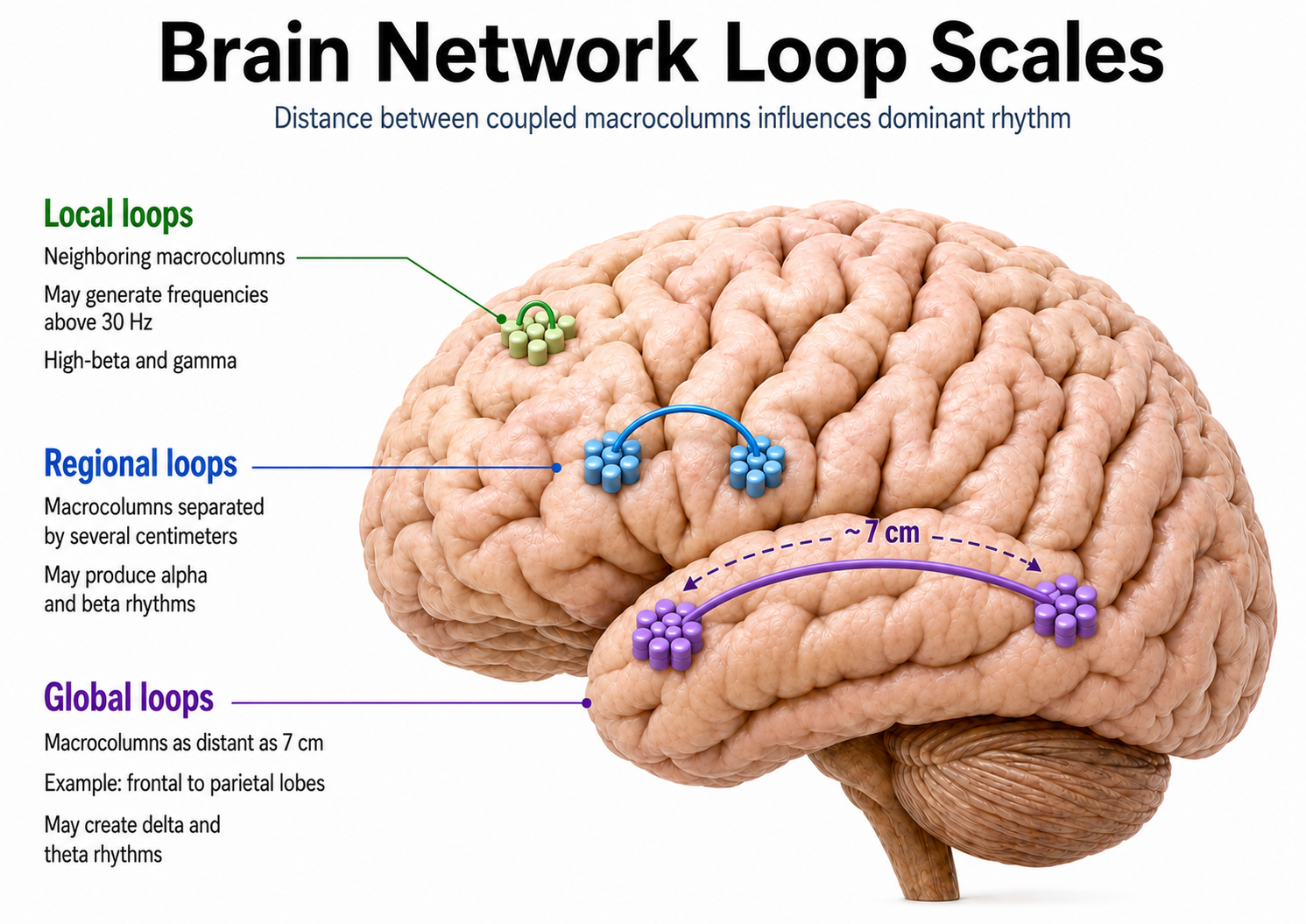
While only 3 percent of linkages are thalamocortical, they greatly influence the EEG by subcortically connecting distant cortical regions and producing most synchronous activity (Steriade, 2005).
Lubar (1997) proposed a violin analogy: the thalamic pacemakers firing at varying frequencies are the strings, and the resonant loops introducing different time delays are the instrument's resonant cavity.
Spindling is a synaptically generated oscillation in a circuit that includes the reticular nuclei (Steriade, 2005). The video of alpha spindling © John S. Anderson.
Different spindle frequencies result from corresponding durations of thalamocortical neuron hyperpolarization. Longer hyperpolarizations associated with EEG synchronized states produce 7-Hz or lower-frequency spindles, while relatively short hyperpolarizations result in 14-Hz spindles (Steriade, 2005). The electrical potentials generated by the thalamus can volume conduct near the speed of light through cerebrospinal fluid (CSF), brain tissue, the skull, and the scalp, so that nearly identical waveforms can simultaneously appear at distant sites (Fisch, 1999; Thompson & Thompson, 2015b). This is an important consideration when interpreting EEG topographic maps. Activity at a given electrode may not originate from the cortex directly beneath it.
Thalamic Generators
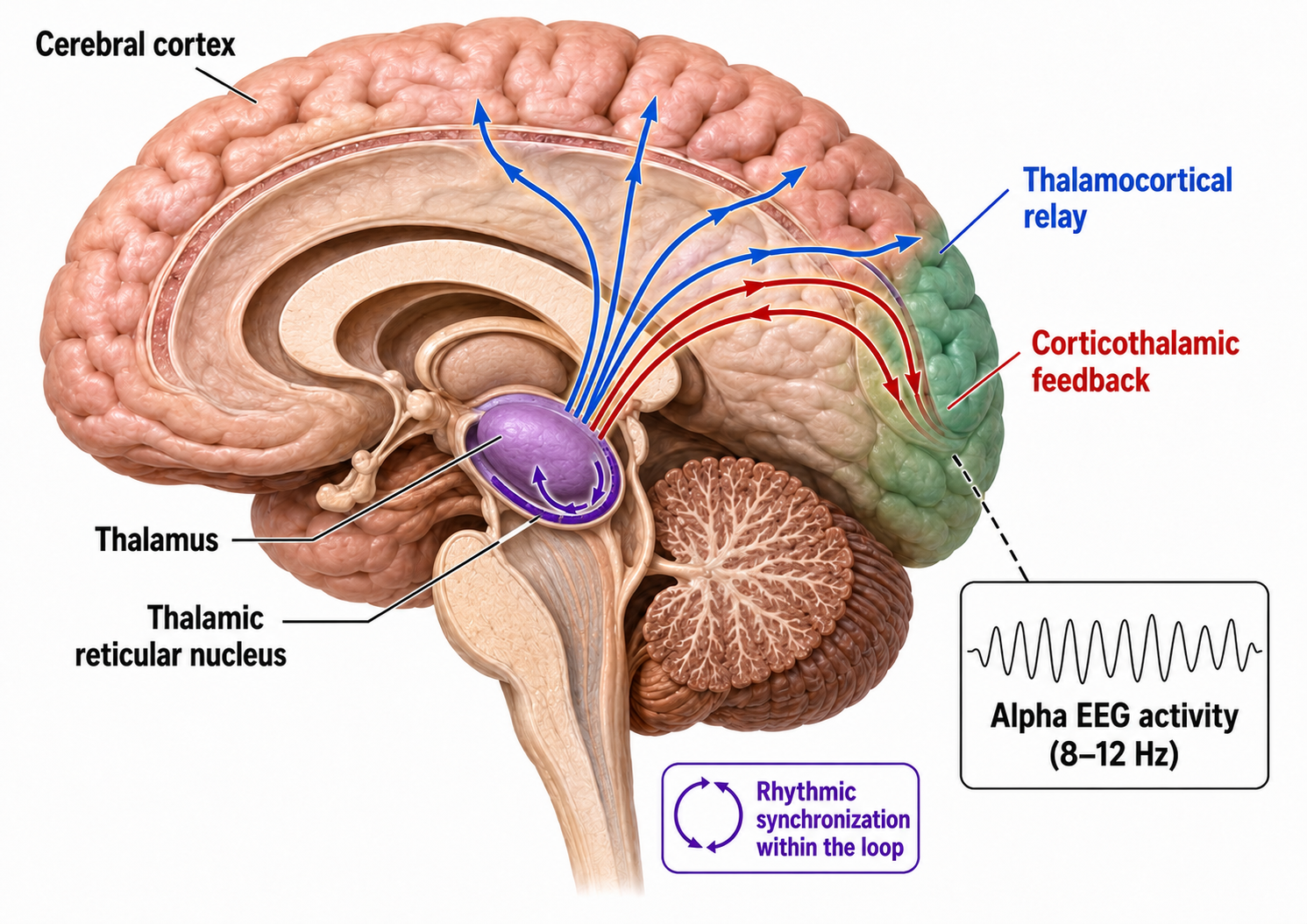
Andersen and Andersson (1968) advanced the facultative pacemaker theory, proposing that thalamic neurons activate both cortical neurons and thalamic inhibitory interneurons via recurrent collaterals. While these thalamocortical neurons only excite a limited number of cortical neurons, the inhibitory interneurons suppress a large pool of thalamocortical relay neurons. When the inhibition ends after about one-tenth of a second, the relay neurons experience rebound excitation, a synchronized depolarization that excites both cortical neurons and another round of thalamic inhibitory interneurons, initiating a new cycle that produces rhythmic EEG activity (Fisch, 1999).
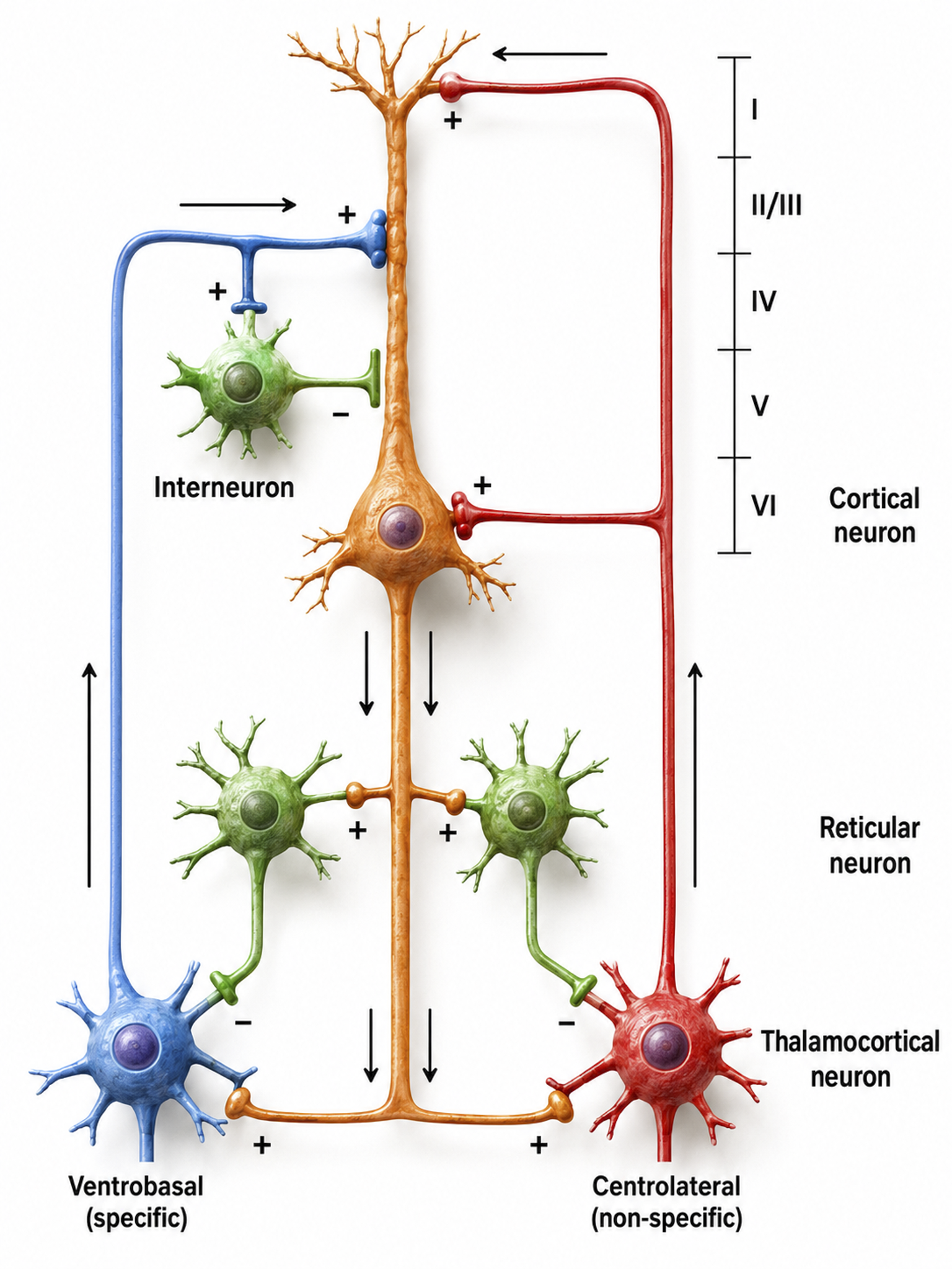
Thalamocortical circuit diagram depicting specific/sensory and non-specific intralaminar thalamocortical systems. Graphic adapted from Zachary Barry and featured in Wikipedia's article Recurrent ThalamoCortical Resonance.
The networking of excitatory and inhibitory thalamic neurons imposes a group rhythm on its members, which is then transmitted to cortical macrocolumns by thalamocortical neurons (Bear, Connors, & Paradiso, 2020). The nucleus reticularis of the thalamus may function as a pacemaker by releasing the inhibitory transmitter GABA at synapses with thalamocortical neurons. When this inhibition ends, these neurons depolarize cortical neurons and thalamic inhibitory interneurons through burst discharges.
As we will see when we reach microanatomy later in this unit, oscillatory activity may involve an interaction between thalamocortical relay neurons (TCR), nucleus reticularis neurons (RE), and interneurons, mediated by diverse neurotransmitters including acetylcholine and GABA. The thalamus is the dominant pacemaker for rhythmic EEG activity, including theta (3-8 Hz), alpha (8-12 Hz), and SMR (13-15 Hz) (Amzica & Lopes da Silva, 2018). For biofeedback practitioners, this means that many of the rhythms you train have their origin not in the cortex beneath the electrode, but in deeper thalamic circuits.
The Locus Coeruleus Inhibits Thalamic Alpha Generators
When we are inattentive, thalamic pacemakers generate the alpha rhythm. When we need to focus attention, we activate the brainstem noradrenergic locus coeruleus, a small but powerful nucleus roughly 15 mm long. The increased release of norepinephrine by this structure focuses attention and abolishes alpha oscillations by suppressing thalamic alpha generators. This may be an underlying mechanism of the phenomenon of alpha blocking.
Although researchers cannot noninvasively monitor locus coeruleus activity in human participants, it correlates with pupil dilation. In human studies, the greater the alpha blocking response and pupil dilation, the better the performance on demanding attention tasks (Dahl et al., 2020; Dahl et al., 2022). Critically, the alpha rhythm is not a cause but a sign that incoming stimulation is too weak to overcome inhibition by the reticular nucleus. EEG activity is not causal; it reflects network activity that has already occurred.
Additional Subcortical Generators
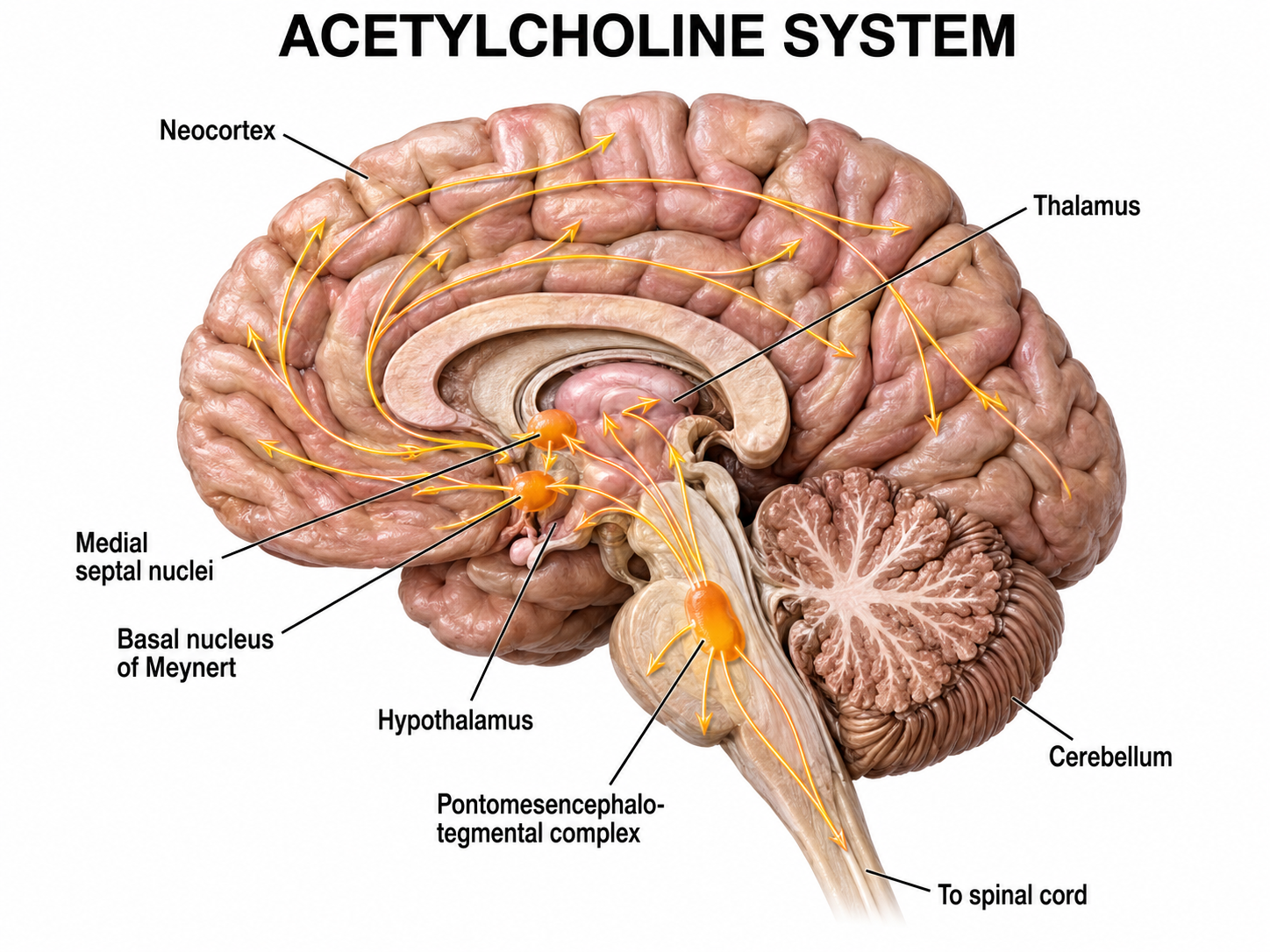
Several subcortical systems desynchronize or disrupt brain rhythms through ascending projections. These include the basal forebrain, reticular formation, locus coeruleus, and raphe systems. These neurons receive information from most sensory systems and cortical regions, and they desynchronize the EEG both directly through synapses on cortical neurons and indirectly through innervation of thalamic pacemakers. Desynchronization shifts pyramidal neurons from burst firing to more continuous single-spike generation (Fisch, 1999).
The cholinergic basal forebrain, located in the ventral frontal lobe and anterior hypothalamus, influences cerebral blood flow and cognitive activity. The basal forebrain comprises several structures, including the basal nucleus of Meynert, medial septal nuclei, substantia innominata, nucleus accumbens, ventral pallidum, and olfactory tubercle. Of these, the basal nucleus of Meynert and the medial septal nuclei are the principal sources of the neurotransmitter acetylcholine.
Graphic adapted from © Vasilisa Tsoy/Shutterstock.
The reticular activating system (RAS) includes a network of roughly 90 nuclei within the central brainstem, extending from the lower medulla to the upper midbrain, that activates the brain to promote attention, consciousness, and wakefulness. This network receives input from ascending sensory tracts (auditory, olfactory, somatosensory, and visual systems) and projects both to the thalamus and diffusely to the cortex, including pathways that bypass the thalamus entirely.
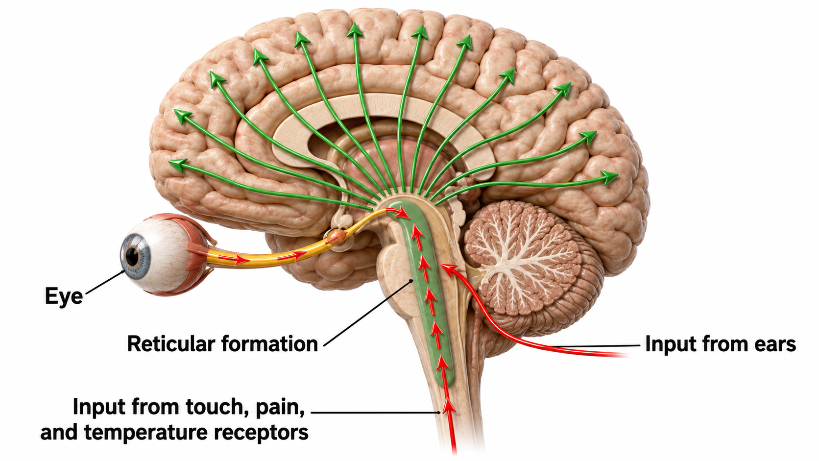
Reticular formation graphic redrawn by minaanandag on Fiverr.com.
The noradrenergic brainstem locus coeruleus system projects to the thalamus, limbic system, and cerebral cortex and contributes to wakefulness and vigilance for salient stimuli. The neurotransmitter norepinephrine (also called noradrenaline) is synthesized chiefly by neurons of the locus coeruleus, a small, pigmented nucleus situated in the dorsal pons along the lateral floor of the fourth ventricle. Its axons project diffusely throughout the neuraxis, reaching the cerebral cortex, hippocampus, amygdala, thalamus, and cerebellum, along with descending fibers to the spinal cord. Through these widespread connections, the locus coeruleus regulates arousal, selective attention, the sleep-wake cycle, and the physiological response to stress.
Within these neurons, norepinephrine is produced through a sequential enzymatic pathway. Tyrosine hydroxylase converts the amino acid tyrosine to L-DOPA, aromatic L-amino acid decarboxylase then yields dopamine, and dopamine β-hydroxylase completes the final step inside synaptic vesicles. Although the locus coeruleus supplies most of the forebrain's norepinephrine, additional noradrenergic populations in the lateral tegmentum and medulla contribute to autonomic and brainstem circuits.
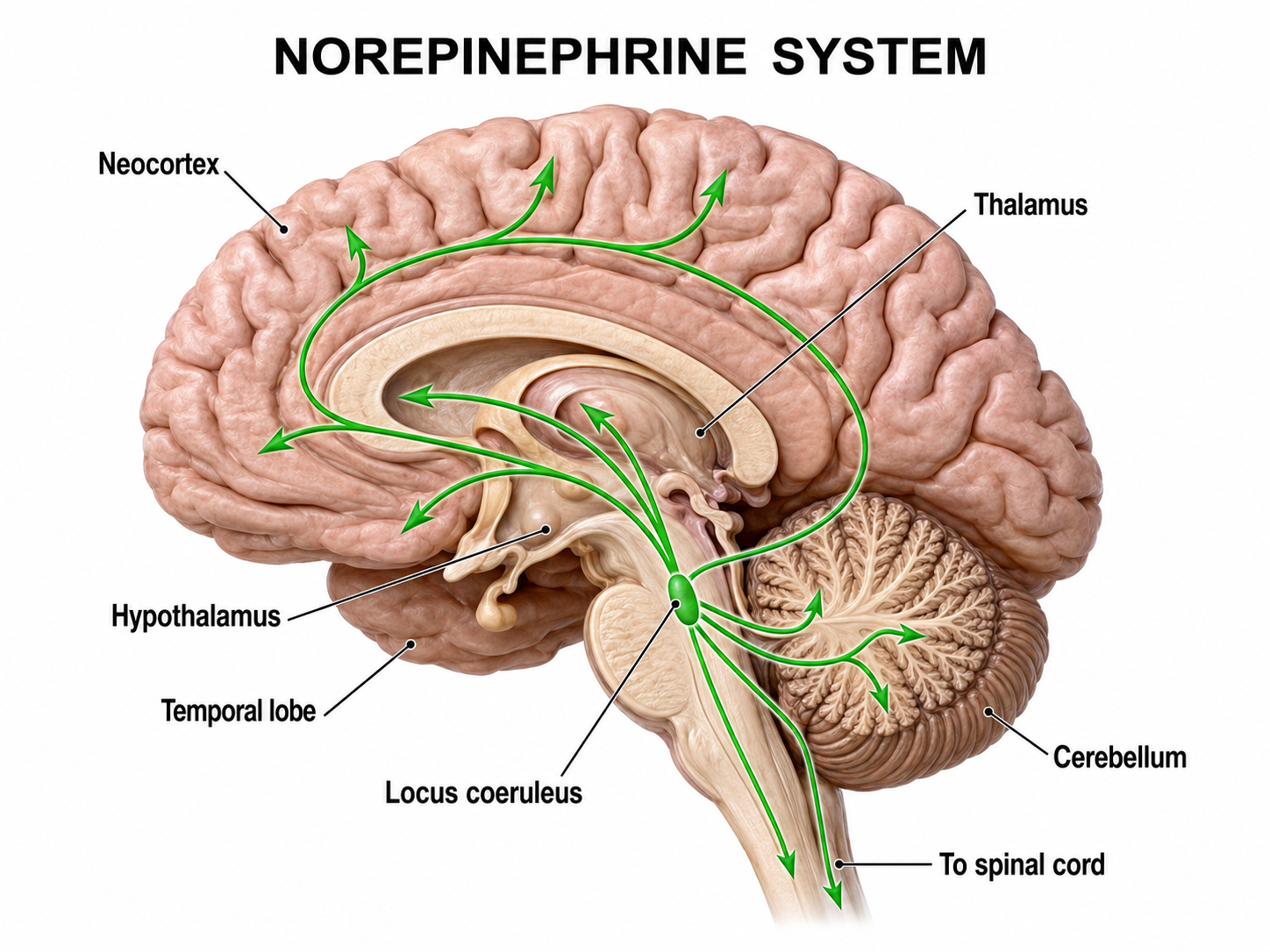
Graphic adapted from © Vasilisa Tsoy/Shutterstock.
Finally, the serotonergic raphe system is a midline network of cell bodies within the brainstem and midbrain that may influence alertness and vigilance through reciprocal connections with the suprachiasmatic nucleus of the hypothalamus (Monti & Jantos, 2008). Together, these subcortical systems determine the brain's overall state of arousal, a critical variable in any biofeedback session.
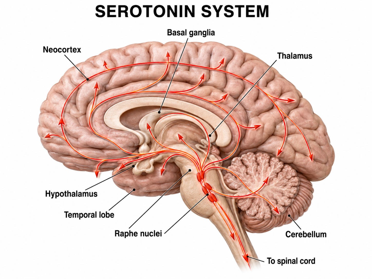
Graphic adapted from © Vasilisa Tsoy/Shutterstock.
Cortical and Subcortical Generators of Specific EEG Rhythms
Slow Cortical Potentials (0-1 Hz)
A slow cortical potential (SCP) is a gradual, sustained shift in the cortex’s baseline voltage that lasts from roughly 300 milliseconds to several seconds and reflects changes in the excitability of large populations of cortical neurons (Birbaumer et al., 1990). Slow cortical potentials (SCPs) have been identified in cortical neurons, the thalamus, and glial cells. Cortical neurons in layers II to VI generate slow oscillations even when the thalamus is removed or when cortical tissue is studied in vitro (in an artificial environment) or in vivo (within a living organism). Thalamic reticular neurons exhibit similar slow spontaneous oscillations when studied in vitro, and synchronized intracortical oscillations may depend on a corticothalamic network that targets these thalamic neurons.
The prevailing account attributes surface-negative SCPs to the synchronous depolarization of pyramidal apical dendrites, with a contribution from glial cells rather than a purely glial origin. Glia contribute by buffering extracellular potassium and by coupling to one another through gap junctions, which are direct electrical connections between glial cells. Glial cells communicate among themselves and with neurons, and their slow oscillations may influence the timing of neuronal firing through their control of potassium ion outflow (Steriade, 2005). These slow oscillations appear to organize the generation of other brain rhythms.
"The concept of a unified corticothalamic network that generates diverse types of brain rhythms grouped by the cortical slow oscillation (Steriade, 2001a,b) is supported by EEG studies in humans" (Mölle et al., 2002). Caton (1875) reported feeble currents of varying direction from electrodes placed on the cortical surface and observed that the surface becomes relatively negative during functional activity. Caton recorded galvanometer deflections rather than calibrated voltages, so his one-page report contains no microvolt figures; the numerical gradients sometimes attributed to him do not appear in it. Underlying "tone" or valence factors determine the firing characteristics of neurons within a network: when SCPs are more positive, reduced cortical neuron firing occurs due to hyperpolarization; when SCPs are more negative, increased firing occurs due to depolarization.
The following 19-channel BioTrace+ /NeXus-32 display of 0.1-1 Hz SCP activity © John S. Anderson.
Perspective on Fast Cortical Potentials
EEG "bands" are somewhat arbitrary ranges of frequencies that have evolved from observation and usage. The following BioTrace+ /NeXus-32 video of raw and spectral EEG displays © John S. Anderson. Frequency is plotted along the horizontal axis, and amplitude is shown on the vertical axis.
While frequency band labels are helpful descriptors, they can also be misleading. Classification of an EEG rhythm is based on context (measurement conditions and EEG activity during the specific epoch), frequency, and waveform morphology. Note, however, that for some purposes it is useful to define individual-specific frequency bands that depart somewhat from the classical definitions. For example, although the individual alpha peak typically sits near 10 Hz, some individuals peak nearer 8 or 9 Hz and others nearer 12 Hz, and this shift can affect the settings selected for neurofeedback (Tarasi & Romei, 2024).
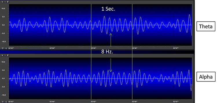
The process of up-training or down-training signal amplitude in one or more of the EEG bands using an EEG is called EEG biofeedback or neurofeedback. Minimum EEG voltages of 20-30 μV are seen in children and adults (Krauss et al., 2011).
Brainwaves Reflect Behavior
The ratio of slow (theta) to faster (beta) brainwaves indicates how alert you are; this is the theta/beta ratio. A higher ratio suggests greater drowsiness or inattention, while a lower ratio reflects focused alertness. This metric is one of the most widely used in neurofeedback assessment.
In the next section, we will examine delta, theta, rhythmic slow-wave, alpha, mu, synchronous "alpha," SMR, beta, high or fast beta, and gamma activity.
Local Versus Global Decision-making
The short time windows of fast oscillators facilitate local integration and decision-making, primarily because of the limitations of axon conduction delays. In contrast, the long time windows of slow oscillators can involve many neurons in large or distant brain areas, favoring complex, global decisions. This principle has direct clinical implications: when you observe a client's EEG dominated by slow activity, it may reflect an over-reliance on global, less differentiated processing at the expense of focused, efficient local integration.
Delta (0.0-4 Hz)
There are two delta rhythms: a slow oscillation under 1 Hz and a traditional 1-4 Hz oscillation. The slow 0.3-0.4 Hz oscillation originates in the neocortex and persists when the thalamus is removed, while thalamocortical neurons generate the 1-4 Hz oscillations observed during human stage-3 sleep. Slow neocortical oscillations may synchronize the thalamic delta rhythm (Steriade, 2005).
Delta activity is generated by cortical neurons when other connections do not activate them and is found predominantly in frontal areas. Delta is associated with sleep and infancy, and during stage 3 (N3) sleep it accompanies the replenishment of astrocyte glycogen stores. Clinicians observe delta in clients diagnosed with ADHD, brain tumors, learning disorders, and traumatic brain injury (TBI). Rhythmic high-amplitude delta is particularly associated with TBI when localized, while diffuse delta may appear in ADHD and learning disorders.
Delta waves are the main EEG activity during infancy, reflecting the immature brain's low-frequency cortical activity. As neural networks develop and mature, faster rhythms like alpha and beta replace delta dominance in waking states. In adults, waking delta activity is minimal but may transiently appear during drowsiness or relaxation.
Normal Amplitudes
Delta should not be present in significant amounts in the awake adult EEG. "Apparent" delta is usually an eye movement artifact, although some delta activity probably occurs in the normal waking adult EEG. In clinical practice, delta bands are inhibited or down-trained but rarely rewarded, though delta desynchronization can be rewarded.
The following 19-channel BioTrace+ /NeXus-32 display of eyes-open 1-4 Hz activity from a 10-year-old male © John S. Anderson.
Theta (4-8 Hz)
Researchers define the boundaries of EEG frequency bands somewhat differently. Although the theta band is sometimes set at 3-8 Hz, most authors place it at 4-8 Hz.
The mechanisms that generate the theta rhythm are poorly understood, and theta's functions differ depending on location and source. Amzica and Lopes da Silva (2018) consider the classic septal/diagonal band pacemaker model incomplete. Hippocampal interneurons exercise top-down control over the hypothetical medial septum pacemaker, and the hypothalamic supramammillary nucleus, with extensive connections to the brainstem, diencephalon, and medial septum, may also pace and modulate hippocampal theta. A non-cholinergic theta source has also been found within the entorhinal cortex of the hippocampus.
Theta is associated with creativity, global synchronization, memory formation, and recall. Increased theta amplitudes correspond with hypo-perfusion (reduced blood flow) and decreased glucose metabolism. Excessive frontal theta is linked with depression, daydreaming, distractibility, and inattention.
Theta/beta (T/B) ratios are developmentally mediated, so any cutoff has to be age-referenced. In Monastra et al. (1999), the control mean fell from roughly 3.0 at ages 6-11 to roughly 1.5 at ages 21-30, and the ADHD thresholds fell in parallel (see the table below). A ratio of 3.0 is therefore unremarkable in a 6-to-11-year-old and markedly elevated in a young adult.
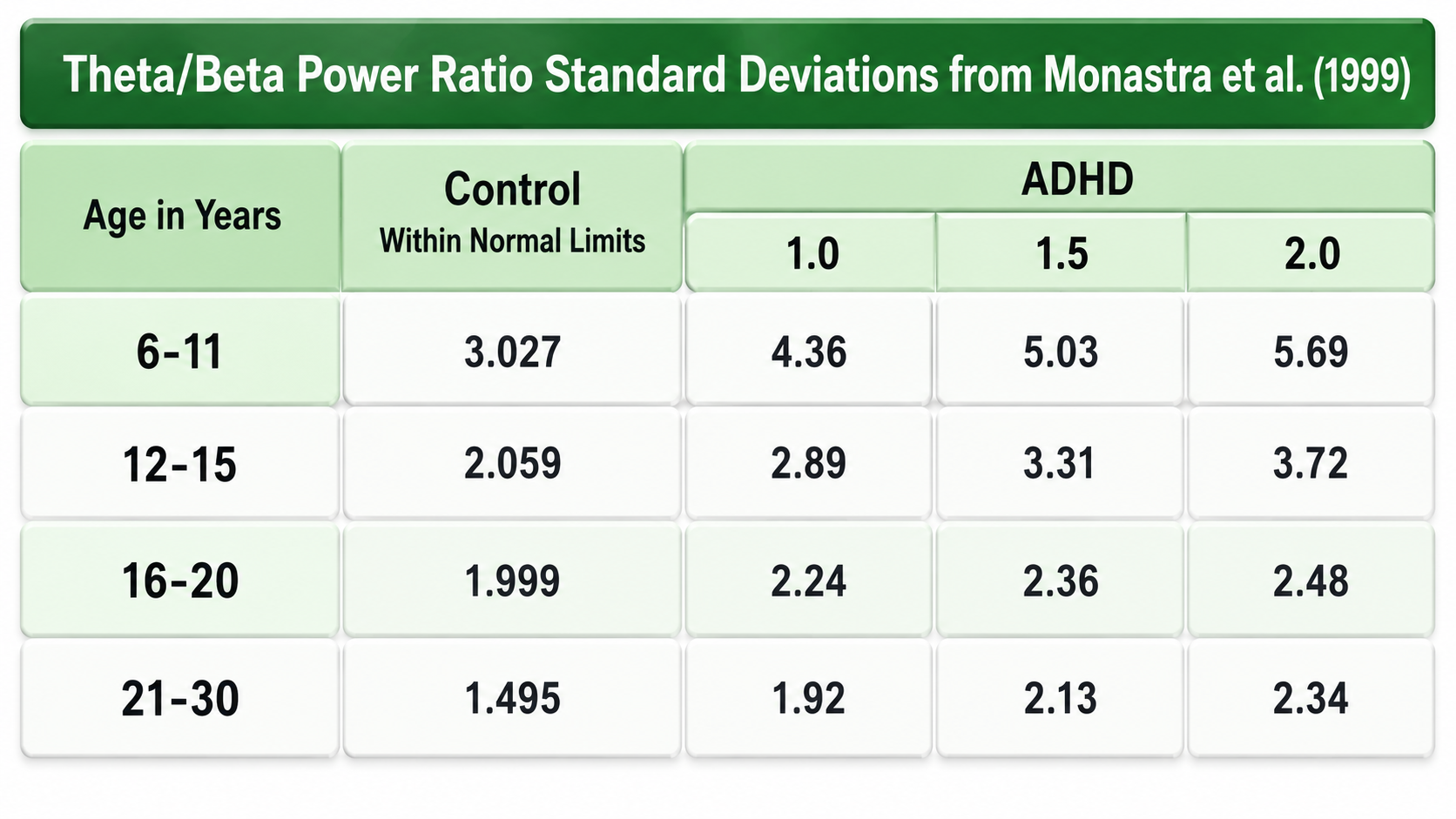
Normal Amplitudes
Theta voltage is age-related in the awake EEG, diminishing from age 8 to 30 with minimal amounts over age 30. A typical 6-7 Hz rhythm in the frontal midline (FCz) is associated with mental activity such as problem-solving and a wide variety of other functions. This rhythm appears to be limbic in origin and is higher in amplitude and more synchronous when processing feedback that an error has occurred. The 4-Hz rhythm is associated with childhood pleasurable experiences and memory searches in adults.
Rhythmic Slow Wave (RSW or Theta)
Clinicians inhibit frontal theta to remediate symptoms and reward posterior RSW in alpha/theta training for addictions, global synchronization, optimal performance, and PTSD. RSW is generally not increased frontally. Clinicians may also train for increases or decreases in phase synchrony. RSW is mainly seen in the frontal-midline (FCz) when awake with eyes open and is generated by the limbic system and thalamus. Depending on location, RSW may represent slowed alpha as thalamic output decreases in frequency.
The following 19-channel BioTrace+ /NeXus-32 display of eyes-open 4-8 Hz activity from a 10-year-old boy © John S. Anderson.
Alpha (8-13 Hz)
The 8-13-Hz alpha rhythm differs from spindle waves in both its source and the activity during which it is observed. Alpha 1 (low alpha) ranges from 8-10 Hz, and alpha 2 (high alpha) from 10-13 Hz (Thompson & Thompson, 2015b). Alpha rhythms depend on the interaction between rhythmic burst firing by a subset of thalamocortical (TC) neurons linked by gap junctions and rhythmic inhibition by widely distributed reticular nucleus neurons (Hughes & Crunelli, 2005). The alpha rhythm is maintained and propagated by cortical networks (Amzica & Lopes da Silva, 2018).
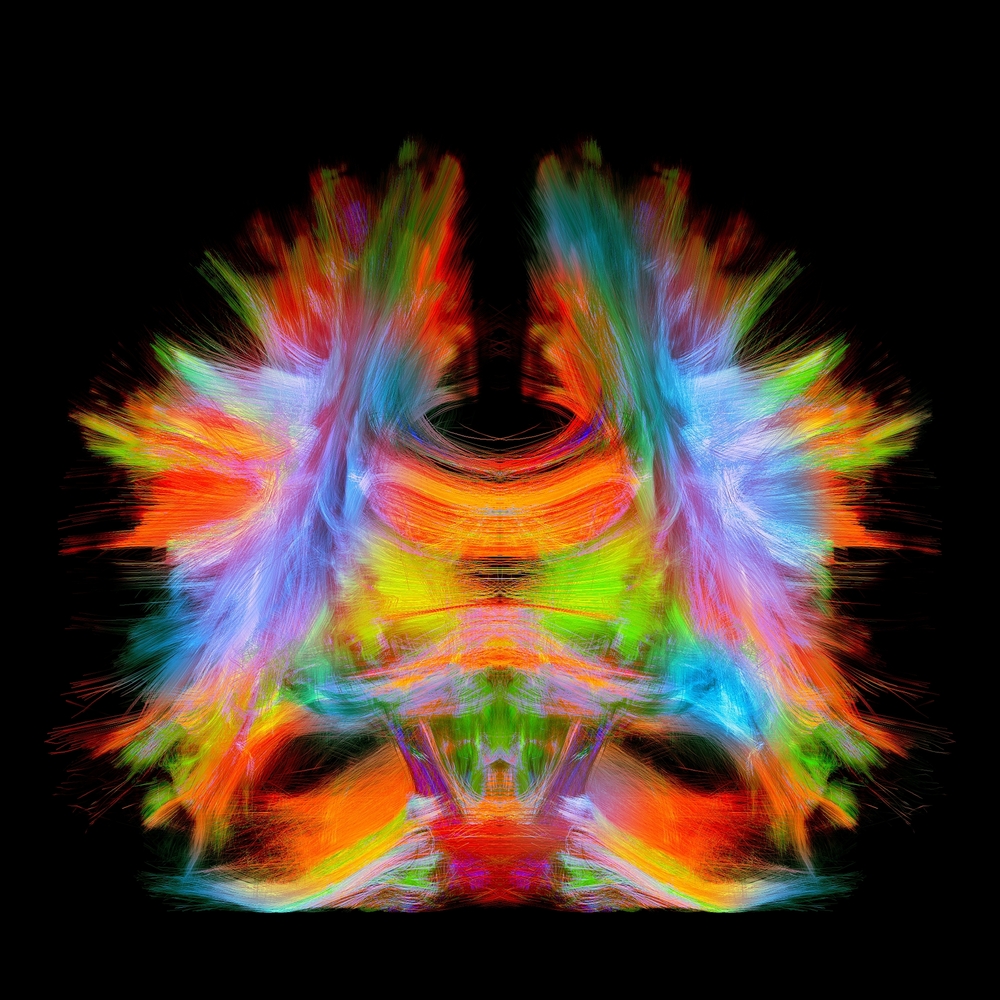
Connectome graphic © Image Source Trading Ltd/Shutterstock.
Researchers have correlated the alpha rhythm with relaxed wakefulness, though there are age- and function-related differences. Spindle waves, in contrast, originate in the thalamus and occur during unconsciousness and stage-2 sleep (Steriade, 2005). Alpha is the dominant rhythm in adults and is located posteriorly.
The 8-10 Hz range is associated with ADHD, daydreaming, fogginess, OCD, and TBI, while frontal asymmetry with excess left frontal alpha is associated with depression. The 10-12 Hz range is seen with inner calm (calm and alert) and meditation. Clinicians train alpha amplitude and phase synchrony up or down depending on location and presenting symptoms.
Posterior Dominant Rhythm (PDR)
The posterior dominant rhythm (PDR) is the posterior alpha rhythm, first visible at about 4 months of age with a frequency of around 4 Hz. Between 3 and 5 years, this rhythm is approximately 8 Hz with amplitudes as high as 100 μV. From 6 to 15 years, it reaches 9 Hz by age 7 and 10 Hz by ages 10-15, with a mean amplitude of 50-60 μV. Girls show a statistically faster maturation of posterior alpha frequency than boys.
From 13 to 21 years, the mean alpha frequency is 10 Hz, and amplitudes decline throughout this period. Faster alpha frequencies are associated with higher IQ and better memory performance. This developmental trajectory illustrates why age-appropriate normative comparisons are essential in clinical qEEG assessment.
The following 19-channel BioTrace+ /NeXus-32 display of the response of the posterior dominant rhythm to eyes opening and closing © John S. Anderson.
Normal Amplitudes
The typical adult alpha peak frequency ranges from 9.5-10.5 Hz, and alpha below 8 Hz is considered abnormal. Alpha frequency and amplitude decline throughout adulthood and into old age, so an older client's slower alpha may be age-appropriate rather than pathological. Most adult amplitudes fall below 60 μV: about 66% of adults show 20-60 μV, 28% show less than 20 μV, and 6% show greater than 60 μV (Schomer & Lopes da Silva, 2011).
Higher alpha amplitudes are observed over the non-dominant (right) hemisphere, a normal pattern known as alpha asymmetry. Most studies show no effect of handedness. Asymmetry is generally no more than 20 μV or 20% of the greater of the two amplitudes (Amzica & Lopes da Silva, 2018). Clinicians should be familiar with these normative values, since departures from them often guide neurofeedback protocol selection.
Causes of Excessive Alpha Amplitudes
Sleep deprivation or metabolic exhaustion can result in high amplitude and slowing of the peak frequency, along with persistent alpha during an eyes-open condition. Meditation practices can cause increased amplitudes and slowing, a faster alpha response to an eyes-closed condition, and persistent alpha in an eyes-open condition.
Marijuana use and abuse can also cause increased amplitudes and slowing, along with persistent alpha in an eyes-open condition, depending on the type of marijuana. These effects can persist for many years following abstinence. Clinicians should consider substance use history when interpreting alpha findings.
The following 19-channel BioTrace+ /NeXus-32 display of eyes-closed 8-12 Hz activity from a 13-year-old girl © John S. Anderson.
Mu Rhythm (7-11 Hz)
While the 7-11-Hz mu rhythm usually overlaps with the alpha range, it is arc-shaped on top and pointed on the bottom of an analog EEG trace (i.e., a wicket rhythm) that differentiates it from the alpha waveform. The mu rhythm is also identified by its location over the sensorimotor strip. It can be recorded at C3 and C4 in a minority of subjects and appears when the person is awake and at rest. Actual, visualized, or observed movement suppresses it (Thompson & Thompson, 2015b). Mu rhythms appear to index sensorimotor cortex activity, and mu suppression has been linked to the mirror neuron system of the premotor and inferior parietal cortex.
These mirror neuron circuits may play a critical role in imitation learning and our ability to understand the actions of others. Mu rhythms facilitate the conversion of visual and auditory input into integrated skill-building functions, and attenuation of the mu rhythm appears to be associated with the activation of this function (Pineda, n.d.). The mu rhythm is highlighted below.
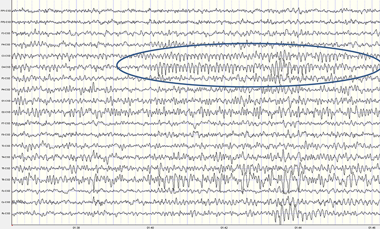
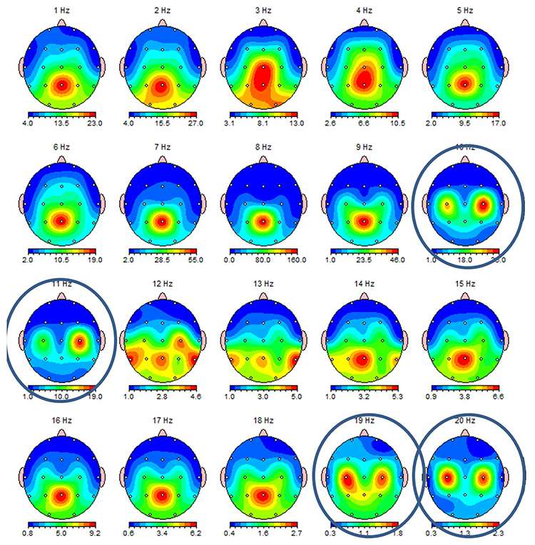
This second example of the mu rhythm shows a classic 10-11 Hz and 19-20 Hz "Owl Eye" presentation. The mu rhythm has an arciform, comb-like shape that produces two spectral peaks in a nearly harmonic relationship: a fundamental near 10 Hz and a beta harmonic near 20 Hz (Tiihonen, Kajola, & Hari, 1989). In the single-hertz topographic maps below, the mu fundamental near 10-11 Hz and its beta component near 19-20 Hz each appear as a pair of left and right central hot spots that look like an owl's eyes on the head map, which is the source of the name. The term is informal field usage rather than a published descriptor.
Synchronous "Alpha"
Various sensory systems (auditory, somatosensory, and visual) produce localized and semi-independent "alpha" activity. However, synchronous, distributed alpha integrates perception and facilitates action across broader cortical networks. Synchronous "alpha" appears to block localized alpha-like patterns such as mu and the posterior rhythm in favor of more broadly distributed network integration during tasks requiring global processing.
Sensorimotor Rhythm (13-15 Hz)
The sensorimotor rhythm (SMR), also called beta 1, is located on the sensorimotor strip (C3, Cz, C4). SMR amplitude increases when the motor circuitry is idle; it increases with stillness and decreases with movement. Deficient SMR may be observed in movement-spectrum complaints like hyperactivity and tics. SMR appears as sleep spindles during stage-2 sleep and is associated with reduced blood perfusion and glucose metabolism in the brain as it transitions to deeper sleep stages.
Clinicians typically reward increased SMR amplitude to calm hyperactivity and during theta/beta ratio training. For practitioners working with athletes or military personnel, SMR training can promote the calm, focused stillness essential for precision tasks like marksmanship or surgical procedures.
The following 19-channel BioTrace+ /NeXus-32 display of 12-15 Hz activity © John S. Anderson.
Beta (over 12 Hz)
Beta consists of rhythmic activity between 12-38 Hz in four ranges: beta 1 (12-15 Hz), beta 2 (15-18 Hz), beta 3 (18-25 Hz), and beta 4 (25-38 Hz). However, writers may define beta subranges somewhat differently.
Beta shows a gradient of activity from occipital to frontal lobes, with more beta occurring frontally where it is associated with focus, analysis, and relaxed thinking (Thompson & Thompson, 2015b). Excessive beta is observed in anxiety, depression (reversed asymmetry), insomnia, OCD, and sleep disorders, while deficient beta is seen in ADHD, cognitive decline, and learning disorders.
Since beta overlaps with the EMG range, clinicians must be careful when up-training this rhythm and should use an EMG inhibit, (a high-beta EEG proxy for EMG), to avoid inadvertently reinforcing muscle artifact. Beta is generated by the brainstem and cortex and is associated with hyper-perfusion and increased glucose metabolism.
Normal 16-20+ Hz Beta Amplitudes
Beta amplitudes are minimal in children up to 12 years, with a significant increase in beta amplitude and organization between 12 and 30 years. Beta is commonly seen in nearly all adults with amplitudes of 20 μV or less. Interhemispheric amplitude asymmetries exceeding 35% are abnormal.
The following 19-channel BioTrace+ /NeXus-32 display of 13-21 Hz activity © John S. Anderson.
Fast or High Beta Rhythms (20-35 Hz)
Fast 20-35-Hz oscillations are generated by activation of the mesencephalic reticular formation. Thalamocortical, rostral thalamic intralaminar, and cortical neurons spontaneously oscillate in this range. This activity is primarily seen in the frontal lobes and is associated with hyper-perfusion and increased glucose metabolism, though persistent excessive activity can lead to metabolic exhaustion.
Fast beta may be associated with peak performance and cognitive processing, particularly with specificity and precision in information processing. However, excessive high beta is associated with alcoholism, anxiety, OCD, rumination, and worry. Clinicians often inhibit high beta activity but rarely reward it. In clinical practice, persistent high beta at frontal sites, particularly if it shows a spindling appearance, is one of the most common EEG signatures of anxiety disorders.
The following 19-channel BioTrace+ /NeXus-32 display of eyes-closed approximately 25 Hz fast beta activity © John S. Anderson.
Gamma Rhythms (28-80 Hz)
Because this unit ends beta at 38 Hz and begins gamma at 28 Hz, the two bands overlap between 28 and 38 Hz; clinical electroencephalography more often ends beta near 30 Hz. Any scalp band edge above roughly 30 Hz is also vulnerable to EMG contamination.
Amzica and Lopes da Silva (2018) concluded that gamma oscillations might speed information distribution and processing. Gamma bursts occur during problem-solving, and the absence of gamma is associated with cognitive deficits and learning disorders. Gamma synchrony is related to cognitive processing and contributes specificity and precision to information processing. Gamma is theorized to serve as a "binding rhythm" that integrates sensory inputs into unified perception and consciousness.
The following 19-channel BioTrace+ /NeXus-32 display of eyes-open 36-44 Hz activity in a 10-year-old boy © John S. Anderson.
Gamma rhythms are linked with SCPs. The following BioTrace+ /NeXus-32 display of SCP and gamma activity © John S. Anderson.
In summary, the brain generates EEG rhythms through an interplay of thalamic pacemakers, cortical resonant loops, and subcortical modulators. Each frequency band reflects distinct physiological processes and clinical states, from delta's association with sleep and injury to gamma's role in cognitive binding. Understanding these generators equips clinicians to make informed decisions about which rhythms to train and why.
Suppose you are training a client to increase sensorimotor rhythm at Cz and notice nearly identical activity appearing at neighboring sites. Remember that the rhythms you train often originate in deeper thalamic circuits rather than in the cortex directly beneath the electrode, and that electrical potentials volume conduct rapidly through tissue, skull, and scalp. As a result, activity recorded at one electrode may not arise from the cortex immediately below it. Keeping the thalamic origin of these rhythms and the reality of volume conduction in mind will make your interpretation of topographic maps more accurate.
The thalamus is the dominant pacemaker for rhythmic EEG activity, including theta, alpha, and the sensorimotor rhythm. Cortical rhythms also arise from synchronized pyramidal neurons in an open-field arrangement and from resonant loops, whose local, regional, and global forms generate progressively lower frequencies as the distance between macrocolumns increases. Subcortical systems, including the basal forebrain, reticular activating system, locus coeruleus, and raphe nuclei, desynchronize and modulate these rhythms to set the brain's level of arousal. Each frequency band reflects distinct states, from delta in sleep and injury to gamma in cognitive binding, with alpha marking relaxed wakefulness and beta marking focus or, in excess, anxiety. Because potentials volume conduct through tissue and skull, activity at a given electrode may not originate from the cortex directly beneath it.
Check Your Understanding
- According to the facultative pacemaker theory, what produces rhythmic EEG activity after thalamic inhibition ends?
- Why do pyramidal neurons, rather than stellate neurons, dominate the scalp EEG?
- Distinguish local, regional, and global resonant loops, and explain how the distance between macrocolumns relates to EEG frequency.
- What happens to the alpha rhythm when the locus coeruleus increases norepinephrine release, and what noninvasive measure correlates with this activity?
- Name the EEG band associated with each of the following: slow-wave sleep, relaxed wakefulness with eyes closed, and calm motor stillness on the sensorimotor strip.
Review Flash Cards on Quizlet
Click the buttons below to review our flash cards for the macroanatomy half of this unit.
Microanatomy: Neurons and Glia
Everything above happens because of cells. Zoom in far enough and the "cerebral symphony" resolves into individual neurons maintaining voltage gradients at considerable metabolic expense, and glial cells that turn out to be far more than passive scaffolding.
Types of Neurons
This section covers the three major types of neurons, sensory, motor, and interneurons, along with the glial cells that work in partnership with them. Understanding these cell types is essential because each plays a distinct role in generating the electrical signals that neurofeedback clinicians measure and train. Neuron graphic © SciePro/Shutterstock.com.
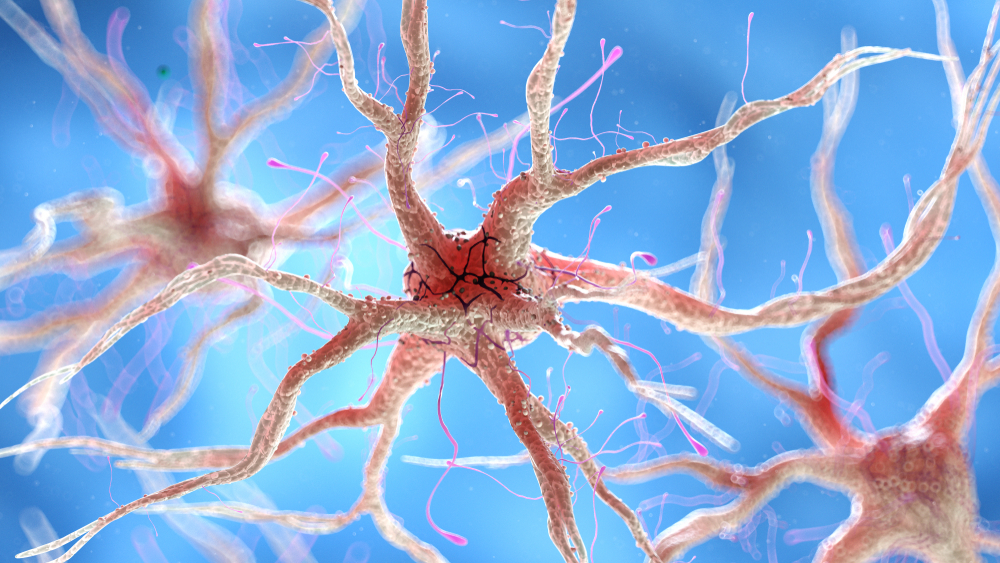
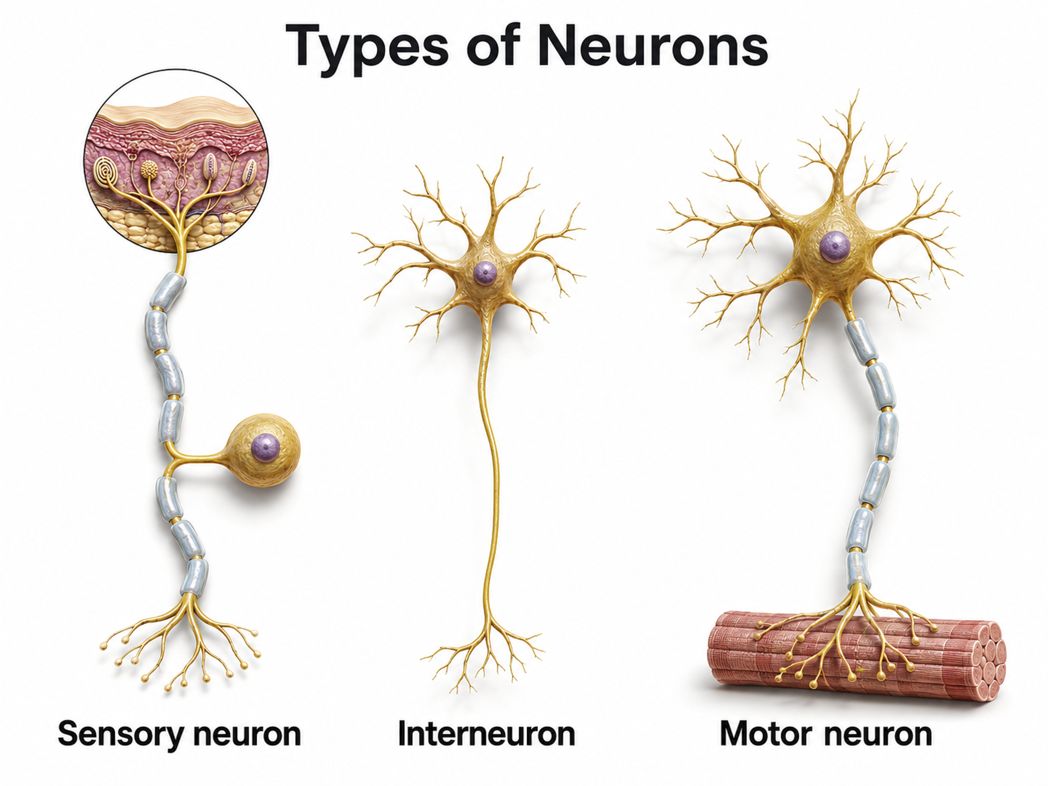
We can divide neurons into three functional categories: sensory, motor, and interneurons. Glial cells like astrocytes and microglia work in partnership with neurons to support communication and maintain the neural environment.
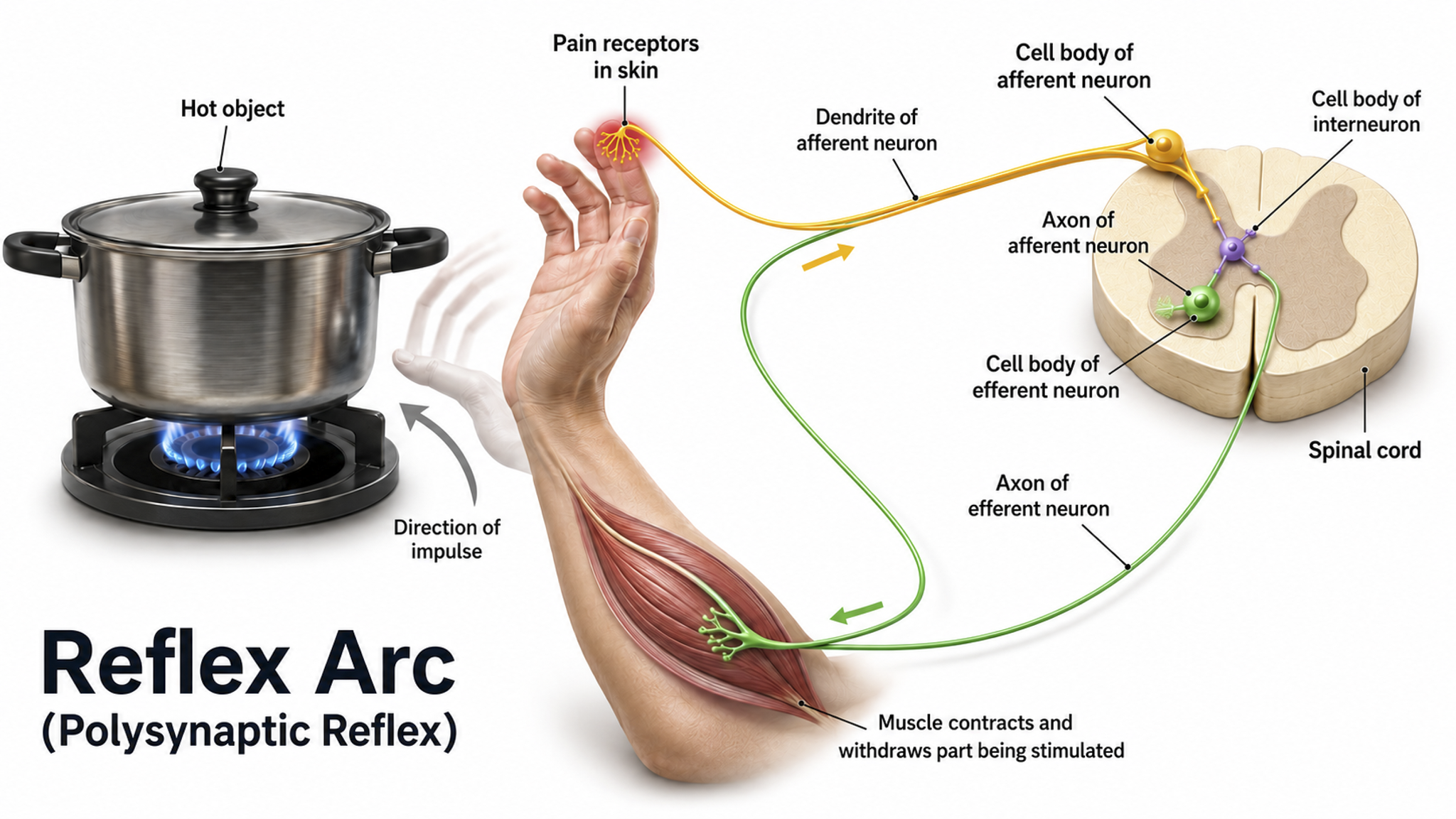
Sensory neurons are specialized for receiving information from the environment and the body. They are called afferent because they transmit sensory information toward the central nervous system (brain and spinal cord). Whether a client feels the warmth of a thermal biofeedback sensor or hears an auditory neurofeedback tone, sensory neurons relay that information centrally for processing. Sensory neuron graphic © TimeLineArtist/Shutterstock.com.
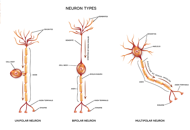
Motor neurons convey commands to glands, muscles, and other neurons. They are called efferent because they carry information toward the periphery. When a client consciously relaxes the frontalis muscle during surface EMG biofeedback, motor neurons translate that intention into reduced muscle tension. Motor and sensory neuron graphic © iso-form llc/Shutterstock.com.
.jpg)
Interneurons provide the integration required for decisions, learning and memory, perception, planning, and movement. They have short processes, analyze incoming information, and distribute their analysis within neural networks. Interneurons are largely confined to the central nervous system, though they also occur in the enteric nervous system and in peripheral autonomic ganglia. Under the broad definition, any neuron that is neither sensory nor motor, they account for the great majority of neurons (Breedlove & Watson, 2023); in the neocortex, local-circuit interneurons make up roughly 20-30% of neurons, the remainder being pyramidal projection cells.
Local interneurons analyze small amounts of information provided by neighboring neurons. Relay interneurons connect networks of local interneurons from separate brain regions, enabling diverse functions like perception, learning, memory, and executive functions such as planning (Carlson & Birkett, 2021). This local-to-global architecture helps explain why neurofeedback training at a single electrode site can influence complex cognitive and emotional processes. Neuron graphic adapted from © Aldona Griskeviciene/Shutterstock.com.
Reflex arc graphic adapted from © SANDIP NEOGI/Shutterstock.com.
Neuron Structure
This section examines the key structural components of neurons, from the cell body to terminal buttons. These structures work together to receive, integrate, and transmit electrical and chemical signals. Understanding neuron anatomy is fundamental to grasping how the EEG signal originates and what neurofeedback clinicians are ultimately measuring.
While neurons have over 200 different designs to perform specialized jobs in the nervous system, they generally share five structures: a cell body or soma, dendrites, an axon hillock, an axon, and terminal buttons.
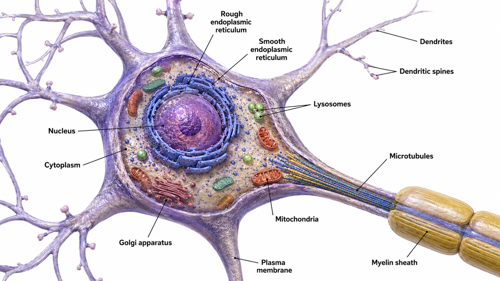
The cell body or soma contains the machinery for the neuron's life processes. It receives and integrates EPSPs and IPSPs, small graded positive and negative changes in membrane potential generated by axons. The cell body of a typical neuron is 20 μm in diameter, and its spherical nucleus, which contains chromosomes comprised of DNA, is 5-10 μm across.
The cell body is the primary location where neurons manufacture proteins (like enzymes, receptors, and ion channels) and peptides (neurotransmitters like oxytocin), though there is increasing evidence of distributed protein manufacturing via local mRNA translation (Nagano & Araki, 2021). Check out the Khan Academy YouTube video, Anatomy of a Neuron. Cell body graphic © MattL_Images/Shutterstock.com.
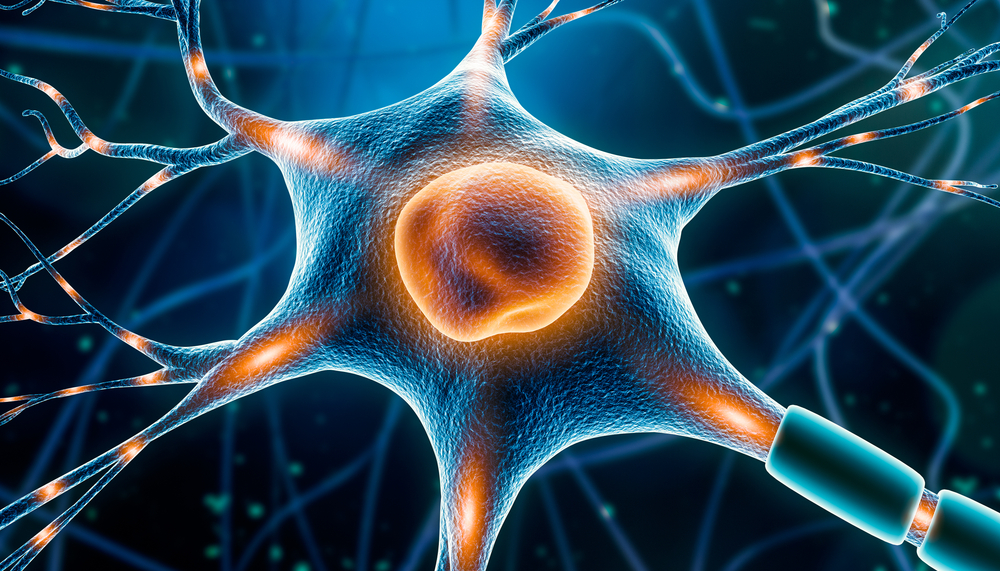
Mitochondria power diverse processes throughout neurons, including opening ion channels, conducting action potentials, releasing and returning neurotransmitters, and transporting proteins. They are directly responsible for EEG signal strength since they fuel the postsynaptic potentials that scalp electrodes detect. Mitochondria are orange in the graphic below © Corona Borealis Studio/Shutterstock.com.
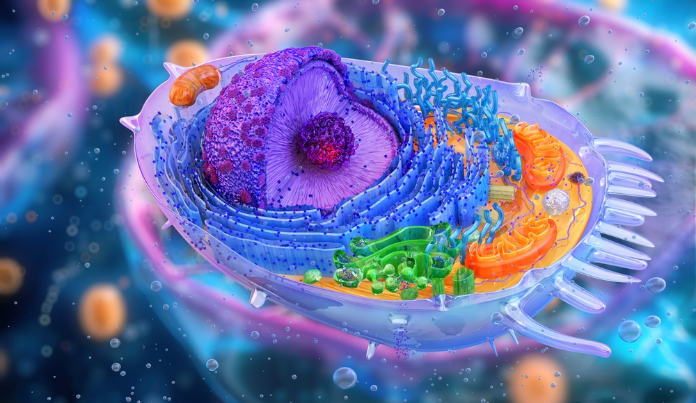
Mitochondria are far more than simple cellular energy factories. In the brain, these organelles are master multitaskers whose involvement in cellular respiration, calcium homeostasis, and reactive oxygen species (ROS) management makes them integral to the brain's demanding physiological environment (Fischer et al., 2020). At their most fundamental level, mitochondria generate the ATP that neurons desperately need. Neurons are constantly sending electrical signals, releasing neurotransmitters, and rebuilding connections, which is why mitochondria cluster at synapses like miniature power plants, ensuring there is always enough fuel for crucial processes like learning and memory formation (Harris et al., 2012).
Mitochondria also serve as cellular calcium regulators, maintaining strict control over calcium levels inside neurons. This calcium management is critical for proper neural signaling; when mitochondria fail at this job, it can trigger a cascade of problems that may lead to neurodegenerative diseases. During intense neural activity, mitochondria buffer incoming calcium waves, preventing toxic buildups while fine-tuning the cellular responses that drive neurotransmitter release and gene expression (Brini et al., 2014). These organelles also walk a delicate tightrope with reactive oxygen species (ROS), which they generate as a byproduct of energy production. In small amounts, ROS serve as important signaling molecules, but excessive production can damage proteins, lipids, and DNA, a particular danger in the brain, which is extremely vulnerable to oxidative damage due to its high oxygen consumption and abundance of lipids (Lin & Beal, 2006).
Mitochondria are also cellular life-and-death decision makers, playing a crucial role in apoptosis (programmed cell death). This process is essential during brain development, helping to sculpt neural circuits by eliminating unnecessary neurons through specific pathways involving cytochrome c release and caspase activation (Green et al., 2011). When damaged, mitochondria can release danger signals called DAMPs (damage-associated molecular patterns), including mitochondrial DNA, which can trigger inflammatory responses that contribute to conditions like multiple sclerosis and chronic traumatic encephalopathy (West et al., 2015). As we age, mitochondrial function gradually declines, leading to decreased ATP production and increased ROS generation, which affects crucial processes like synaptic function and memory formation (Lopez-Otin et al., 2013).
The dynamic nature of mitochondria is particularly fascinating in the context of brain plasticity. These organelles constantly undergo fusion and fission, processes that allow them to adapt to changing energy demands and repair damage. These dynamics are especially important in maintaining synaptic plasticity, the foundation of learning and memory that makes neurofeedback training possible. When these processes malfunction, it can lead to developmental disorders including autism spectrum disorder and intellectual disabilities (López-Doménech et al., 2016). For neurofeedback clinicians, understanding mitochondrial function helps explain why factors like sleep quality, exercise, and nutrition can profoundly influence a client's capacity for neural learning during training.
Dendrites are branched structures designed to receive messages from other neurons via axodendritic synapses (junctions between axons and dendrites) and send messages to other neurons via dendrodendritic synapses (junctions between the dendrites of two neurons). Dendrites receive thousands of synaptic contacts and have specialized proteins called receptors for neurotransmitters released into the synaptic cleft (Bear, Connors, & Paradiso, 2026).
A neuron's dendrites are called a dendritic tree, and each extension is called a dendritic branch.
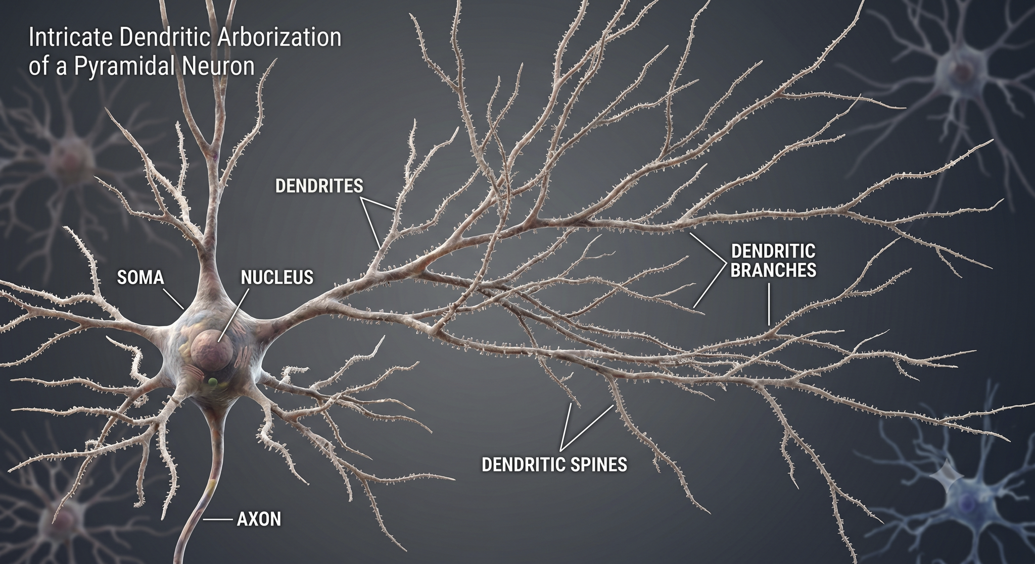
Biological psychologists classify neurons based on whether their dendrites feature spines. Dendritic spines are protrusions on the dendrite shaft where axons typically form axodendritic synapses. These tiny structures are critically important in neurofeedback because they are a primary site of synaptic plasticity; they can grow, shrink, or change shape in response to experience, including neurofeedback training. Graphic © Jose Luis Calvo/Shutterstock.com.
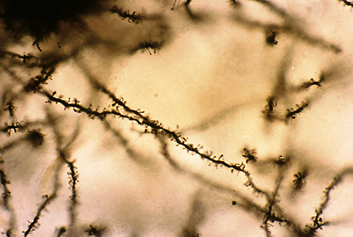
Spiny neurons have dendritic spines, while aspinous neurons do not (Bear, Connors, & Paradiso, 2026).
During learning, spines' number, size, and shape may change to adjust the space for receptors (neuroplasticity).
An axon is a cylindrical structure only found in neurons that is specialized for distributing information within the central and peripheral nervous systems. Axons range from 1 to 25 µm in diameter and 0.1 mm to more than a meter in length. Most neurons are interneurons, and many have axons and dendrites short enough that they do not extend beyond their local cell cluster. Axons usually branch repeatedly, and each branch is called an axon collateral.
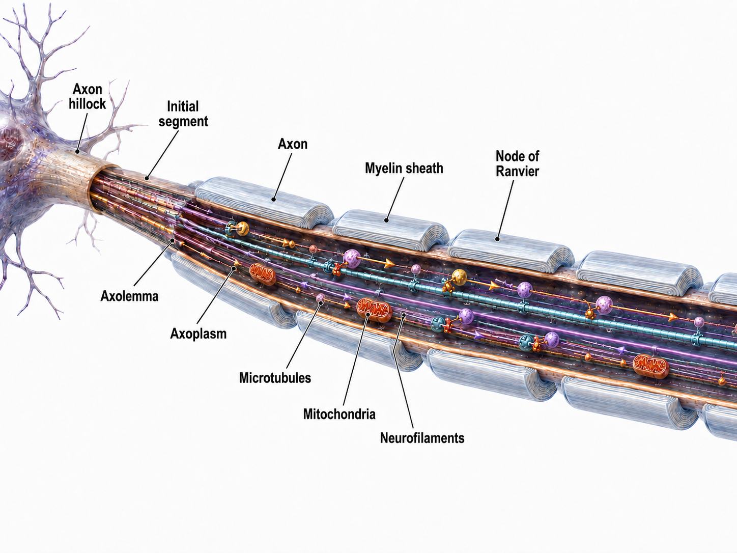
Axons transmit action potentials toward a neuron's terminal buttons. Using microtubules, an axon also bidirectionally transports molecules between the cell body and terminal buttons.
An axon hillock is a swelling of the cell body where the axon begins. Think of it as the neuron's decision point; it integrates all incoming signals to determine whether to fire. The middle of an axon is the axon proper, and the end is the axon terminal (Bear, Connors, & Paradiso, 2026).
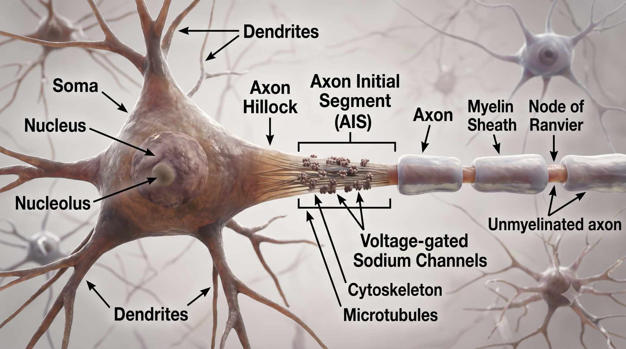
The axon hillock sums EPSPs and IPSPs over milliseconds to generate an action potential.
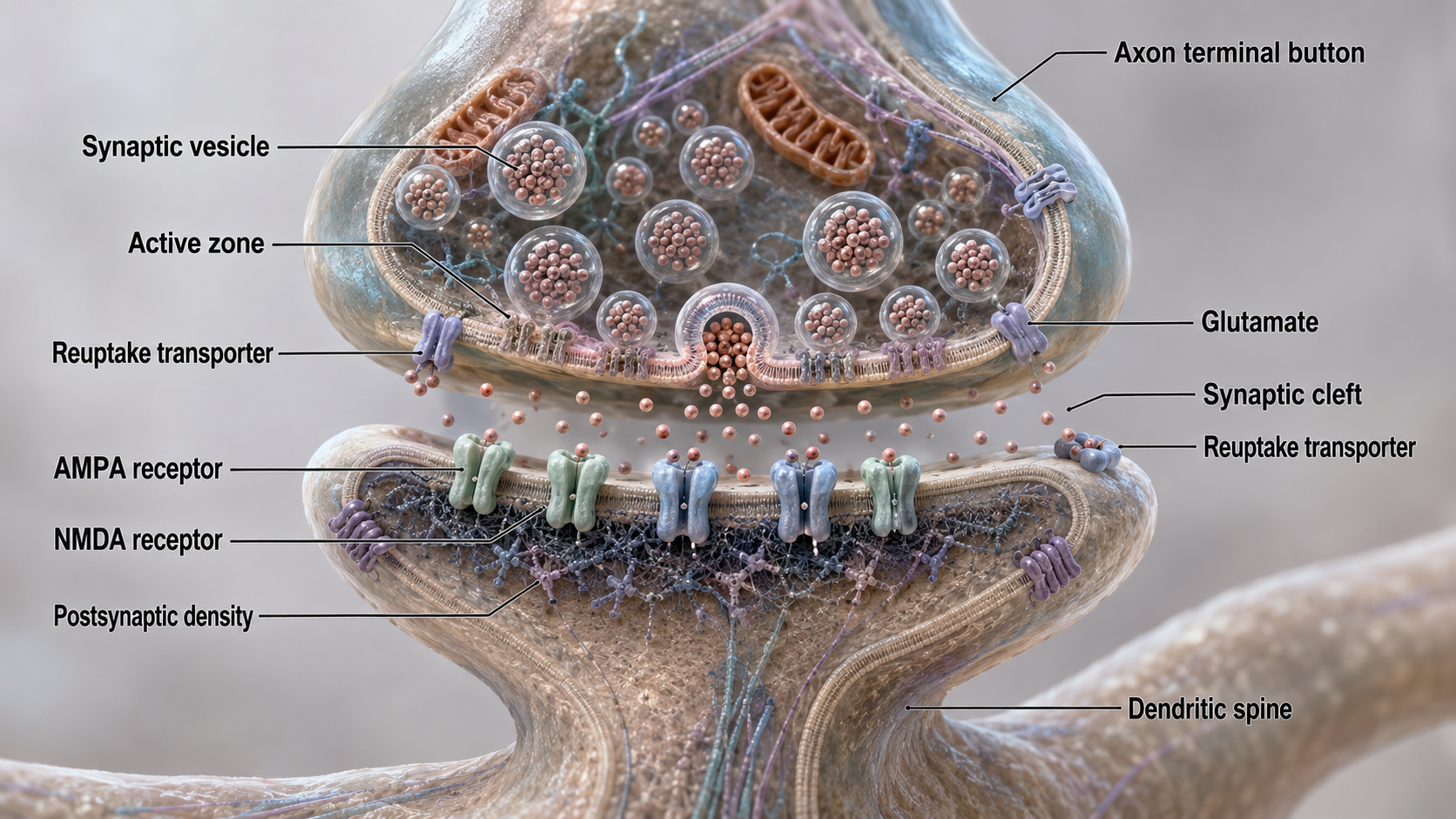
Axon terminals are buds located on the ends of axon branches that form synapses and release neurochemicals to other neurons. Axon terminals contain vesicles that store neurotransmitters for release when an action potential arrives. Their presynaptic membrane may have reuptake transporters that return neurotransmitters (NTs) from the synapse or extracellular space for repackaging.
Types of Glial Cells
This section covers the four main categories of glial cells: astrocytes, microglia, oligodendrocytes, and Schwann cells. Once dismissed as mere scaffolding, glial cells are now recognized as active partners in neural processing. Their role in modulating neuron excitability is directly relevant to understanding EEG generation and the mechanisms behind neurofeedback.
While there are hundreds of types of neurons, there are only four main categories of glial cells (astrocytes, microglia, oligodendrocytes, and Schwann cells).
Old school view: glial cells mainly provide structural support (glia is derived from the Greek for glue).
New school view: glial cells help neurons process information, including modulating neuron excitability.
Check out the YouTube video, Neurology - Glial Cells, White Matter and Gray Matter.
Astrocytes are star-shaped glial cells in the central nervous system that perform vital functions reaching well beyond structural support. Astrocyte endfeet form junctions with capillaries comprising part of the protective blood-brain barrier, and they regulate blood flow to neurons, delivering stored glucose during peak metabolic demand. Schummers et al. (2008) showed that cortical astrocytes carry tuned sensory responses and shape the hemodynamic signal, one route by which they take part in this coupling. Astrocyte graphic © Kateryna Kon/Shutterstock.com.
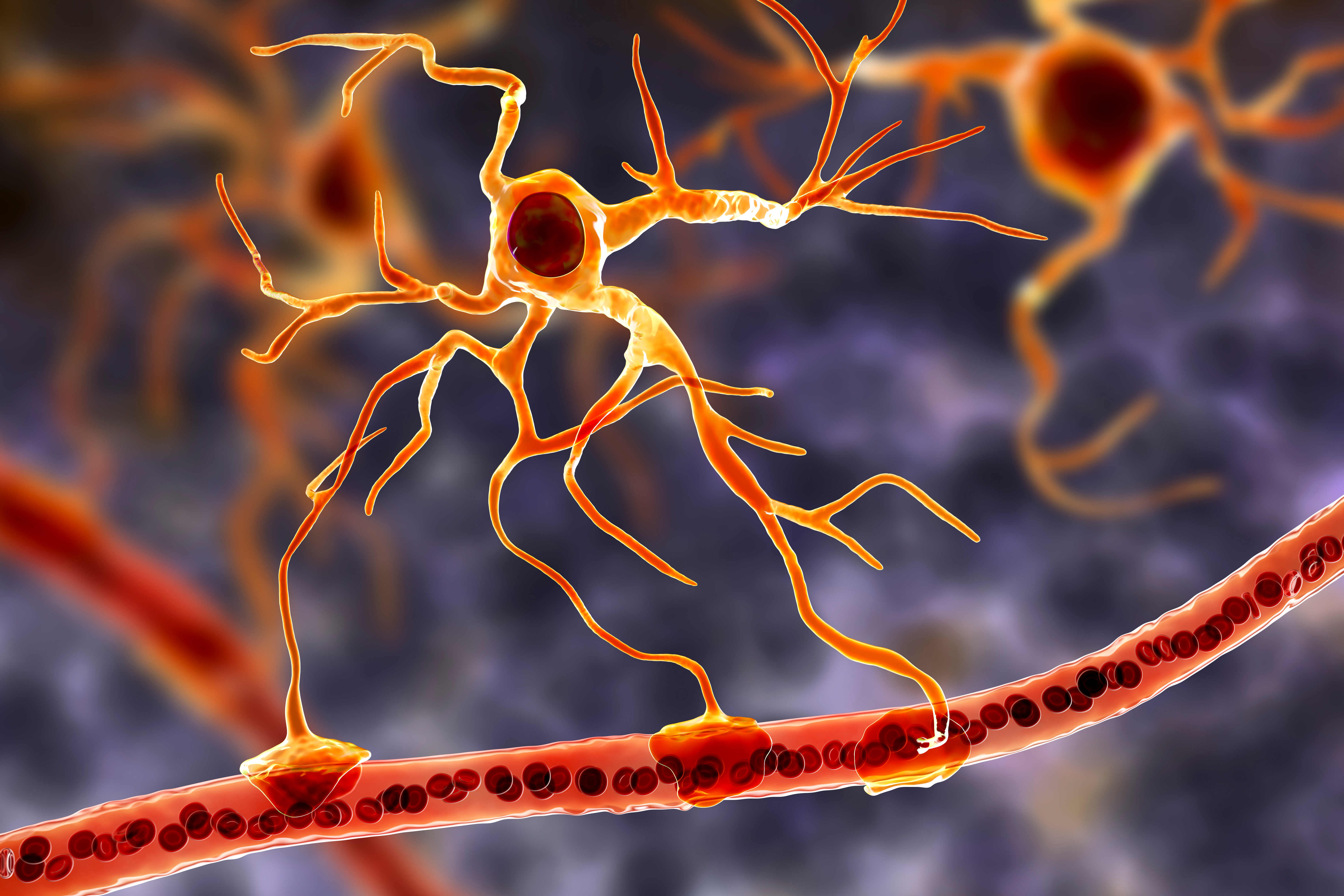
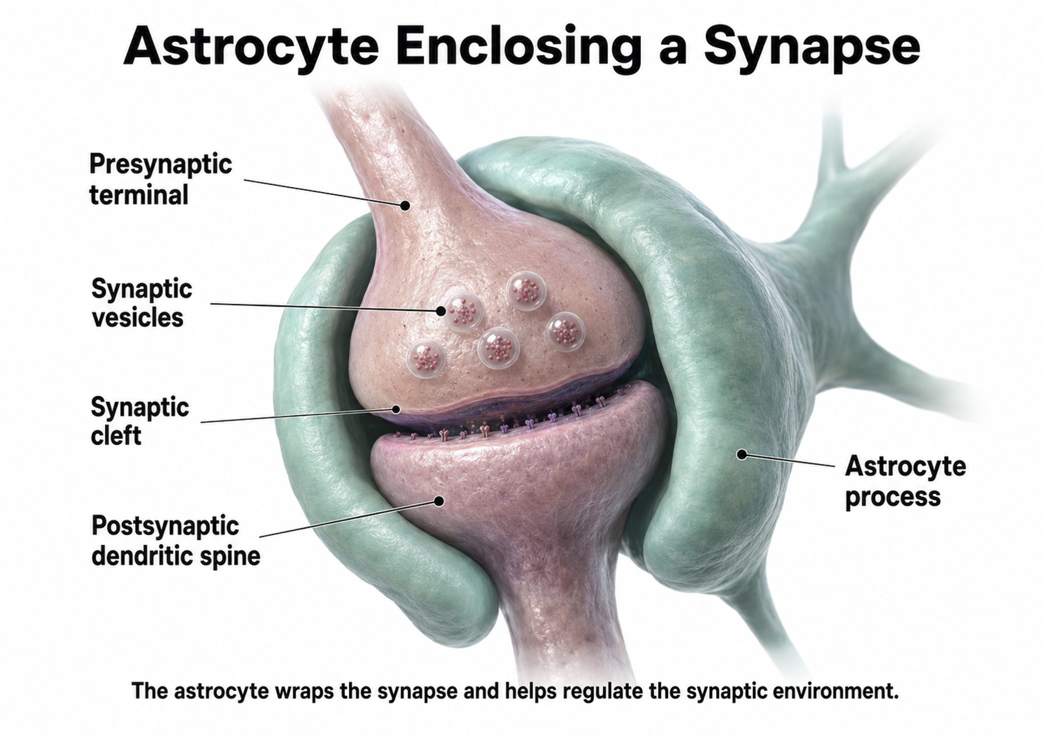
Astrocytes enclose synapses, determine where synapses can form by releasing specialized molecules, regulate synapse maturation, bidirectionally communicate with synapses, prune surplus synapses, help neurons regulate brain microcirculation, and eavesdrop on nearby synapse activity (Breedlove & Watson, 2023; Parri & Crunelli, 2003; Shan et al., 2021). Astrocytes also transport amino acid NTs (e.g., GABA and glutamate) from the synaptic cleft.
Astrocytes are theorized to participate in gliotransmission between neurons and each other (Eroglu & Barres, 2010; Perea et al., 2009). However, gliotransmission remains controversial.
. . . the physiological role of gliotransmission is highly debatable . . . as gliotransmitter release has been reliably demonstrated only in vitro in cultures and brain slice experiments that are often accompanied by manipulations (e.g., high frequency stimulation) which can affect astrocytic channels or receptors leading to impaired signaling cascades. This experimental design imposes questions about the existence of gliotransmission . . . and whether it plays a physiological role in the brain . . . (Buskila et al., 2019).
The presynaptic and postsynaptic neurons and astrocytes comprise a tripartite synapse, a three-part communication unit that expands our understanding of how neural signals are generated and modified. This concept is important for neurofeedback because it means the EEG signal reflects not just neuron-to-neuron communication, but a more complex interaction involving glial modulation.
An essential role of astrocytes is regulating the chemical content of this extracellular space. For example, astrocytes envelop synaptic junctions in the brain, thereby restricting the spread of neurotransmitter molecules that have been released. Astrocytes also have special proteins in their membranes that actively remove many neurotransmitters from the synaptic cleft. A recent and unexpected discovery is that astrocytic membranes also possess neurotransmitter receptors that, like the receptors on neurons, can trigger electrical and biochemical events inside the glial cell (Bear et al., 2020, p. 49).
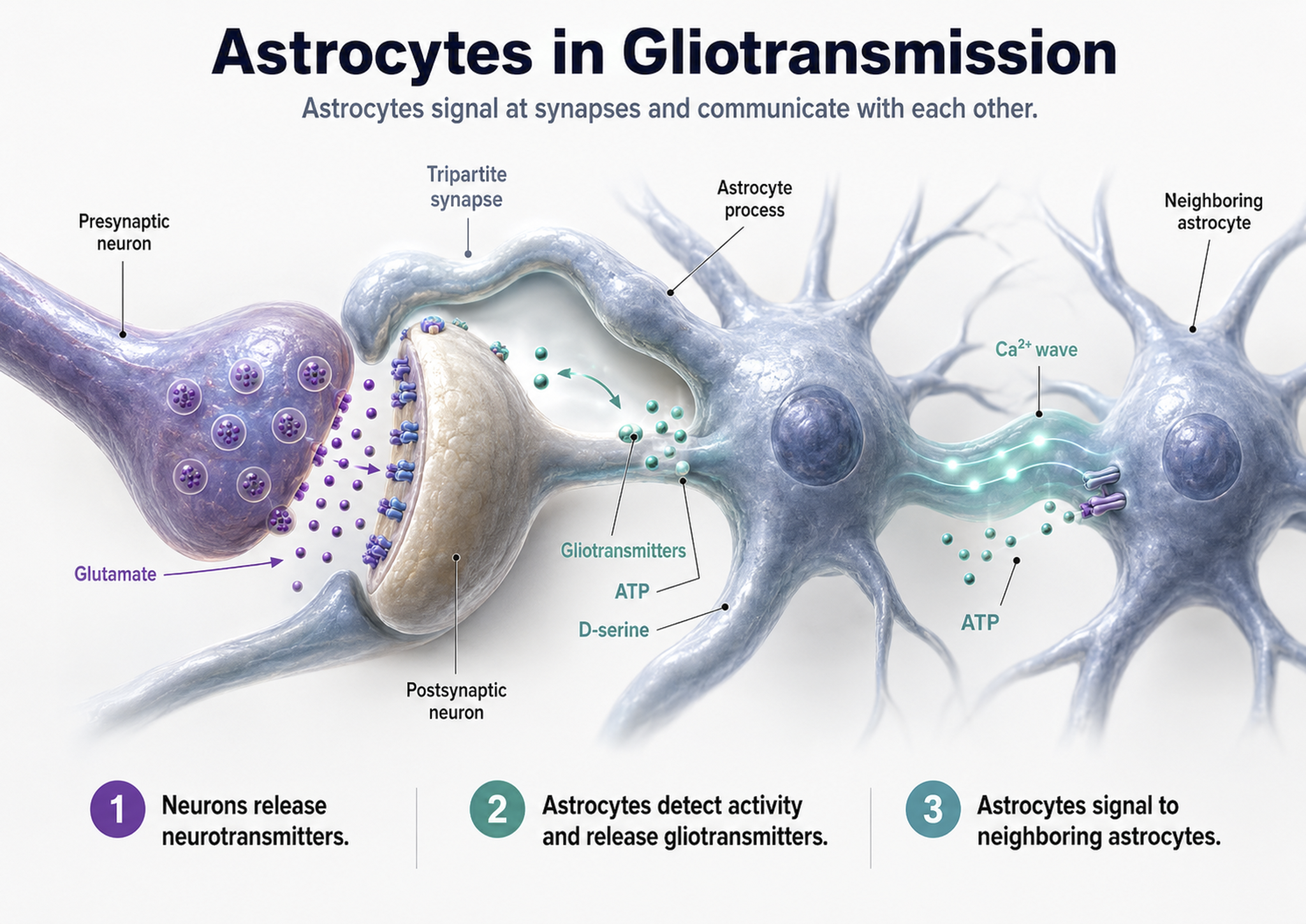
Astrocyte glutamate release may be essential for hippocampal long-term depression (LTD), a long-lasting reduction in transmission strength, and long-term memory modulation (Navarrete et al., 2019). Astrocyte calcium and brain-derived neurotrophic factor (BDNF) release appear critical for late-phase hippocampal long-term potentiation (LTP), a long-lasting increase in transmission strength, and long-term memory regulation (Liu et al., 2022).
Astrocytes communicate with each other through gap junctions (Bennett et al., 2003). They may also contribute to brainwaves by regulating synapses via these gap junctions and calcium signaling.
These capabilities allow astrocytes to regulate neuronal excitability via glutamate uptake, gliotransmission and tight control of the extracellular positively-charged potassium (K+) levels via a process termed K+ clearance. Spatio-temporal synchrony of activity across neuronal and astrocytic networks, both locally and distributed across cortical regions, underpins brain states and thereby behavioral states, and it is becoming apparent that astrocytes play an important role in the development and maintenance of neural activity underlying these complex behavioral states (Buskila et al., 2019).
Microglia
Microscopic microglial cells participate in the immune response and are the brain's resident defense force. They scavenge and engulf diverse materials (phagocytosis), release cytotoxins to control infection, present antigens to T-cells, remove branches from neurons near damaged tissue to aid regrowth (synaptic stripping), promote tissue repair, and if they are persistently activated, they can promote chronic neuroinflammation in the CNS that amplifies neurodegeneration. They assist synaptic remodeling by removing unnecessary synapses. Finally, microglia cross the blood-brain barrier to promote homeostasis (Bear, Connors, & Paradiso, 2026). Graphic © Juan Gaertner/Shutterstock.com.
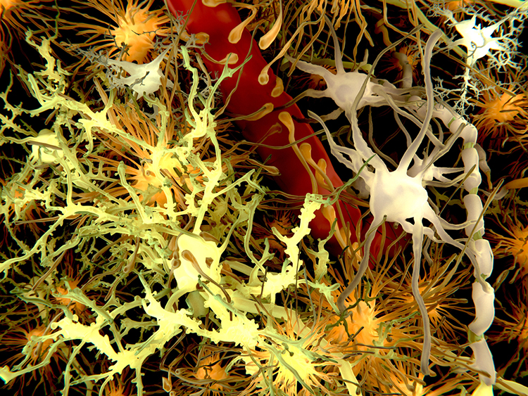
Description: yellow = neurons, orange = astrocytes, grey = oligodendrocytes, white = microglia.
Oligodendrocytes, which are smaller than astrocytes, form up to 50 segments of myelin that only insulate adjacent axons within the brain and spinal cord of the central nervous system.
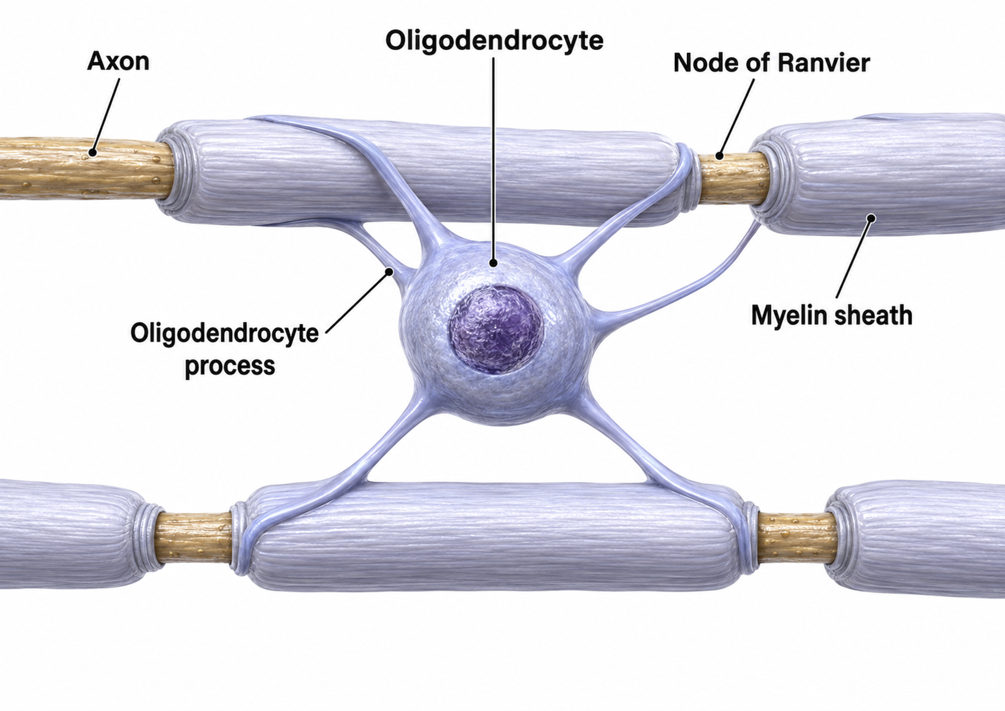
Oligodendrocytes block axonal regeneration by releasing growth inhibitory proteins, which partly explains the minimal functional recovery in the CNS following spinal cord damage.
Multiple sclerosis, a demyelinating disease, destroys oligodendrocytes, disrupting neural communication in ways that clinicians can sometimes detect through QEEG assessment.
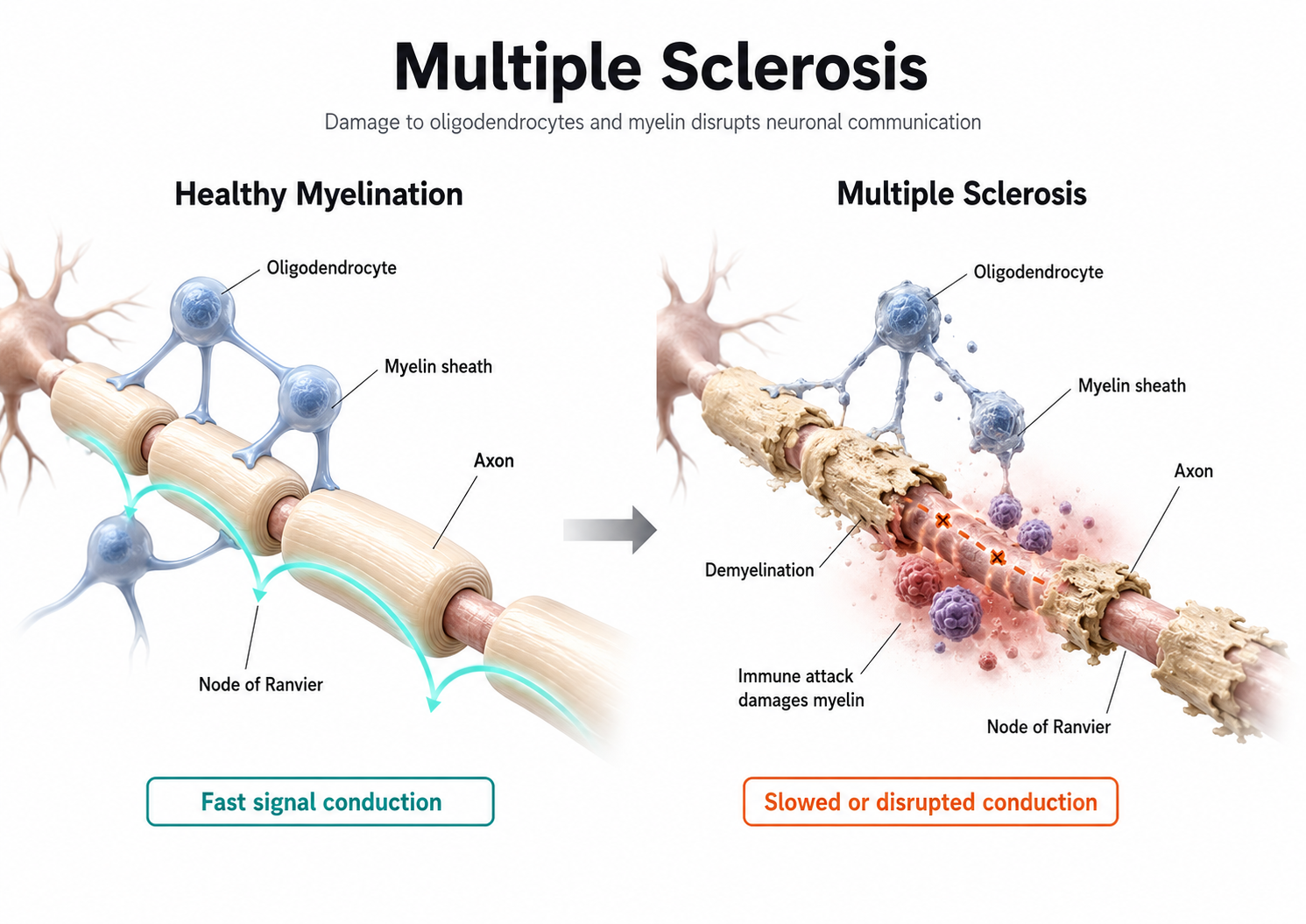
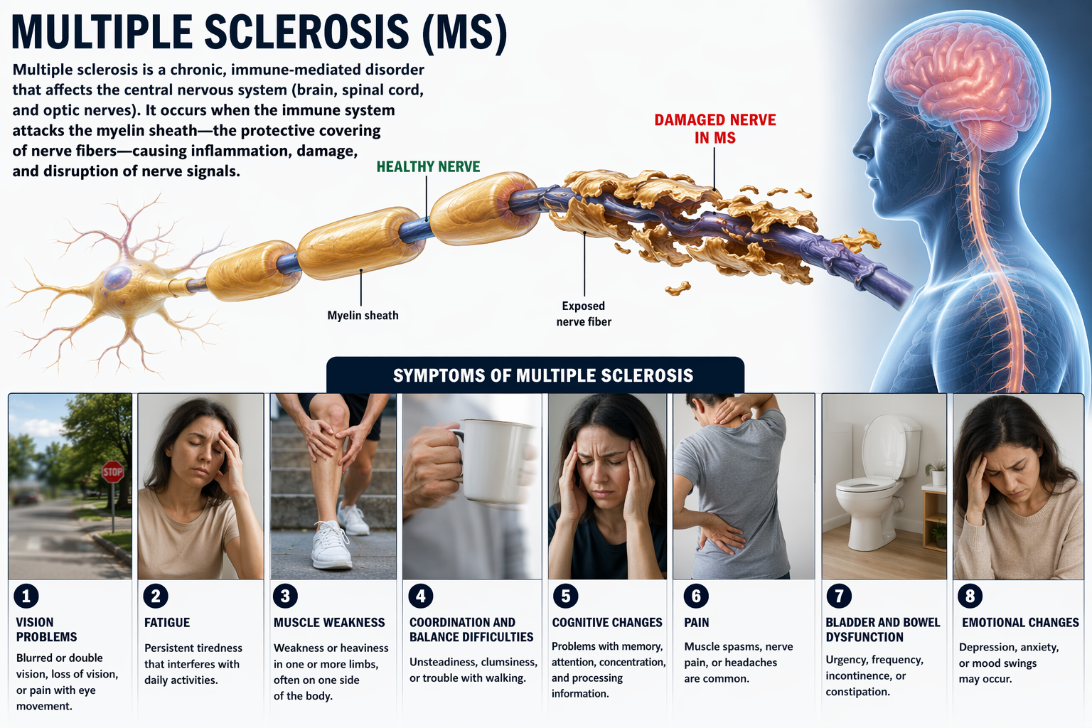
Schwann cells provide myelin for single peripheral nervous system (PNS) axons and facilitate axonal regeneration following damage (Breedlove & Watson, 2023).
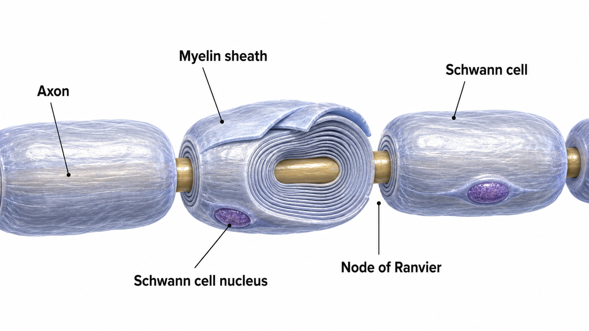
The Resting Membrane Potential
This section explains the resting membrane potential, the steady electrical charge a neuron maintains when it is not signaling. Understanding the resting potential is foundational because every postsynaptic potential, action potential, and ultimately the EEG itself is a deviation from this baseline. When clinicians watch a neurofeedback display, they are watching the summed consequences of millions of neurons departing from and returning to this resting state.
The resting membrane potential is the voltage difference across a neuron's membrane at rest, typically about -70 mV, with the inside of the cell more negative than the outside. This charge separation exists because ions are distributed unequally across the membrane. Sodium (Na+) and chloride (Cl-) ions are concentrated outside the neuron, while potassium (K+) ions and large negatively charged proteins are concentrated inside (Bear, Connors, & Paradiso, 2026). This uneven distribution is the ion concentration gradient that stores the energy driving neural signaling.
Two forces hold the resting potential in place: selective permeability and active transport. Selective permeability means the resting membrane is far more permeable to potassium than to sodium because potassium leak channels stay open. Potassium therefore drifts out of the cell down its concentration gradient, leaving the interior more negative until the growing electrical attraction pulling potassium back inward balances the chemical push driving it outward. The membrane voltage at which these two forces cancel for a given ion is its equilibrium potential, and because the resting membrane is dominated by potassium, the resting potential sits close to potassium's equilibrium value (Breedlove & Watson, 2023).
Left alone, the small constant leak of ions would gradually erase these gradients. The sodium-potassium pump prevents this by using ATP to exchange three sodium ions pushed out of the cell for every two potassium ions brought in, continuously restoring the gradients that the resting potential depends on. This relentless transport is one reason neurons are such heavy energy consumers and why the mitochondria described earlier cluster where this demand is greatest (Breedlove & Watson, 2023).
Because the resting potential is actively maintained, anything that limits a neuron's energy supply degrades it. Hypoxia, ischemia, and severe hypoglycemia starve the sodium-potassium pump, the gradients run down, and cortical signaling slows. This is the physiological reason such states produce diffuse slowing in the EEG, a pattern clinicians may encounter when a client's medical status, medication, or substance use compromises cortical metabolism.
Excitatory and Inhibitory Postsynaptic Potentials
This section explains the small graded potentials, EPSPs and IPSPs, that are the primary source of the EEG signal recorded at the scalp. Understanding these potentials is essential because they, not action potentials, are what neurofeedback clinicians measure during neurofeedback sessions.
Graded positive and negative changes in membrane potential, called excitatory postsynaptic potentials and inhibitory postsynaptic potentials, are essential to the EEG and communication among neurons.
An excitatory postsynaptic potential (EPSP) is a subthreshold depolarization that makes the membrane potential more positive and pushes the neuron toward its excitation threshold. EPSPs are produced when neurotransmitters bind to receptors and cause positive sodium ions to enter the cell. At a single synapse, a postsynaptic membrane may have tens to thousands of transmitter-gated ion channels, and the amount of transmitter released determines how many of these channels will be activated. The size of an EPSP will be a multiple of the number of vesicles, each containing several thousand transmitter molecules.
An inhibitory postsynaptic potential (IPSP) is a hyperpolarization that makes the membrane potential more negative and pushes the neuron away from its excitation threshold. At most inhibitory synapses, IPSPs are produced when neurotransmitters like GABA or glycine bind to receptors and cause negative chloride ions to enter the cell. When an inhibitory synapse is closer to the soma than an excitatory synapse, it can counteract positive current flow and decrease the size of the EPSP, a mechanism called shunting inhibition (Bear, Connors, & Paradiso, 2026). The balance between EPSPs and IPSPs at any moment determines whether a neuron fires, making this push-pull dynamic the fundamental language of the EEG.
Integrating Postsynaptic Potentials
Integration is the summation of EPSPs and IPSPs at the unmyelinated axon hillock, the neuron's decision-making junction.
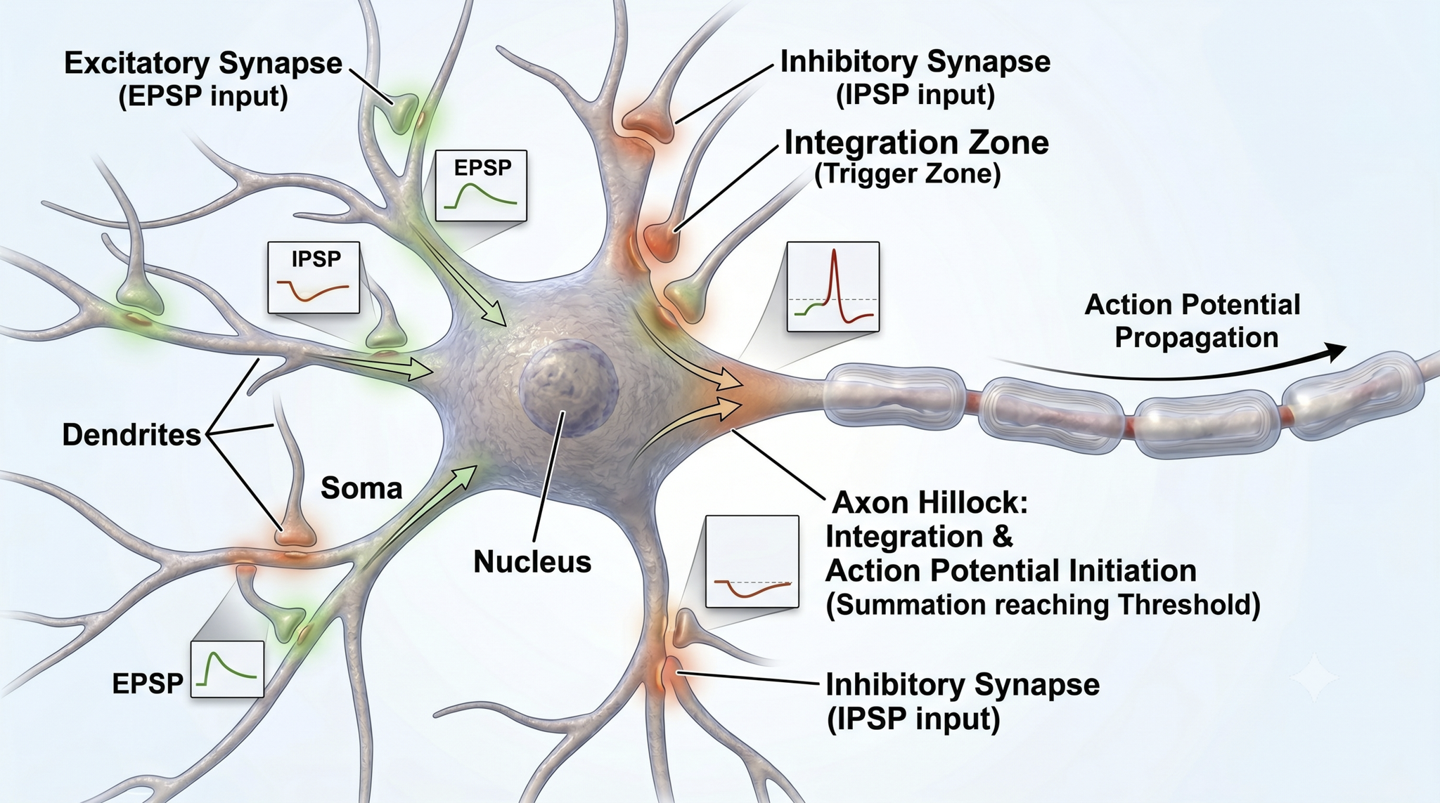
The axon hillock of a postsynaptic neuron uses two methods to sum EPSPs and IPSPs: spatial and temporal summation.
In spatial summation, the axon hillock sums the simultaneous postsynaptic potentials (PSPs) from thousands of synapses on dendrites.
In temporal summation, the axon hillock adds the PSPs from presynaptic neurons that repeatedly fire within a 1-15-ms time window.
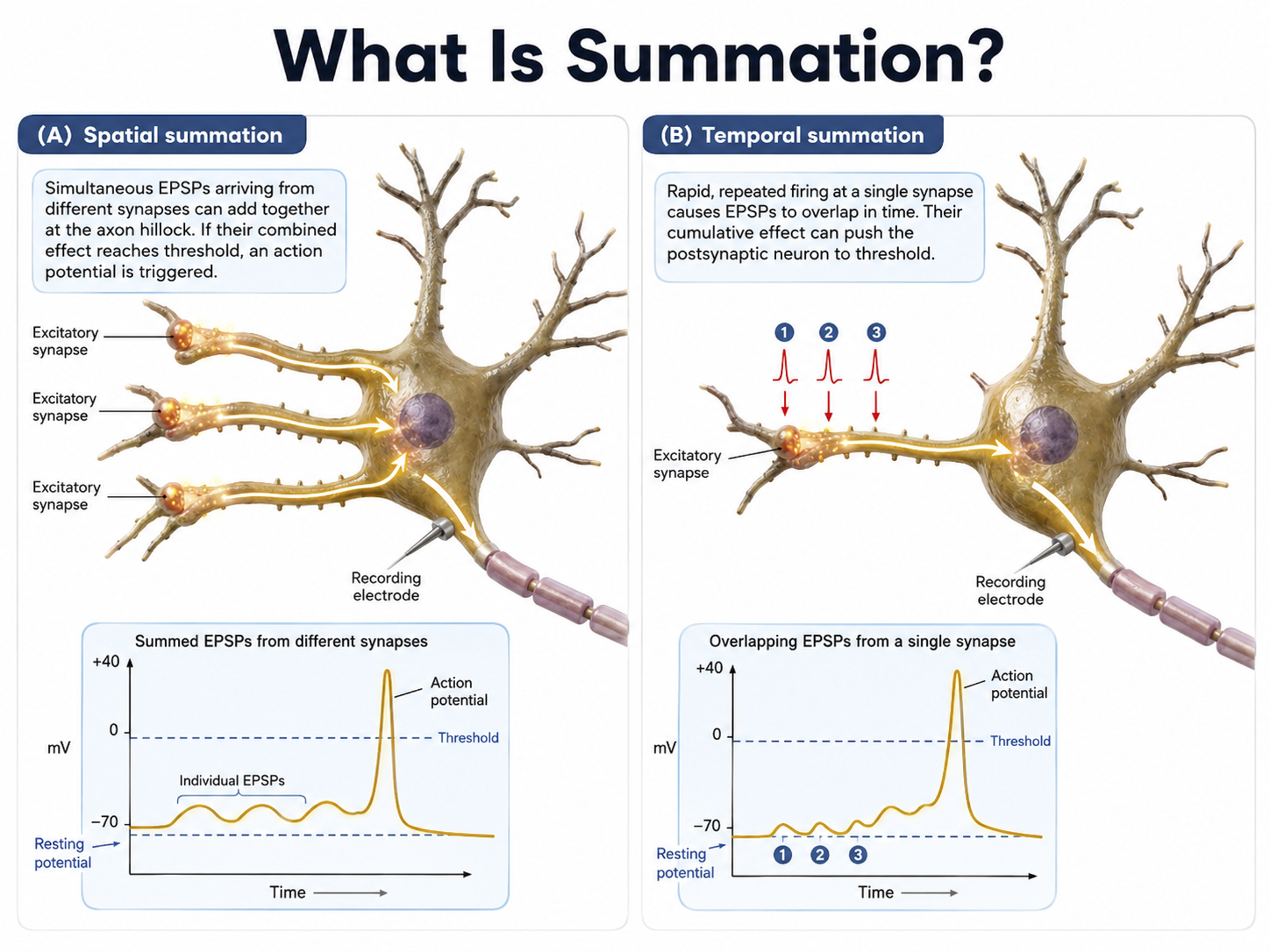
Each EPSP depolarizes the axon hillock by about 0.5 mV. If there were no competing IPSPs, it would take about 30 EPSPs to trigger an action potential. Each IPSP hyperpolarizes the axon hillock by about 0.5 mV. If the summated EPSPs and IPSPs move the axon hillock from a resting potential of -70 mV to a threshold of excitation of -55 mV, sodium channels in the axon hillock membrane open, and an action potential propagates down the axon. Graphic adapted from © 2003 Josephine Wilson.
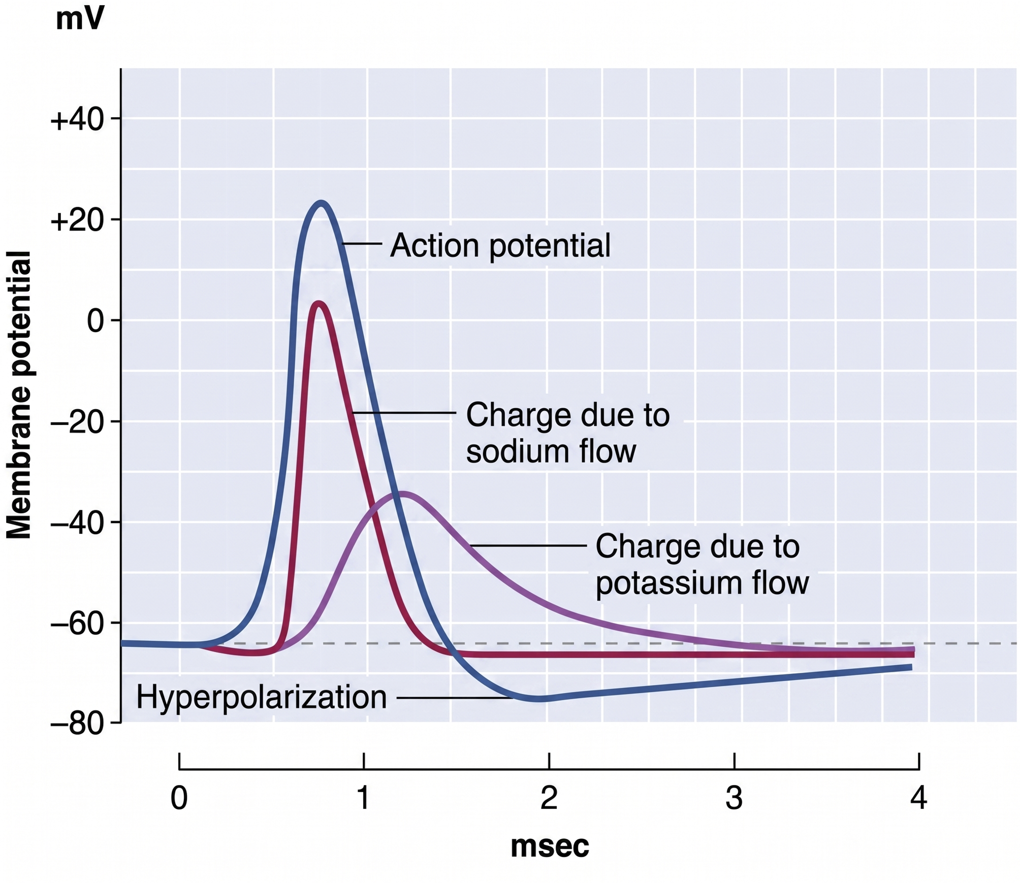
Check out the YouTube video, Best Action Potential Explanation.
Action Potentials
This section covers how action potentials transmit signals along axons and explains two key principles, the all-or-none law and the rate law, along with the two modes of conduction: unmyelinated and myelinated. Understanding conduction speed and efficiency explains why demyelinating diseases like multiple sclerosis are so devastating.
An action potential is a brief electrical impulse that transmits information from the axon hillock to the terminal button. This wave of positive charge only travels in one direction because the preceding segment is refractory due to the closing of its sodium channels. An action potential takes 1-2 ms from the point the axon hillock reaches its threshold to its repolarization to a negative resting potential.
The action potential unfolds as a rapid, self-reinforcing sequence of ion movements through voltage-gated ion channels, channels that open and close in response to changes in membrane voltage. When summation drives the axon hillock to threshold, voltage-gated sodium channels open and sodium rushes in, depolarizing the membrane toward +30 mV in the rising phase. Within about a millisecond these sodium channels inactivate while voltage-gated potassium channels open, allowing potassium to leave and repolarize the membrane in the falling phase. A brief overshoot of potassium efflux produces a short hyperpolarization before the resting potential is restored (Bear, Connors, & Paradiso, 2026).
Because the sodium channels remain inactivated during the absolute refractory period, the membrane cannot fire again no matter how strong the input, which forces the impulse to travel in one direction and sets an upper limit on firing rate. During the following relative refractory period, a stronger-than-usual input is required to fire the neuron, the mechanism that allows the rate law described below to encode stimulus intensity (Breedlove & Watson, 2023).
Action potentials travel down axons, which branch multiple times and terminate at synapses. The all-or-none law and rate law describe action potential transmission. The all-or-none law states that once an action potential is triggered in an axon, it is propagated, without decrement, to the end of the axon.
The rate law states that neurons represent the intensity of a stimulus by variation in the rate of axon firing. More intense stimuli shorten the interval before a neuron can fire again, allowing it to fire more rapidly; an intense stimulus can cause a neuron to fire every 2 or 3 ms, while a weak stimulus might lengthen the time lag to every 4 or 5 ms. Action potential graphic © extender_01/Shutterstock.com.
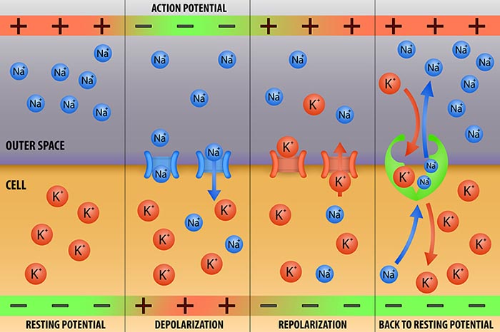
We can compare action potential conduction to the movement of water through a leaky garden hose.
Garden hose: water can take two paths, inside the hose or through holes in its wall, and the majority of the water will flow where movement is easiest. For a small-diameter hose with many large holes, most of the water will travel through the leaks. Conversely, for a large-diameter hose with only a few small holes, the bulk of the water will remain inside.
Axon: positive charge can take two paths, inside the axon or through pores in its membrane. Like water, a positive charge will take the path of least resistance. For a small-diameter axon with many open sodium ion channels, the majority of the current will exit the axonal membrane to the extracellular fluid. Small diameter, unmyelinated axons transmit action potentials without weakening since sodium ion channels constantly regenerate this signal. This method is slow because the signal travels step-by-step, small segment by small segment, and waits for sodium channels to admit enough positive ions to reach the excitation threshold.
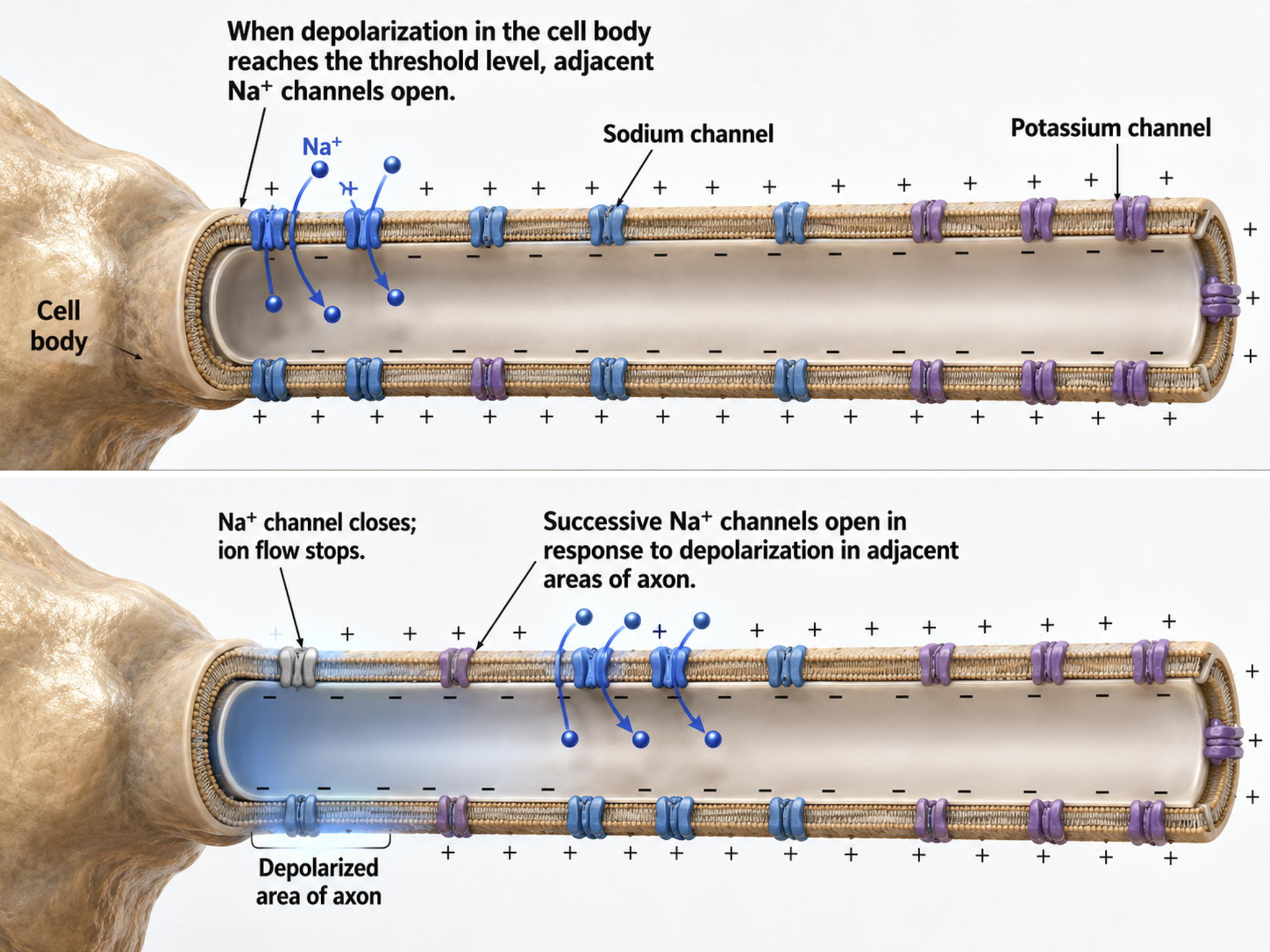
This method also consumes considerable energy since sodium-potassium transporters, powered by ATP, are located across the axon membrane to exchange three sodium for two potassium ions.
Conversely, for a large-diameter axon with few open ion channels, the bulk of the current will remain inside the axon's interior. Wider spacing between adjacent ion channels means that the action potential can depolarize a longer axon segment, which increases conduction velocity (Bear, Connors, & Paradiso, 2026).
Medium-to-large diameter myelinated axons transmit action potentials using a method called saltatory conduction. Each segment of insulating myelin is almost 1-mm long, while the gaps between segments, called nodes of Ranvier, are 1 to 2 thousandths of a millimeter. An action potential weakens under each myelinated segment (cable properties) and is then regenerated at each Ranvier node. The destruction of this insulation by demyelinating diseases like multiple sclerosis (MS) can be devastating because it disrupts neuron-to-neuron communication.
Saltatory conduction can be 200 times faster because the action potential jumps from node to node in 1-mm steps, instead of steps that are a thousand times smaller. This method is also more energy-efficient because sodium-potassium transporters are only needed at the nodes of Ranvier, where ion exchange is possible. These transporters account for about 40% of a neuron's energy expenditure (Breedlove & Watson, 2023; Garrett, 2003).
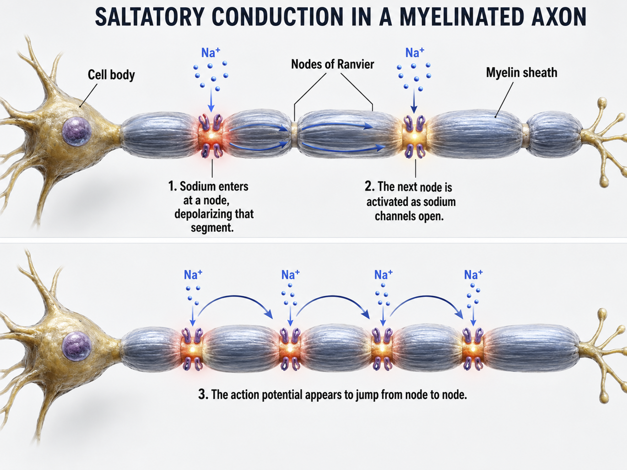
In summary, the type of neuron, the presence or absence of myelin, and the integrity of that myelin sheath all determine how quickly and efficiently neural signals travel, factors that directly affect the EEG patterns clinicians observe during assessment and training.
Neurons come in three functional classes, sensory, motor, and interneurons, and they work in partnership with glial cells that actively modulate signaling rather than merely supporting it. Each neuron maintains a resting membrane potential of about -70 mV that the sodium-potassium pump preserves at high metabolic cost. Graded excitatory and inhibitory postsynaptic potentials summate at the axon hillock, and when they reach the threshold of excitation an all-or-none action potential travels down the axon. Myelination and axon diameter determine how quickly and efficiently that signal propagates, which is why demyelinating disease disrupts communication. These postsynaptic potentials, not the action potentials, are the primary source of the scalp EEG.
Check Your Understanding
- How do sensory neurons, motor neurons, and interneurons differ in the direction and purpose of the information they carry?
- Why does the sodium-potassium pump make neurons such heavy energy consumers, and what happens to the EEG when a neuron's energy supply is compromised?
- How do spatial and temporal summation at the axon hillock determine whether a neuron reaches its threshold of excitation?
- Why does saltatory conduction in myelinated axons increase both speed and energy efficiency compared with conduction in unmyelinated axons?
- Why does the scalp EEG reflect postsynaptic potentials rather than action potentials?
Synaptic Transmission
This section explains how neurons communicate across chemical synapses, including neurotransmitter co-release, extra-synaptic transmission, modulation, and the major neurotransmitter families and pathways. These mechanisms are central to understanding how the brain generates the electrical activity that neurofeedback clinicians measure and train.
Neurons communicate through the release of over 200 neurochemicals and ions. Axon terminal buttons release neurochemicals across a 20-50-nm fluid-filled gap between presynaptic and postsynaptic structures called a synaptic cleft and into the extracellular fluid surrounding the neuron (Bear et al., 2020). Chemical synapses produce short-duration (millisecond) and long-duration (seconds to days) changes in the nervous system. Synapse animation without sound © 3Dme Creative Studio/Shutterstock.com.
Chemical synapses are functionally asymmetrical because the presynaptic neuron sends a chemical message and the postsynaptic neuron receives it. They are structurally asymmetrical because the presynaptic element (axon) contains vesicles containing NTs, and the postsynaptic element (dendrite) does not. NT release from a terminal button is called exocytosis (Breedlove & Watson, 2023). Synapse graphic adapted from © SciePro/Shutterstock.com.
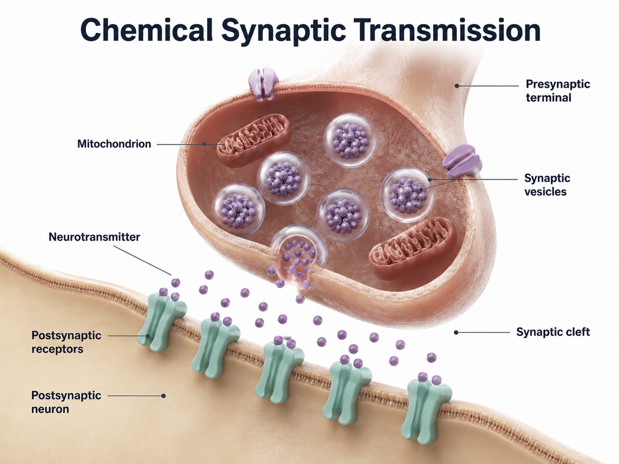
In the graphic below, an axon terminal button releases NTs into the synaptic cleft. NTs briefly bind to receptors on a dendritic spine through reversible, noncovalent interactions and then disengage after they initiate small graded potential changes (e.g., EPSPs or IPSPs) or more diverse, gradual, and long-lived actions (e.g., creating second messengers inside the target neuron). For clinicians, this distinction matters: fast ionotropic effects contribute to the moment-to-moment EEG, while slower metabotropic effects drive the long-term changes associated with neurofeedback training outcomes.
Ionotropic receptors open an ion pore directly. When a neurotransmitter binds to the receptor, the channel opens and ions flow rapidly across the membrane, producing fast and short-lived effects. Metabotropic receptors act more slowly. Instead of opening a channel themselves, they trigger a sequence of metabolic steps that activate a second messenger system inside the neuron, producing slower and longer-lasting changes. Chemical synapse graphic © nobeastsofierce/Shutterstock.com.
.jpg)
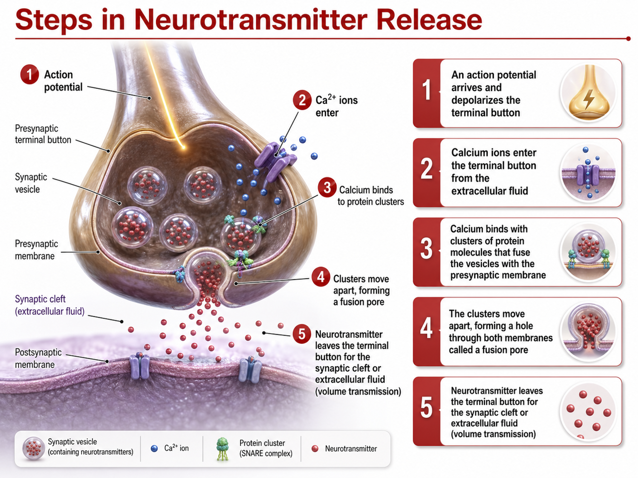
Neurotransmitter Co-Release
Old-school view: according to Dale's law, a neuron can only release one NT at a synapse.
New-school view: neurons can release a classical NT and a peptide.
Dale's law proposed that a neuron releases only one NT. However, researchers have found increasing evidence of NT co-release (Svensson et al., 2019). A neuron can store different NTs in separate types of vesicles (Hökfelt et al., 2003). Neurons can also store multiple NTs in the same vesicles (e.g., ATP and glutamate), although they may not release them simultaneously (Merighi et al., 2011; Xia et al., 2009). Co-release adds a layer of complexity to synaptic communication; a single neuron can send multiple chemical messages, producing nuanced effects on its postsynaptic partners.
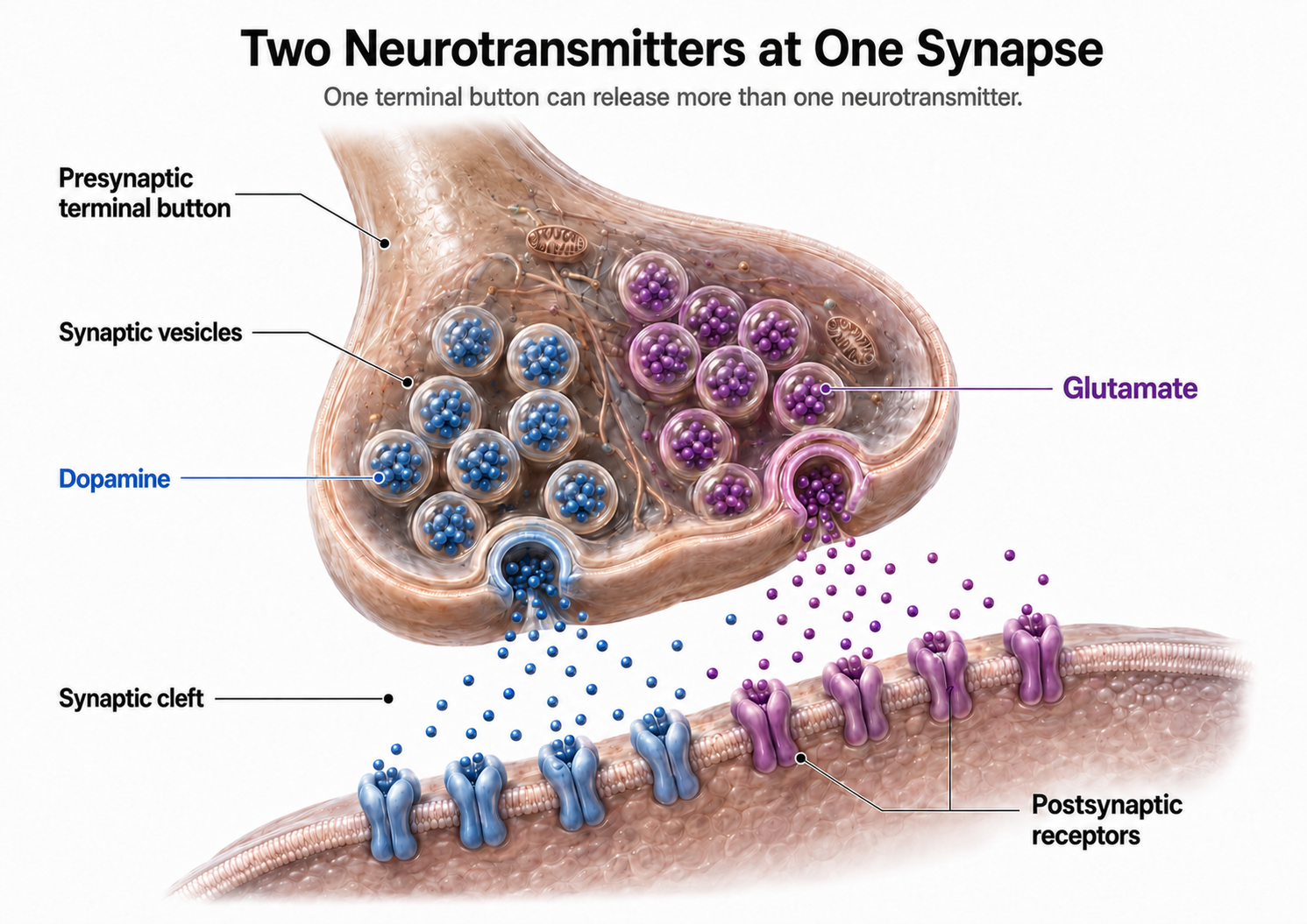
Extra-Synaptic Transmission: Think Outside the Cleft
This section covers three mechanisms by which neurons release neurotransmitters outside of classical synapses: volume transmission, axonal varicosities, and retrograde transmission. These extra-synaptic pathways help explain how neurotransmitters influence broad regions of the brain, not just the neurons immediately across a synaptic cleft.
Neurons release NTs outside of classical synapses. These mechanisms include release from terminal buttons into the extracellular space, axonal varicosities, and retrograde transmission.
Old-school view: axon terminals only release NTs into the synaptic cleft.
New-school view: NT release also occurs outside of the synaptic cleft. Axonal varicosities (swellings in axon walls), dendrites, and the terminal button can release NTs into the extracellular space. Graphic © 3Dme Creative Studio/Shutterstock.com.
Volume Transmission
Volume transmission involves NT release and eventual binding to a receptor outside the synaptic cleft (Coggan et al., 2005). Do not confuse this process with volume conduction, which is the spread of an electrical signal when measured at some distance from its source. Graphic adapted from the American Scientist.
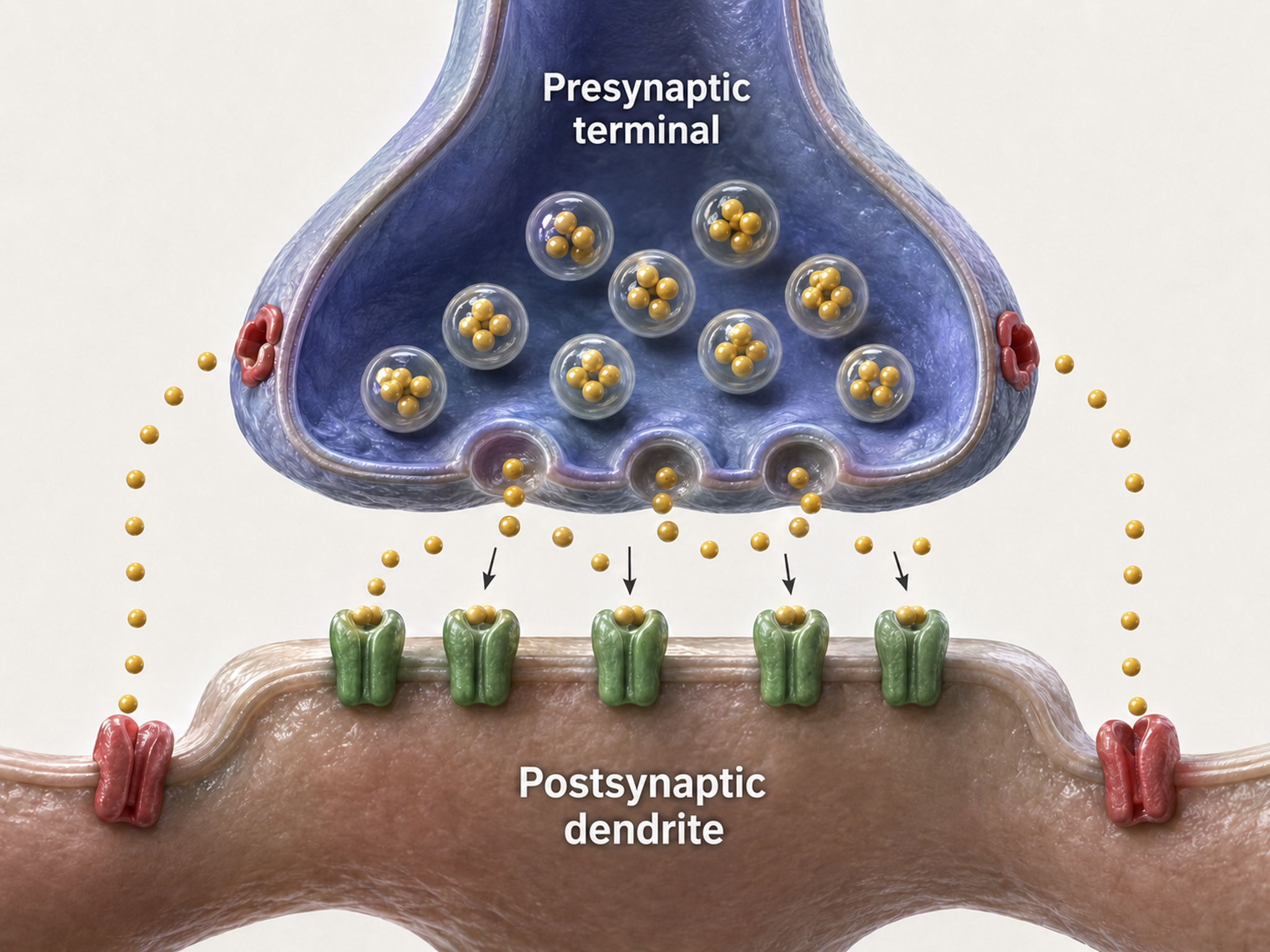
Axonal Varicosities
Axons can release NTs into the extracellular space through varicosities (swellings) along their length, analogous to drip irrigation (Breedlove & Watson, 2023).
Most neurons that release norepinephrine do not do so through terminal buttons on the ends of axonal branches. Instead, they usually release them through axonal varicosities, beadlike swellings of the axonal branches (Carlson & Birkett, 2019, pp. 82-83).
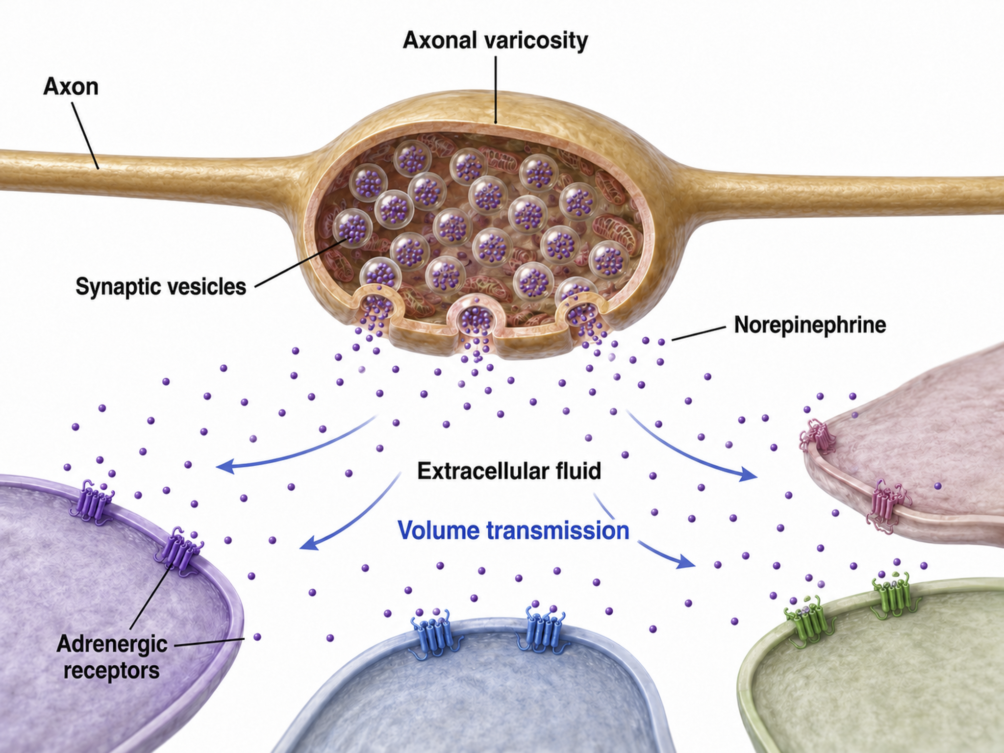
Retrograde Transmission
In retrograde transmission, a presynaptic neuron sends a chemical message to the postsynaptic neuron. In response, the postsynaptic neuron synthesizes and distributes an endocannabinoid (e.g., anandamide) or gas (e.g., nitric oxide, or NO) to the presynaptic neuron and its immediate active neighbors. Neurons synthesize these NTs on demand since they cannot be contained by vesicles. This backward signaling allows the postsynaptic neuron to fine-tune the messages it receives, a feedback loop that plays an important role in synaptic plasticity.
. . . this gaseous signal has a range of influence that extends well beyond the cell of origin, diffusing a few tens of micrometers from its site of production before it is degraded. This property makes NO a potentially useful agent for coordinating the activities of multiple cells in a localized region and may mediate certain forms of synaptic plasticity that spread within small networks of neurons (Purves, 2017, pp. 142-143).
Retrograde NTs can bind to membrane-bound receptors or diffuse into the target cell, initiating second messenger production to adjust synaptic efficiency in learning and memory (Breedlove & Watson, 2023).
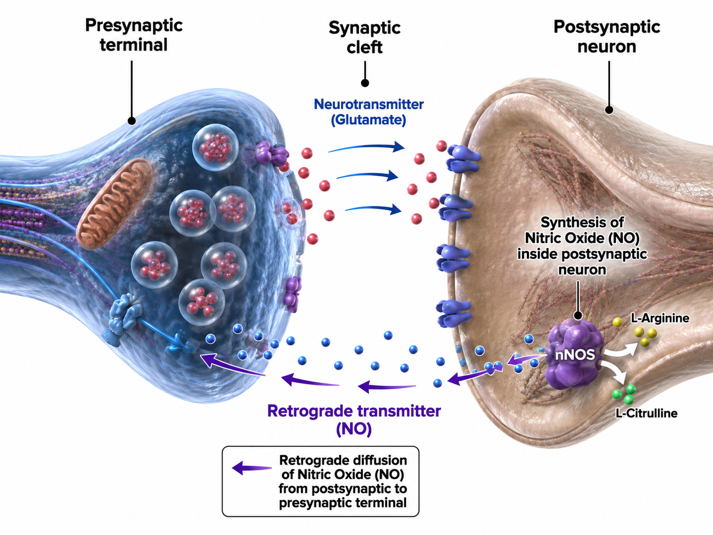
Modulation
This section explains how the nervous system fine-tunes its signals through modulation of neurotransmitter release and receptor activity. Modulation is analogous to a volume control knob on a stereo preamplifier rather than a simple on/off switch; it provides analog adjustment rather than digital switching.
We will consider two of countless modulation mechanisms: modulation of NT release and modulation of NT action at its receptor.
Neurotransmitter Release Modulation
Axons can influence the amount of NTs released when an action potential arrives at an axon terminal through axoaxonic synapses (junctions between two axons).
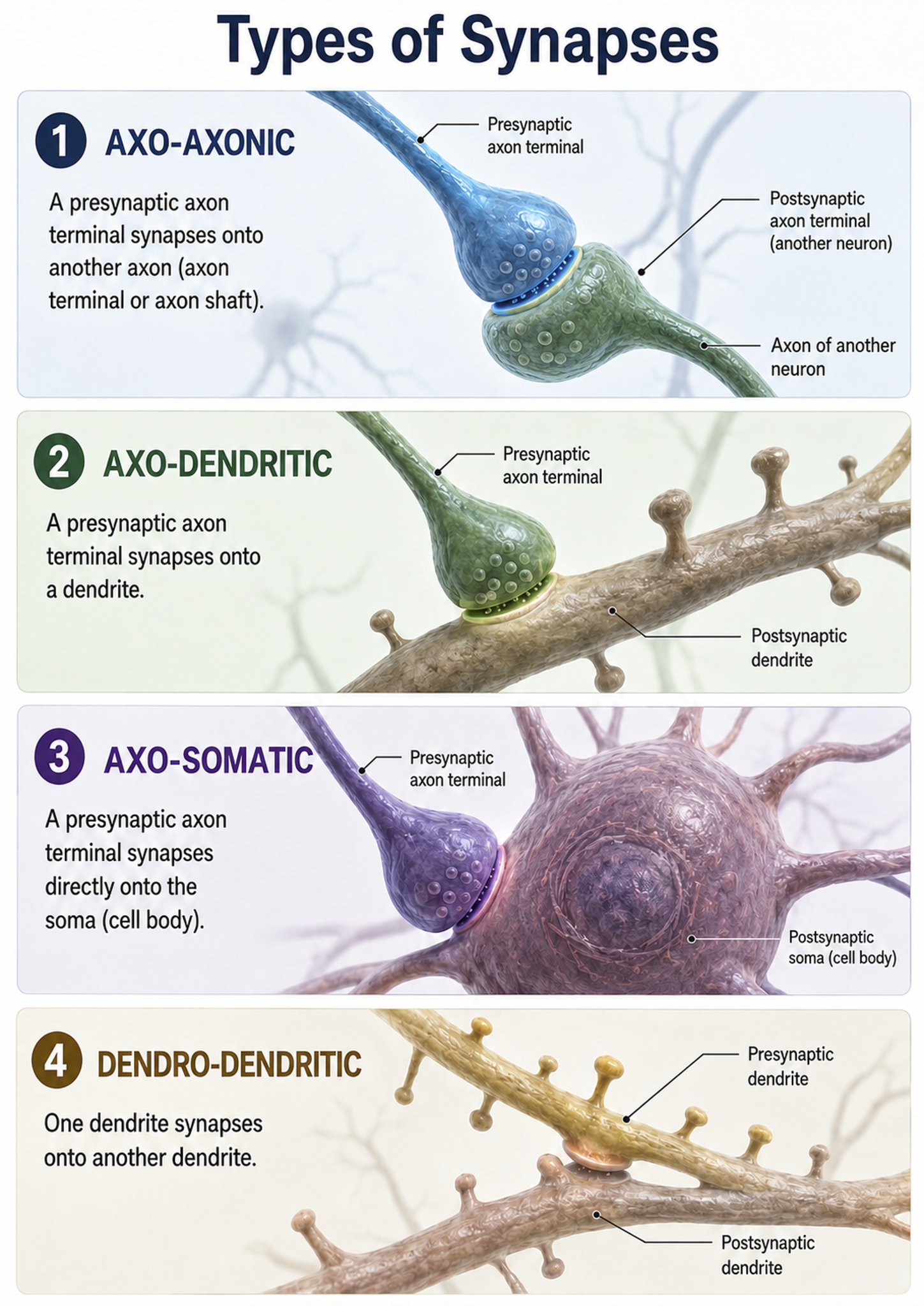
Axoaxonic synapses do not affect the generation of an action potential, only the amount of neurotransmitter distributed. In presynaptic facilitation, a neuron increases the presynaptic neuron's neurotransmitter release by delivering a neurotransmitter that increases calcium ion entry into its terminal button. In presynaptic inhibition, a neuron decreases neurotransmitter release by reducing calcium ion entry. These modulatory effects are confined to a single synapse (Breedlove & Watson, 2023).
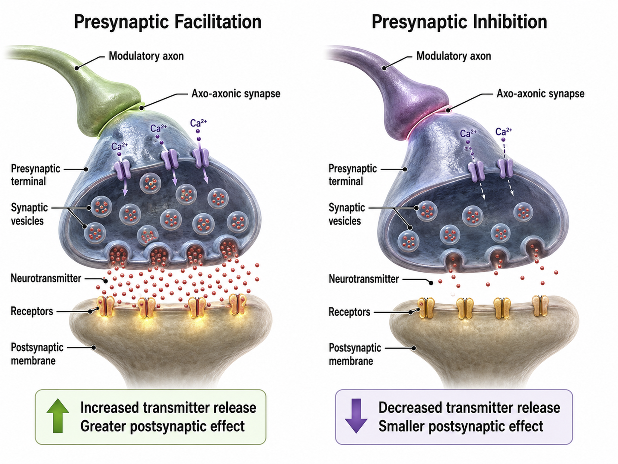
Autoreceptors Modulate Neurotransmitter Release
Autoreceptors are metabotropic receptors on the presynaptic membrane that function as a built-in feedback system. When NTs released into the synaptic cleft bind to autoreceptors, this hyperpolarizes the axon terminal button so it will release less NT when the next action potential arrives. This self-regulation prevents excessive neurotransmitter release, a mechanism that many psychotropic medications exploit.
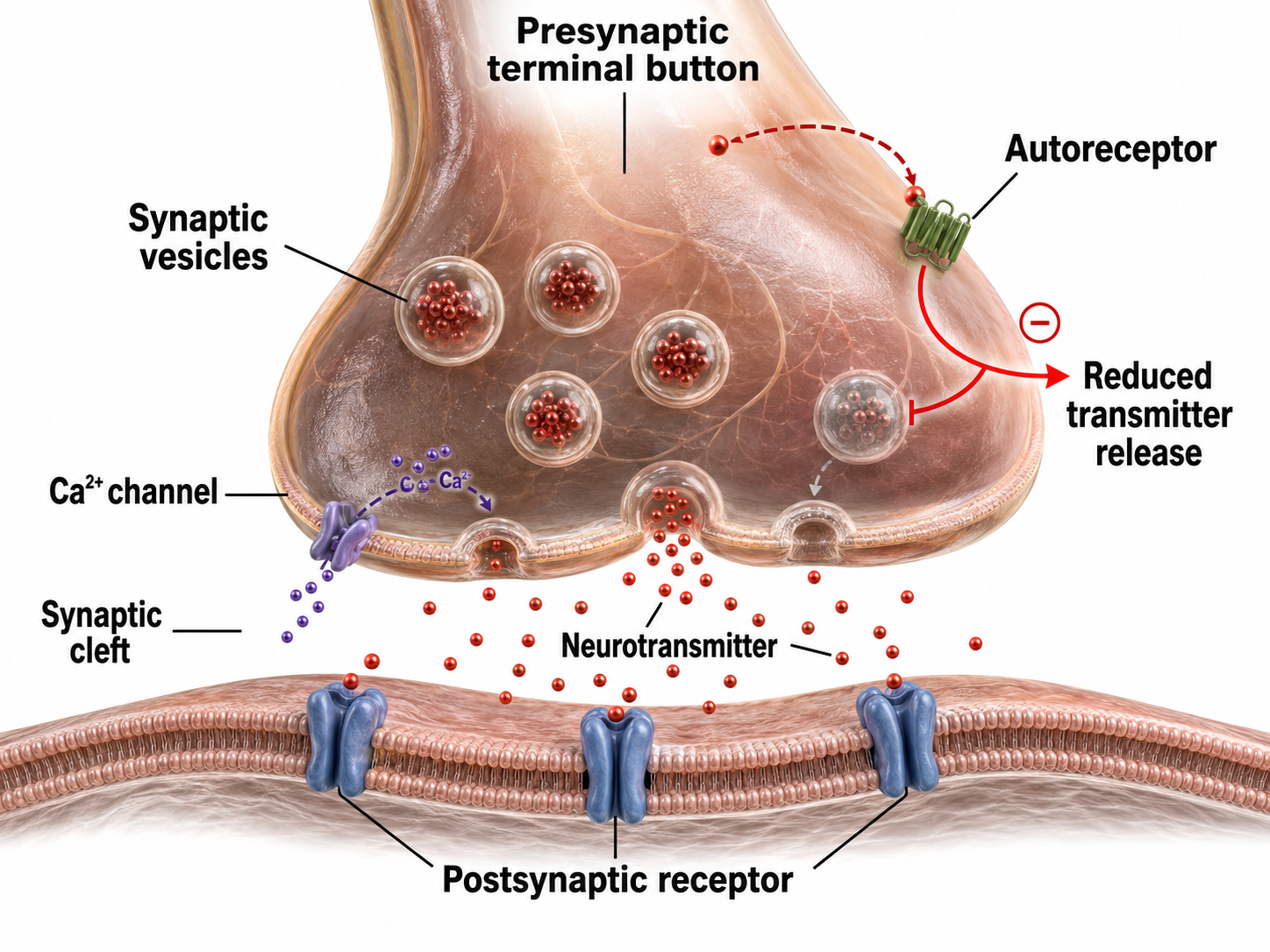
Neuromodulators Adjust Neurotransmitter Action
A postsynaptic neurotransmitter receptor contains two functionally distinct binding sites. The orthosteric site is the primary location where the neurotransmitter binds, and it behaves like an on-off switch that activates the receptor. The allosteric site is a separate, secondary location where modulators bind. Rather than switching the receptor on or off, it acts like a volume control, adjusting how strongly the receptor responds when a neurotransmitter occupies the orthosteric site.
Receptors contain binding sites for drugs like alcohol, ions, or endogenous molecules. When alcohol binds to its allosteric site, it strengthens GABA's binding at its orthosteric site, causing greater chloride entry into the neuron and increasing its hyperpolarization. Ingesting multiple CNS depressants (e.g., alcohol and barbiturates) can yield dangerous additive effects, amplifying GABA's action to a level that can depress or stop breathing. This is why clinicians conducting neurofeedback assessments should always inquire about substance use; CNS depressants will alter the EEG patterns observed during recording.
Types of Neurotransmitters
While the actual number of NTs is not known, more than 200 molecules have been identified. Each neurotransmitter may have multiple receptors, and a NT's effect, excitatory or inhibitory, depends on its interaction with specific receptors. The same NT can produce opposite results at different receptor subtypes (Breedlove & Watson, 2023).
The principal NT families include amino acid neurotransmitters (GABA, glutamate), amine neurotransmitters (acetylcholine, dopamine, serotonin), peptide neurotransmitters, also called neuropeptides (oxytocin, vasopressin), gas neurotransmitters or gasotransmitters (nitric oxide, carbon monoxide, hydrogen sulfide), purine neurotransmitters (ATP, adenosine), and lipid neurotransmitters (anandamide and 2-arachidonoylglycerol, or 2-AG). We adapted the table below from Breedlove and Watson (2023).
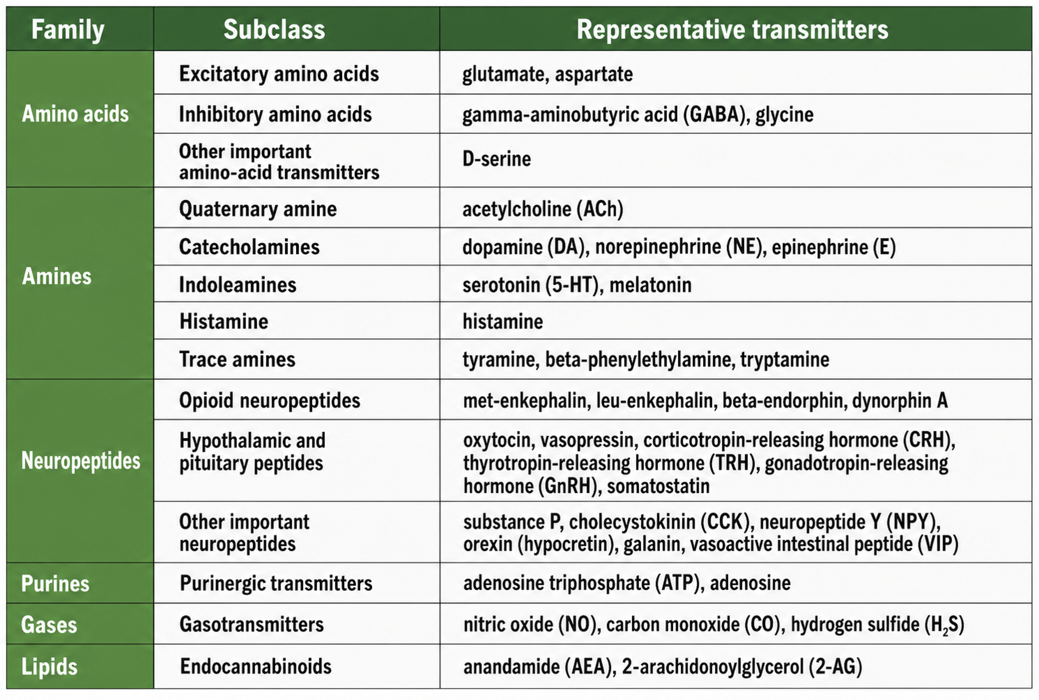
Neurotransmitter Pathways
Researchers have identified distinct pathways for acetylcholine, dopamine, norepinephrine, and serotonin. Understanding these pathways is critical for neurofeedback practitioners because they explain how specific neurotransmitter systems influence the EEG patterns targeted in training protocols. The reproduced diagrams are adapted from © Vasilisa Tsoy/Shutterstock.com.
Cholinergic pathways
Cholinergic cell bodies and their projections originate in the basal forebrain and brainstem. Cholinergic pathways are involved in arousal, attention, memory, motivation, muscle contraction, and sleep.
Dopaminergic pathways
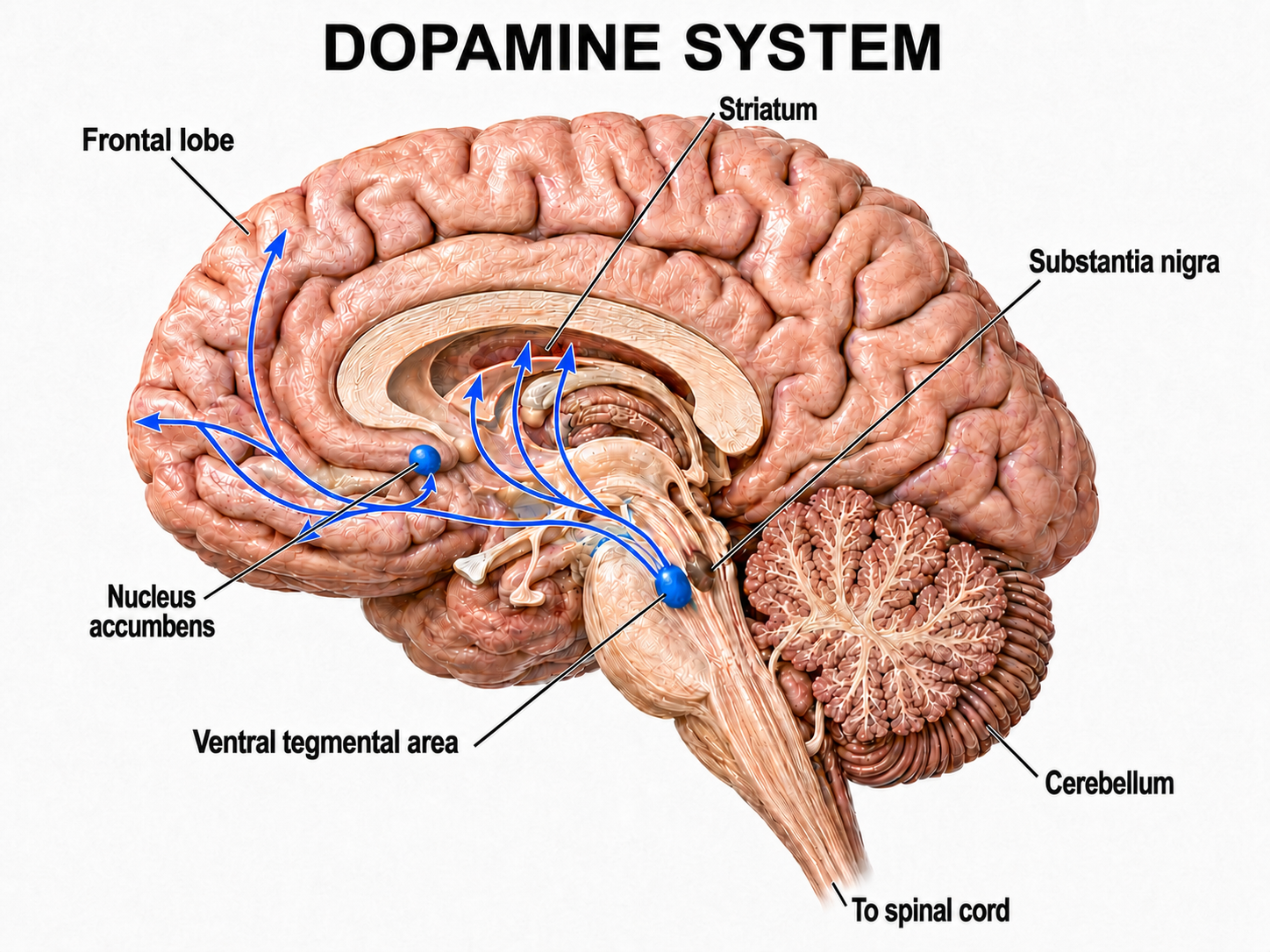
Two major dopaminergic pathways originate in the midbrain: the mesostriatal and mesolimbocortical pathways. Dopaminergic pathways are involved in addiction, motor control, and salience (reward- and threat-based motivation). These pathways are particularly relevant in neurofeedback because the dopamine reward system is engaged when clients receive positive feedback during training.
Noradrenergic pathways
The noradrenergic pathways originate in the locus coeruleus of the dorsal pons and in the lateral tegmental cell groups of the pons and medulla. Noradrenergic pathways are involved in arousal, attention, memory, vigilance, sleep, and mobilizing the brain and body for action, including the fight-or-flight response. Dysregulation of these pathways is commonly seen in PTSD and anxiety disorders treated with neurofeedback.
Serotonergic pathways
The serotonergic pathways originate in the brainstem and midbrain raphe nuclei. Serotonergic pathways are involved in appetite, mood, and sleep.
Termination of Neurotransmitter Action
Following exocytosis, which is NT release from a presynaptic terminal vesicle, NT action is terminated by two main mechanisms: reuptake and enzymatic degradation. In reuptake, reuptake transporters located in the presynaptic terminal and astrocytes that enclose the synapse return NT molecules to the presynaptic neuron. This is the mechanism that SSRI antidepressants block; by inhibiting serotonin reuptake, they prolong serotonin's action in the synaptic cleft. Astrocytes remove glutamate from the synapse.
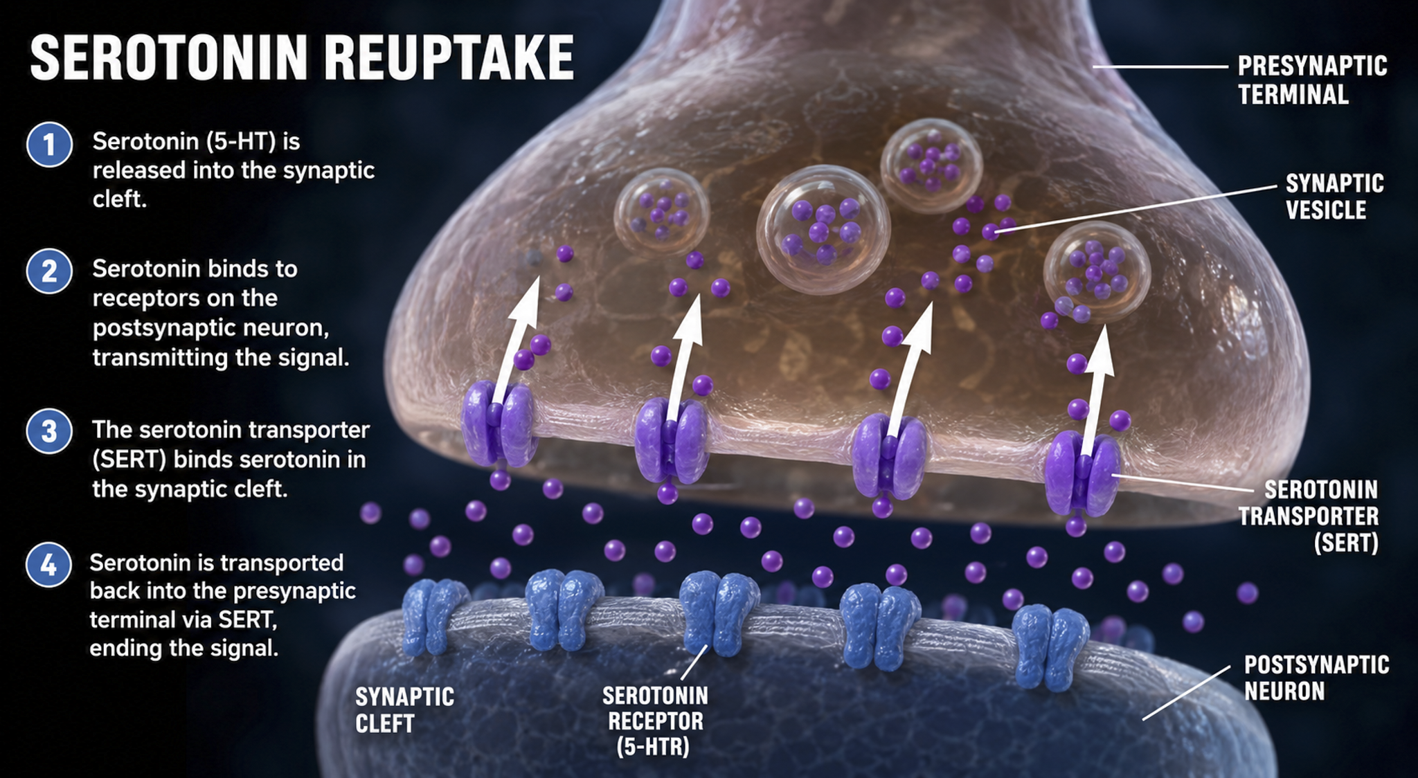
In enzymatic degradation, enzymes located in the synaptic cleft and the cytoplasm of the presynaptic neuron's terminal button split neurotransmitter molecules apart (e.g., acetylcholine).
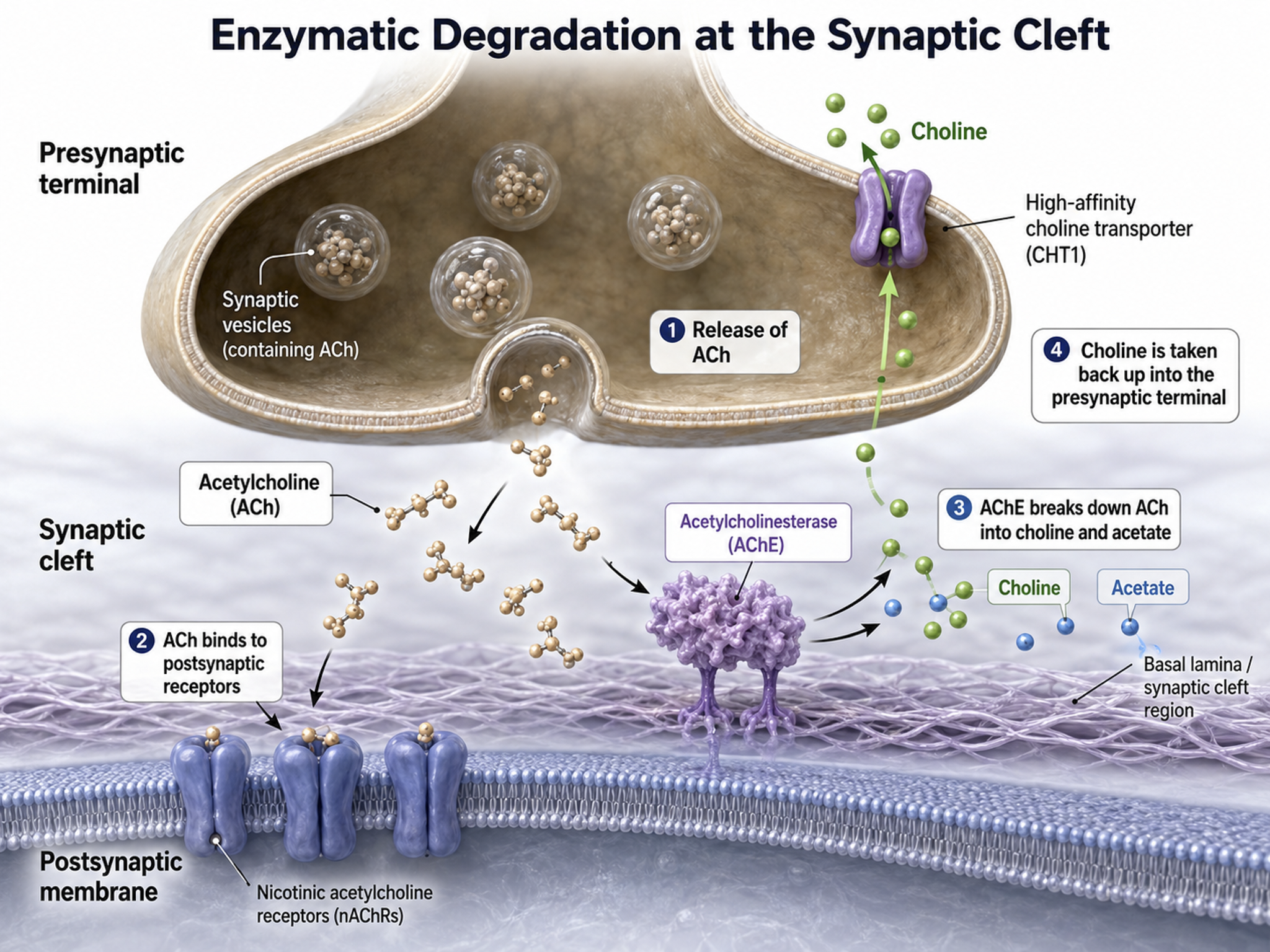
Electrical Synapses
Electrical synapses communicate information across gap junctions between adjacent membranes using ions. Gap junctions are narrow spaces between two cells bridged by connexons (protein channels) that allow ions near-instantaneous travel. Gap junction illustration adapted from © VectorMine/Shutterstock.com.
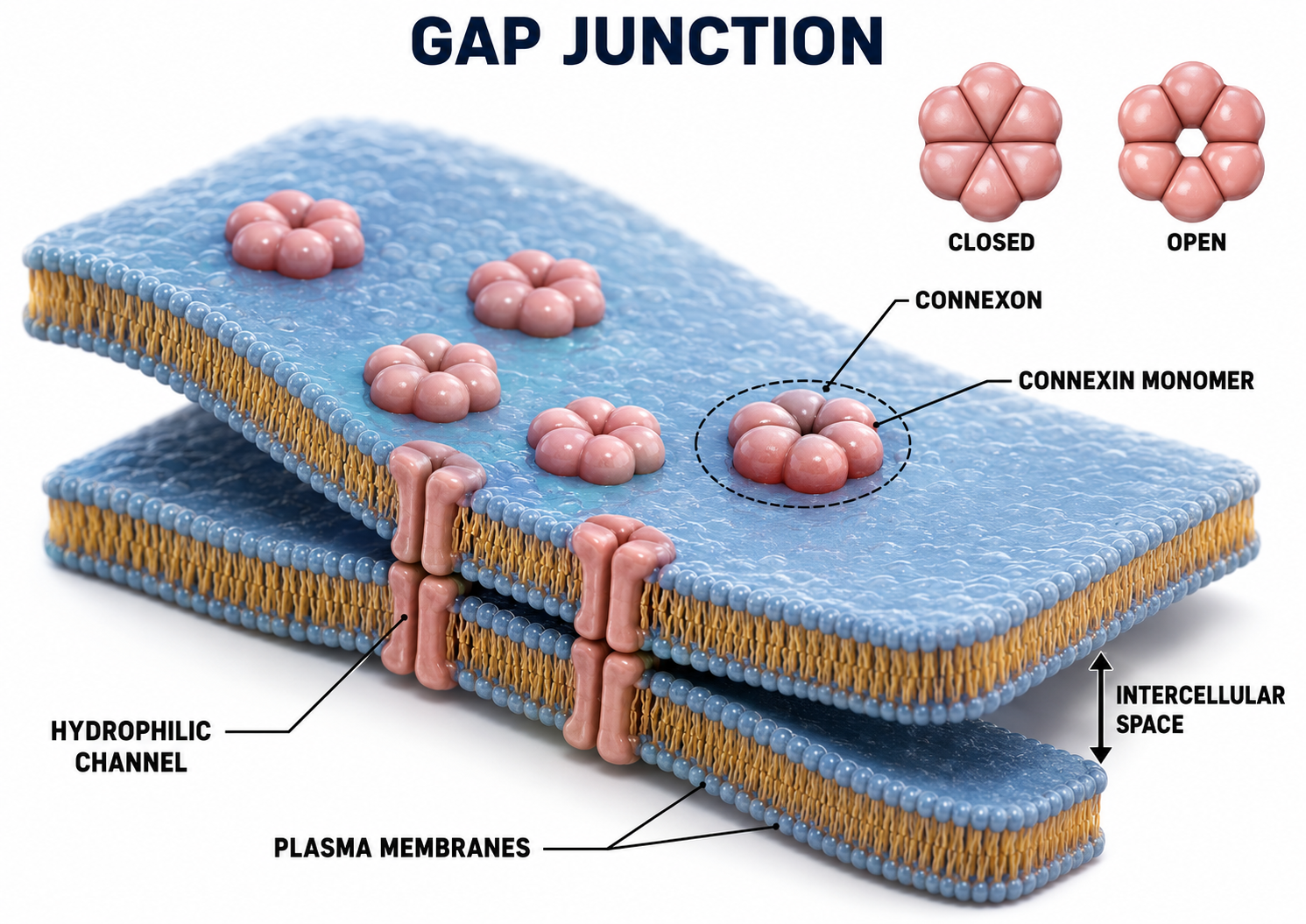
Electrical synapses are generally symmetrical. Ions flow across a 3-nm gap junction into the more negatively charged neuron as long as the gap junction remains open, meaning that whether neurons are presynaptic or postsynaptic depends on their respective charges. When two neurons are electrically coupled, an action potential in one induces a postsynaptic potential (PSP) in the paired neuron.
Transmission across electrical synapses is nearly instantaneous, compared with the roughly 0.5-1-ms synaptic delay at chemical synapses. The rapid information transmission that characterizes electrical synapses enables large circuits of neurons to synchronize their activity and fire simultaneously, a property that is directly relevant to the synchronized oscillations observed in the EEG.
Studies in recent years have revealed that electrical synapses are common in every part of the mammalian CNS. When two neurons are electrically coupled, an action potential in the presynaptic neuron causes a small amount of ionic current to flow across the gap junction channels into the other neuron. This current causes an electrically mediated postsynaptic potential (PSP) in the second neuron. Note that, because most electrical synapses are bidirectional, when that second neuron generates an action potential, it will in turn induce a PSP in the first neuron (Bear et al., 2020, p. 113).
Neurons that secrete hormones use electrical synapses to release their chemical messengers simultaneously. Neonatal brains may use gap junctions to activate many neurons at once. Image of long, fibrous astrocyte processes using Golgi's silver chromate technique © Jose Luis Calvo/Shutterstock.com.
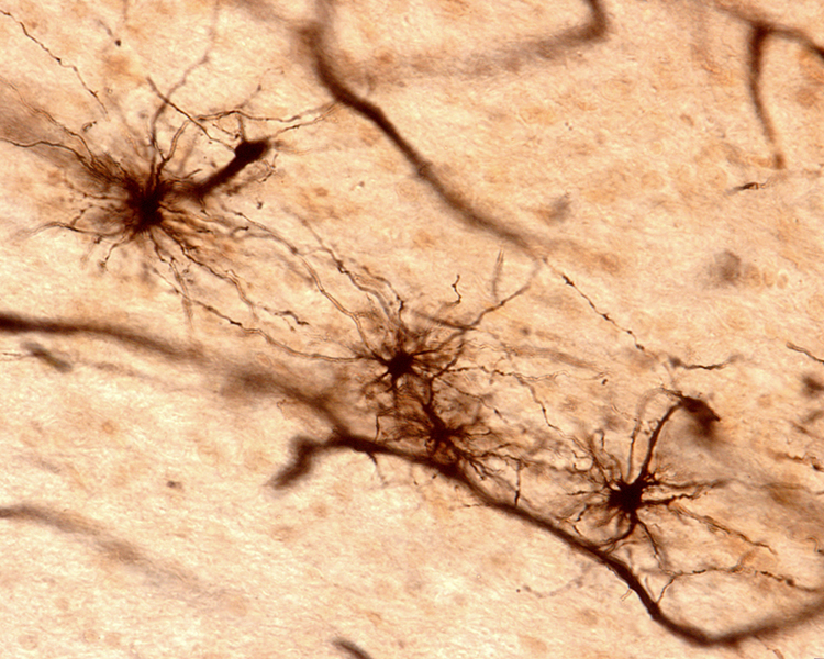
Gap junctions may be a preliminary step toward developing chemical synapses between these neurons, eventually replacing their electrical synapses. Prenatally and postnatally, gap junctions enable nearby neurons to coordinate their development by sharing electrical and chemical communications (Bear, Connors, & Paradiso, 2026; Breedlove & Watson, 2023).
Old school view: synapses are either electrical or chemical.
New school view: synapses can be both electrical and chemical.
Neurons communicate chiefly through chemical synapses, releasing neurotransmitters from vesicles by exocytosis across the synaptic cleft to bind ionotropic receptors for fast effects or metabotropic receptors for slower, longer-lasting ones. Communication is not limited to the cleft, because volume transmission, axonal varicosities, and retrograde signaling let neurotransmitters influence broader regions. The nervous system fine-tunes these signals through modulation of both transmitter release and receptor sensitivity, acting more like a volume control than an on-off switch. More than two hundred neurotransmitters have been identified, and a given transmitter can be excitatory or inhibitory depending on the receptor it activates. Electrical synapses complement chemical ones by allowing near-instantaneous, synchronized firing across coupled neurons.
Check Your Understanding
- How do ionotropic and metabotropic receptors differ in their speed and duration of action, and why does that distinction matter for the EEG?
- What are three ways neurons release neurotransmitters outside of a classical synapse, and why is this significant?
- How do presynaptic facilitation and presynaptic inhibition change synaptic communication without altering the action potential itself?
- Why can the same neurotransmitter produce opposite effects at different synapses?
- How does transmission across electrical synapses differ from transmission across chemical synapses?
Discoveries Since Graduate School
This section highlights major neuroscience discoveries that have reshaped our understanding of neural communication since many clinicians completed their training. These advances, from adult neurogenesis to the mirror neuron system, have direct implications for how we conceptualize and deliver neurofeedback interventions.
Neuroscientists have learned a great deal more about neuron-to-neuron communication since graduate school. The most important findings are that the adult brain creates new neurons, silent synapses may mediate neuroplasticity in adulthood, the lymphatic system extends to the brain, neuronal networks exhibit mirroring properties, and neurons can release more than one NT, release NTs outside of a synapse, conduct two-way conversations, modulate NT release and action, talk to astrocytes that enclose synapses, and electrically communicate almost instantaneously.
Neurogenesis
Neuroscience has challenged the long-held doctrine that the adult human brain does not create new neurons. Neurogenesis, the creation of new neurons in adults, was first demonstrated in the human hippocampus by Eriksson et al. (1998), and recent evidence favors its persistence there, but the question is still actively contested rather than settled. Adult neurogenesis in the olfactory bulb is well established in rodents (Lim & Alvarez-Buylla, 2016) but is not supported by carbon dating of human tissue. Neurogenesis elsewhere in the adult human brain remains more controversial still.
Animal research has yielded evidence of functionally significant neurogenesis in the amygdala, caudate nucleus and putamen (striatum), cortex, hypothalamus, and substantia nigra (Jurkowski et al., 2020). The neurogenesis graphic by Rebeca Cuesta is licensed under the Creative Commons Attribution-Share Alike 4.0 International license.
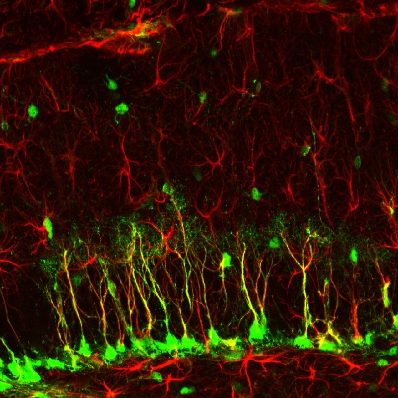
Silent Synapses
Silent synapses are inactive due to the absence of glutamate AMPA receptors. Researchers studying adult mice discovered these synapses on the ends of threadlike filopodia, thin, exploratory projections that extend from neurons. The simultaneous firing of two neurons connected by a silent synapse causes missing AMPA receptors to appear on the filopodia cell membrane and remodel it to resemble a dendritic spine (Vardalaki et al., 2022).
The next step is to determine whether the adult human brain also contains silent synapses. If it does, they represent a potential target for increasing cognitive flexibility in the elderly and may help explain how neurofeedback training can produce changes even in mature brains. Filopodia photomicrograph by Aurea D. Sousa and Richard E. Cheney under the Creative Commons Attribution-Share Alike 4.0 International license.
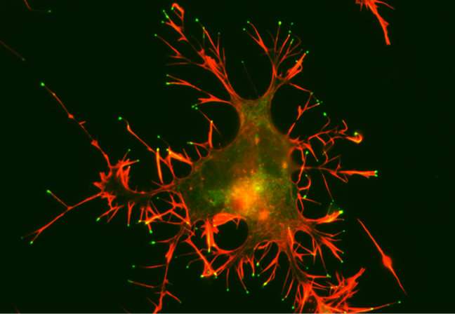
A CAD cell (a neuronal cell line) expressing GFP-Myo10 (green) was stained for actin filaments (red) to visualize the slender cellular protrusions known as filopodia. Overexpressing Myo10 induces large numbers of filopodia and is responsible for the unusually large number of filopodia on this cell.
Holly Barker (2022) writing for The Scientist, explained:
The study may explain how the brain is able to learn new things without having to sacrifice existing connections, the researchers say. The ability of the brain to use different synapses 'solves the plasticity versus flexibility dilemma,' says Harnett. If all the brain's synapses are flexible, then you can't preserve old information. But if they're all stable, then it is difficult to learn new things, he says. Instead, the brain employs both: spiny synapses for stability and filopodia for flexibility.But instead of distinct categories, Harnett's group is beginning to think about dendritic projections as existing on a continuum, from filopodia on one end to mature spines at the other. 'It is a spectrum of maturity, strength, and plasticity,' says study author Dimitra Vardalaki, a PhD candidate in Harnett's lab.
The discovery of silent synapses in adult mice is a very recent development (Vardalaki et al., 2022). Because these dormant synapses sit on filopodia and can be activated without disturbing established connections, they may let the brain learn new information while preserving old memories. If the adult human brain contains them as well, they offer a plausible cellular route by which neurofeedback could reshape mature circuits. Confirming their presence in humans is an active and important research question.
Mirror Neuron System
Researchers discovered primate neurons with both motor and visual properties in the premotor cortex. The mirror properties are due to a neuron's connections and not its construction. The cortex graphic adapted from © Vasilisa Tsoy/Shutterstock.com.
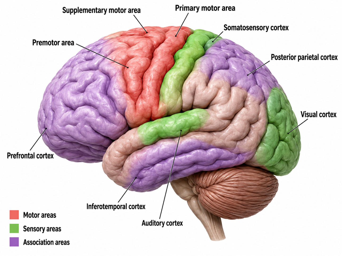
These mirror neurons fired when primates grasped and manipulated objects, and when another primate or human performed the same action (di Pellegrino et al., 1992; Rizzolatti & Craighero, 2004). Mirroring extends across species, including facial expressions.
Molenberghs et al. (2011) unexpectedly found neurons with mirroring properties in the cerebellum, limbic system, and primary visual cortex. Graphic adapted from Wikimedia Commons.
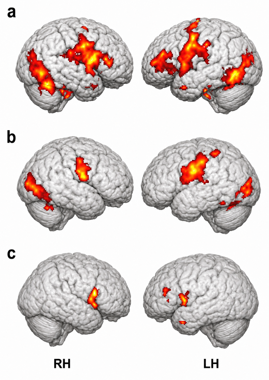
The authors proposed that a core network is responsible for observing and executing movements. The nervous system recruits additional areas to perform non-motor affective, auditory, and somatosensory functions.
Mirror neurons look like other neurons when examined using a microscope. Their mirror properties emerge from their connections within sensory, motor, and emotional systems. Perhaps most mirror neurons may be tuned by experience (Catmur, Walsh, & Heyes, 2007).
The mirror neuron system (MNS) appears to encode the goal of a motor act and its component movements, whether a model manipulates an object or mimes the action. The MNS encodes the actions of others and stores them to predict their future actions (Rajmohan & Mohandas, 2007).
Soon after birth, an immature mirror neuron system may allow babies to imitate their parents' mouth movements, like thrusting out the tongue.
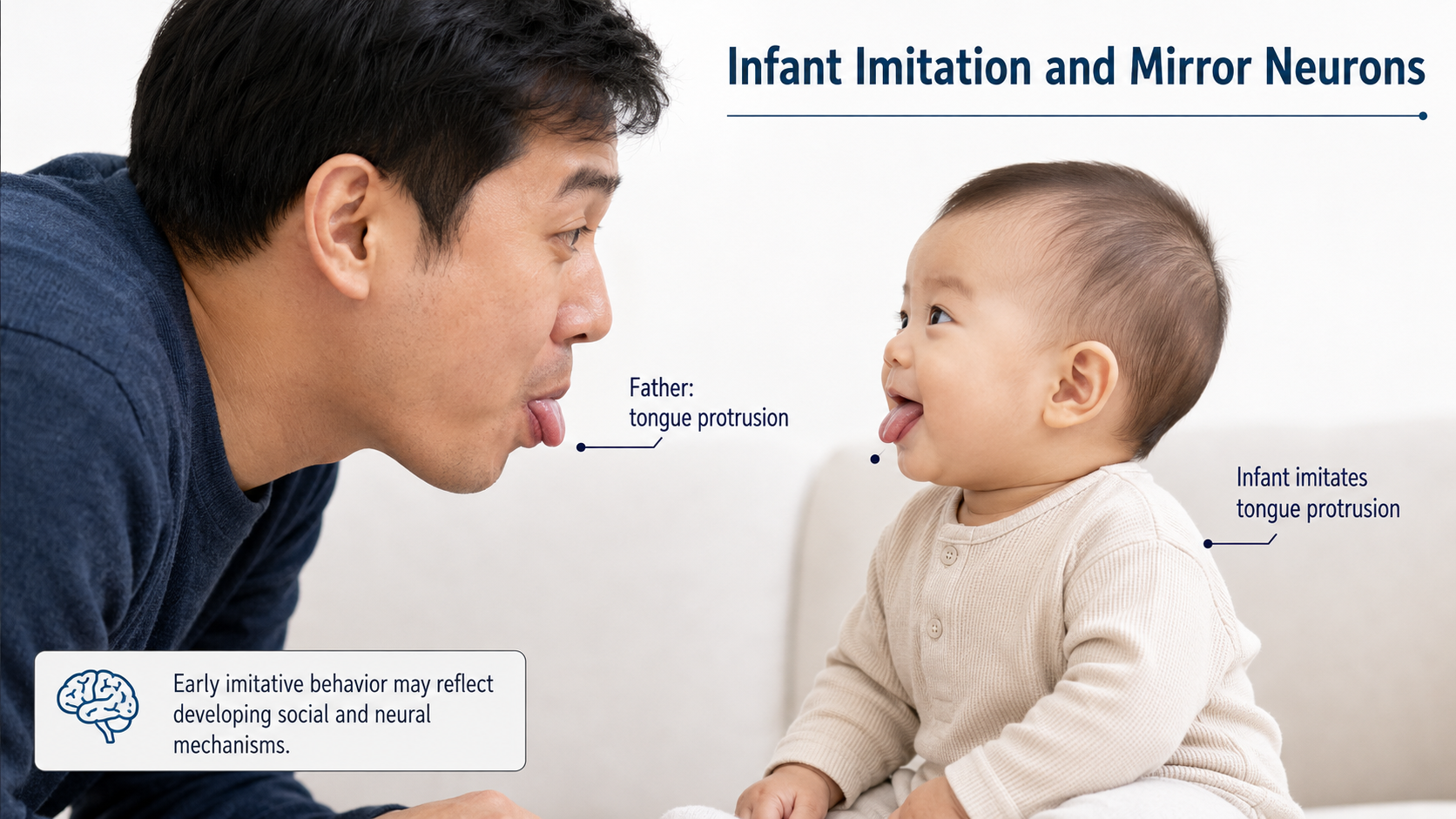
Ramachandran (2011) has called the mirror neurons activated when they observe others' movements "monkey see, monkey do neurons." He calls mirror neurons activated by others' emotional displays "Gandhi neurons." Check out Ramachandran's TED Talk, The Neurons that Shaped Civilization.
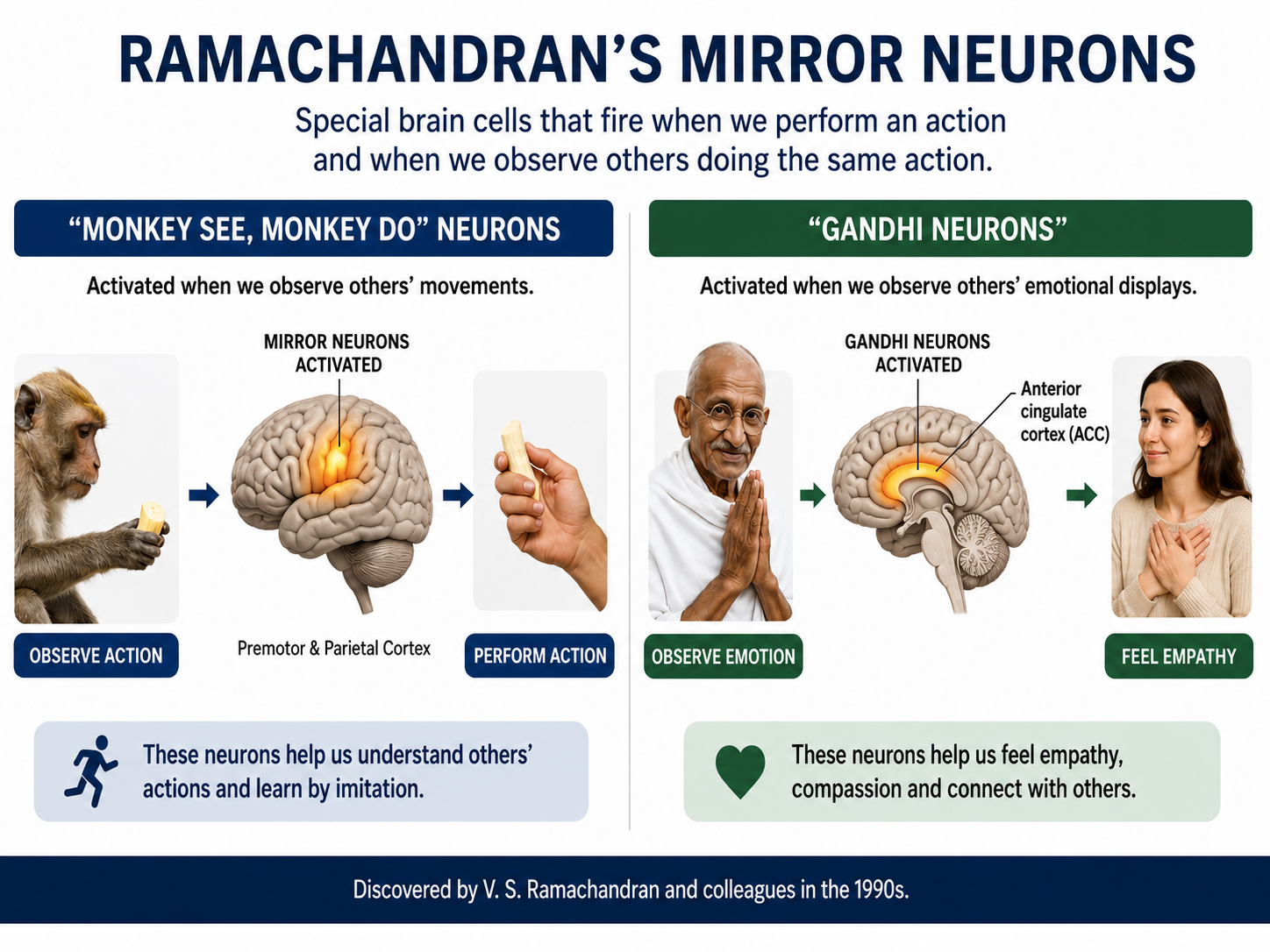
Investigators have speculated that the human MNS may mediate empathy, imitation learning, language, social cognition, and theory of mind (Buccino et al., 2006; Rajmohan & Mohandas, 2007; Schmidt et al., 2021).
Rizzolatti and Sinigaglia (2008) hypothesized that the primary role of the MNS is to help us understand others' intentions, which allows us to achieve empathy. When we observe others' facial expressions of emotion, visual information may be directly transmitted to mirror neurons in the insula, producing the visceral changes that color our emotions.
In autism, mirror neurons may not fire when observing other individuals performing actions. This may help explain deficits in empathy, social skills, language, and the development of a theory of mind (Enticott et al., 2012).
Heyes and Catmur (2022) summarized the current state of our knowledge about the MNS.
For action understanding, multivoxel pattern analysis, patient studies, and brain stimulation suggest that mirror-neuron brain areas contribute to low-level processing of observed actions (e.g., distinguishing types of grip) but not to high-level action interpretation (e.g., inferring actors' intentions). In the area of speech perception, although it remains unclear whether mirror neurons play a specific, causal role in speech perception, there is compelling evidence for the involvement of the motor system in the discrimination of speech in perceptually noisy conditions. For imitation, there is strong evidence from patient, brain-stimulation, and brain-imaging studies that mirror-neuron brain areas play a causal role in copying of body movement topography. In the area of autism, studies using behavioral and neurological measures have tried and failed to find evidence supporting the 'broken-mirror theory' of autism.
Cortical Architecture
This section describes the organization of the cerebral cortex, including its gray and white matter, convolutions, and layered structure. Understanding cortical architecture is essential because the arrangement and orientation of cortical neurons determines what the scalp EEG can and cannot detect.
While no one has counted the neurons in the human nervous system directly, the best current estimate is that an adult brain contains about 86 billion neurons (Azevedo et al., 2009; see also Voytek, 2013). Cortical neurons average several thousand synapses each; the brain-wide average is lower, because most neurons are small cerebellar granule cells with only a few inputs. The once-standard claim that glia outnumber neurons ten to one has been retracted.
Isotropic-fractionator counts give a glia-to-neuron ratio below 1:1 and fewer than 100 billion glial cells in the human brain (Azevedo et al., 2009; von Bartheld et al., 2016). Glial somas are considerably smaller than neuronal somas, roughly 6 to 10 μm in diameter (Hammond, 1996), although a single astrocyte's processes span a far larger territory. Animation © nmlfd/iStockphoto.com.
The cerebral cortex comprises neuronal cell bodies, glial cells, and blood vessels. Beneath the neocortex lie myelinated nerves (white matter), unmyelinated fibers, and glial cells.
The cerebral cortex covers the cerebral hemispheres and consists of gray and white matter. Gray (or grey) matter, which looks grayish brown, comprises cell bodies. White matter gains its opaque white color from myelinated axons. The cerebral cortex is shown below.
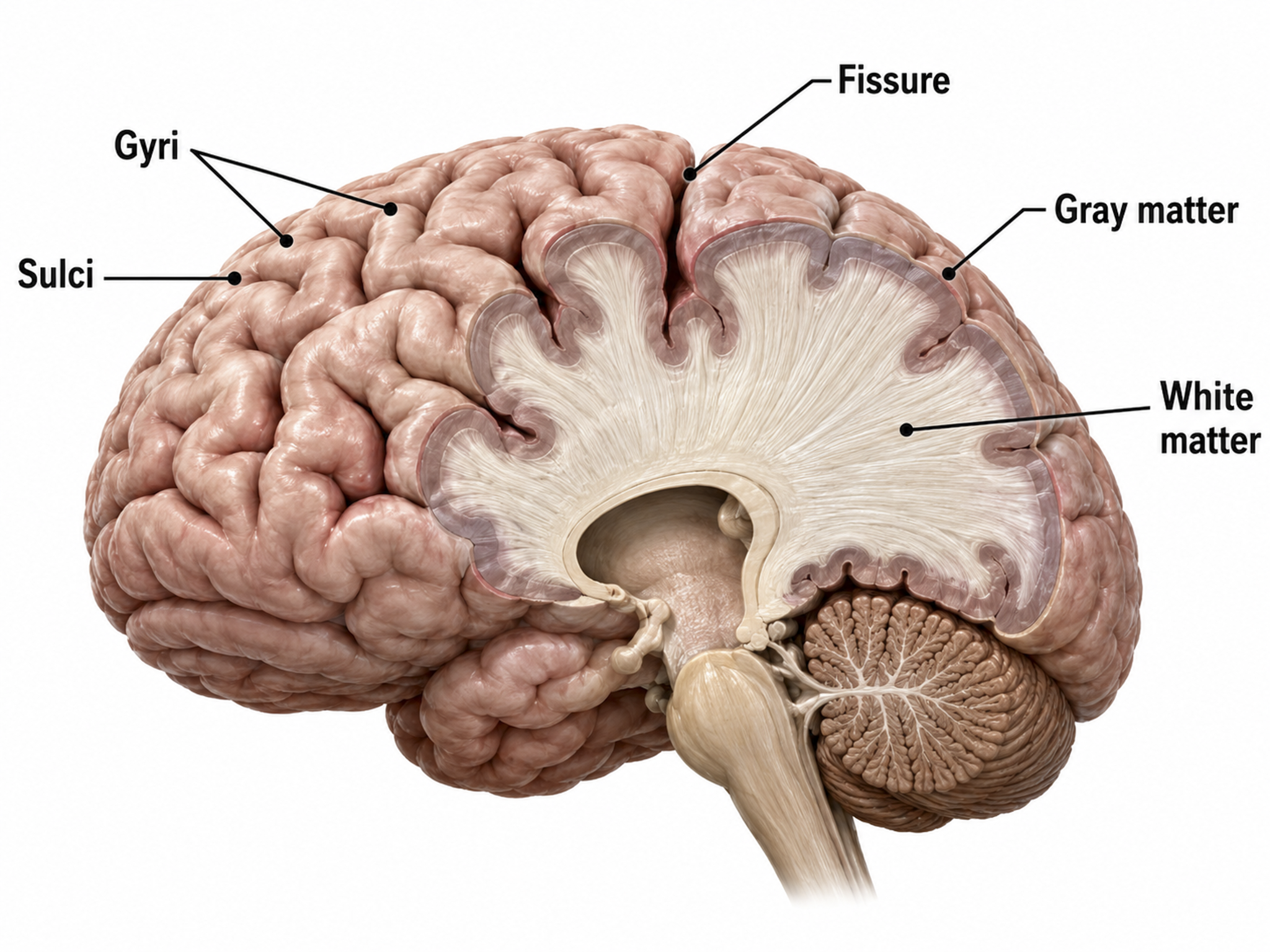
The convolutions of the cerebral cortex bury about two-thirds of its surface area within the sulci and maximize the amount of cortical tissue housed within the skull. Cerebral cortical convolutions include sulci, which are shallow grooves in the surface of the cerebral hemisphere (central sulcus), fissures, which are deep grooves (lateral fissure), and gyri, which are ridges of cortex demarcated by sulci or fissures (precentral gyrus) (Carlson & Birkett, 2021).
There are two main types of cortex: neocortex and allocortex.
The neocortex or isocortex consists of six layers, 1 to 4.5 mm thick and averaging about 2.5 mm, with a total surface area usually given as roughly 2,000 to 2,500 cm2 and white matter underneath. Layers I-III receive corticocortical afferent fibers that connect the left and right hemispheres. Layer III is the main source of corticocortical efferent fibers.
Layer IV is the primary destination of thalamocortical afferents and intra-hemispheric corticocortical afferents. Layer V is the primary origin of efferent fibers that target subcortical structures that have motor functions. Layer VI projects corticothalamic efferent fibers to the thalamus, which together with the thalamocortical afferents, creates a dynamic and reciprocal relationship between these two structures (Creutzfeldt, 1995).
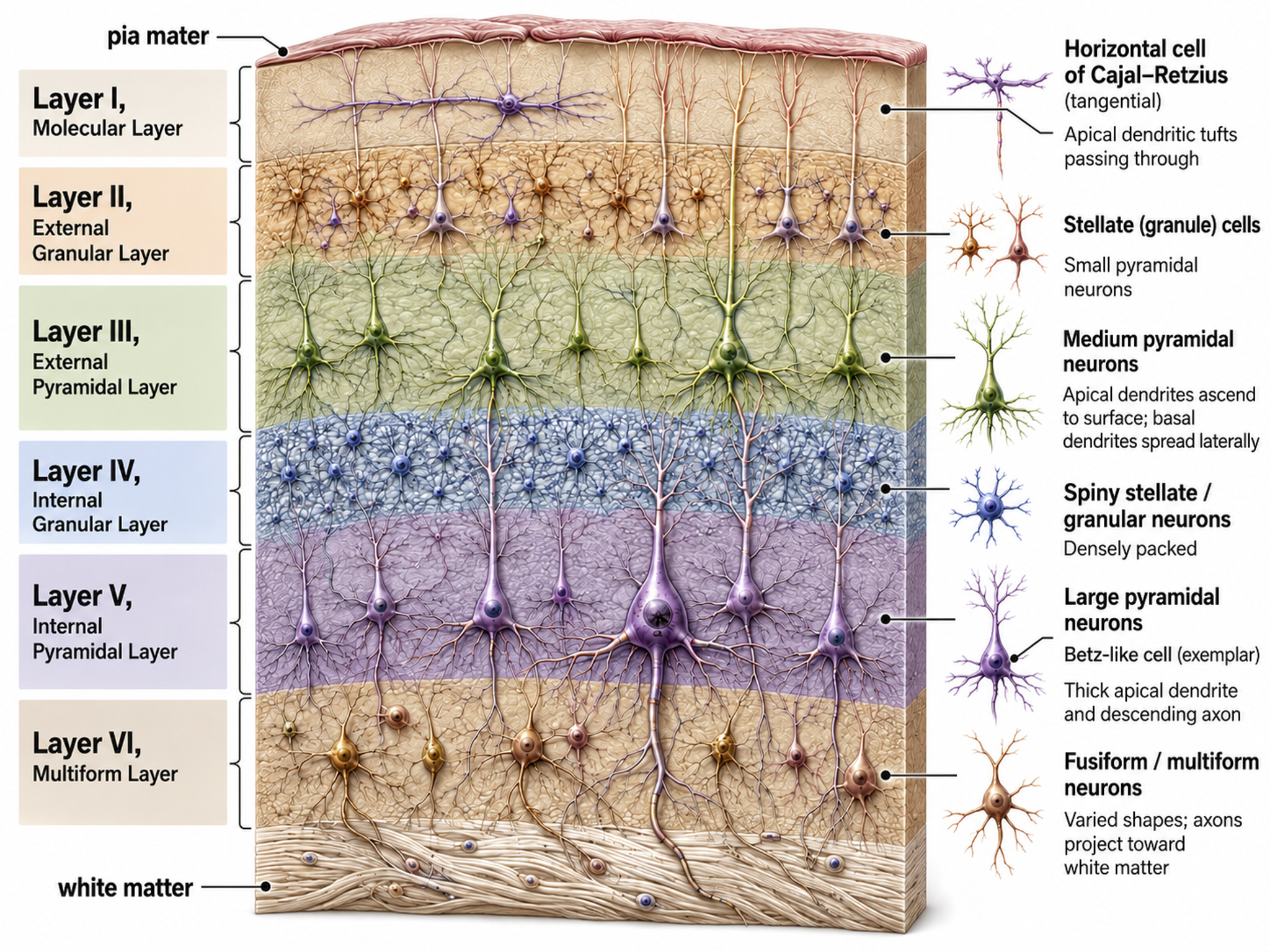
Allocortex, which means other cortex, usually has between three or four layers, compared with the neocortex's six layers. The allocortex has less volume than the neocortex and comprises the olfactory system and hippocampus.
A transitional region between the neocortex and allocortex is called the paralimbic cortex.
For a basic overview of the cortex, watch the Khan Academy video Cerebral Cortex.
Neurons in the Cortex
We can classify cerebral cortical neurons by whether their dendrites display spines. Spiny neurons, which have either pyramidal or stellate (star-like)-shaped cell bodies, are usually excitatory. While all pyramidal cells are spiny neurons, stellate cells can be spiny or aspinous (Bear, Connors, & Paradiso, 2026). Pyramidal neurons are especially important for neurofeedback because their parallel alignment and perpendicular orientation to the cortical surface make them the primary generators of the EEG signal detected at the scalp.
The graphic below shows spiny and aspinous dendrites.
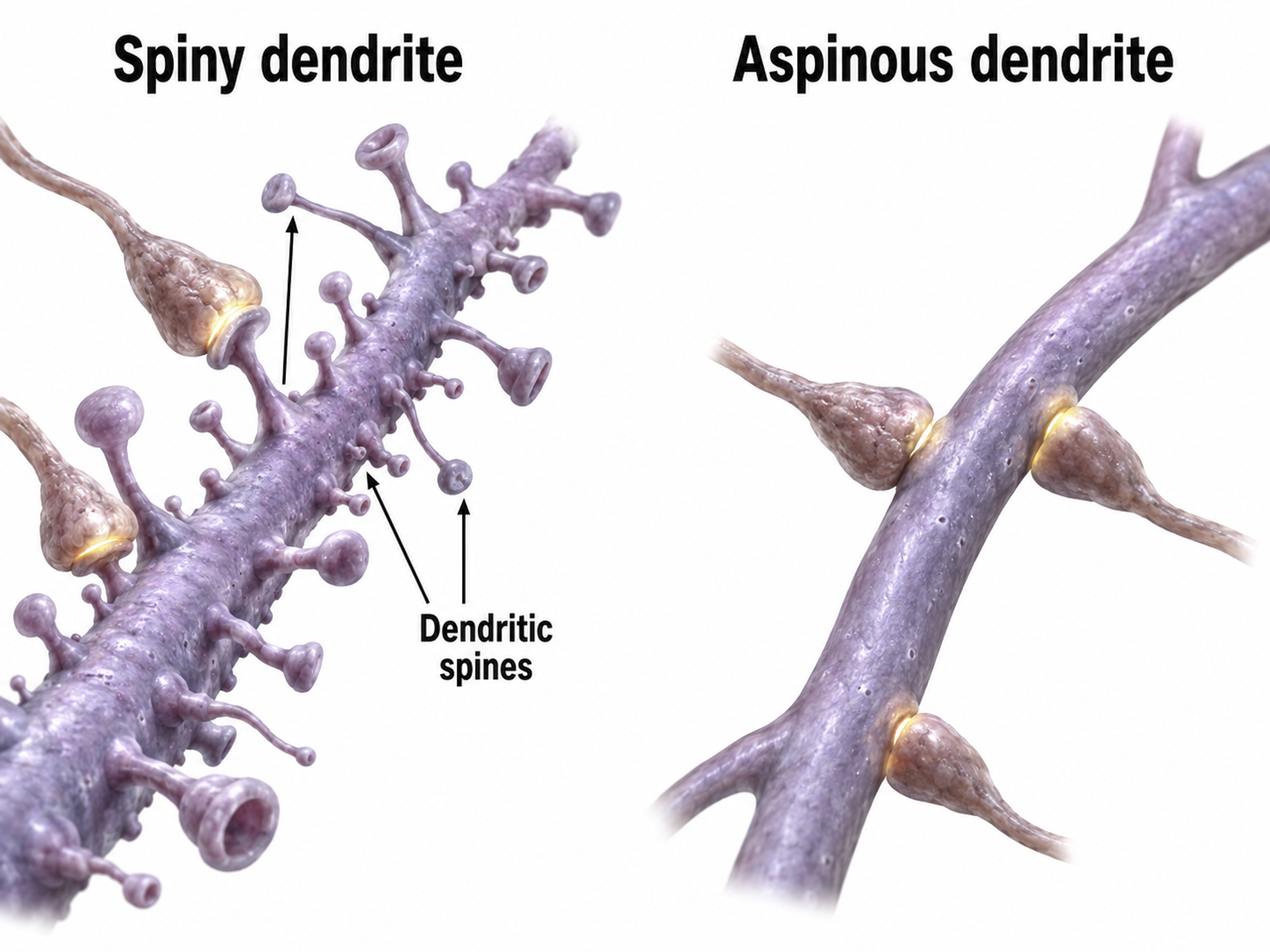
Dr. John C. Fiala, and Dr. Kristen M. Harris created this reconstruction of a dendritic spine. Creative Commons Attribution-Share Alike 3.0.
_and_massive_aggregation_of_polyribosomes_(black)..jpg)
There are many types of aspinous (smooth) neurons which are believed to be inhibitory.
The adult brain is far more plastic than the textbooks many of us trained on suggested. Neurogenesis continues in the adult hippocampus, silent synapses sitting on filopodia offer a route to learning new material without overwriting old memories, and mirror neuron systems link observation to action. The cortex is organized into six layers and into vertical columns and macrocolumns, with layer IV receiving thalamic input and layer V sending output to subcortical targets. Pyramidal neurons dominate the scalp EEG because their aligned apical dendrites form open fields whose potentials summate rather than cancel.
Check Your Understanding
- Where does adult neurogenesis occur, and why is that region clinically interesting?
- What is a silent synapse, and why might silent synapses matter for neurofeedback?
- Which cortical layer receives thalamic input, and which layer projects to subcortical structures?
- Why does the columnar organization of the cortex help explain why synchronized activity is visible at the scalp?
Review Flash Cards on Quizlet
Click the buttons below to review our flash cards for the microanatomy half of this unit.
Cutting-Edge Topics in qEEG Research
Several findings in this unit are recent enough that they have not reached most textbooks. Each one changes something practical about how you think about the brain you are training.
Meningeal Lymphatics and Glymphatic Clearance
The discovery that the dural venous sinuses house meningeal lymphatic vessels overturned the long-held belief that the central nervous system lacks lymphatic drainage (Aspelund et al., 2015; Louveau et al., 2015). Working together with the glymphatic system, which clears wastes such as amyloid-beta, tau, and alpha-synuclein mainly during slow-wave sleep, this anatomy links cerebral venous drainage to waste clearance and immune surveillance. For practitioners, it reinforces why sleep quality belongs in every client assessment.
The Neurovascular Unit and Blood-Brain Barrier
Rather than a passive wall of endothelial cells, the blood-brain barrier is now understood as a coordinated neurovascular unit that also includes pericytes, astrocyte endfeet, microglia, and the surrounding basement membrane (Abbott et al., 2010; Ding et al., 2025). This tightly regulated selectivity protects neural tissue but excludes most small-molecule and nearly all large-molecule therapeutics, which makes the barrier a central obstacle in treating neurological and psychiatric disorders (Daneman & Prat, 2015).
Locus Coeruleus, Pupillometry, and Attention
Although the locus coeruleus cannot be monitored noninvasively, its activity correlates with pupil dilation, which gives researchers an indirect window on noradrenergic arousal (Dahl et al., 2020; Dahl et al., 2022). Greater alpha blocking and pupil dilation predict better performance on demanding attention tasks, connecting a small brainstem nucleus to the alpha dynamics that clinicians train.
Silent Synapses in the Adult Brain
The discovery of silent synapses in adult mice is very recent (Vardalaki et al., 2022). Because these dormant synapses sit on filopodia and can be activated without disturbing established connections, they may let the brain learn new information while preserving old memories. If the adult human brain contains them as well, they offer a plausible cellular route by which neurofeedback could reshape mature circuits, and confirming their presence in humans is an active research question.
Individualized Frequency Bands
Classical band boundaries are useful but not absolute. The individual alpha peak typically sits near 10 Hz, but some individuals peak nearer 8 or 9 Hz and others nearer 12 Hz, so some clinicians define individualized frequency bands that depart from the classical definitions, and this shift can change the settings chosen for neurofeedback (Tarasi & Romei, 2024).
Assignment
Now that you have completed this unit, work through the following. First, explain why the scalp EEG reflects postsynaptic potentials from cortical pyramidal neurons rather than action potentials, and why at least six square centimeters of synchronized cortex are required to produce a reliable scalp signal. Second, trace the path a signal takes from a thalamic pacemaker to your amplifier, naming each tissue layer it crosses and describing what each one does to the signal.
Then return to Marcus, the client from the chapter overview whose map showed excess frontal theta and a slow posterior dominant rhythm. Using what you now know about generators, explain two anatomically plausible accounts of that pattern, and describe what additional information you would gather before choosing a training approach.
Glossary
absolute refractory period: the brief interval during and just after an action potential when inactivated sodium channels make it impossible to fire a second action potential regardless of stimulus strength.
acetylcholine (ACh): a neurotransmitter released by all autonomic preganglionic neurons, parasympathetic postganglionic neurons, sympathetic sudomotor neurons, and somatic motor neurons.
action potential: a rapid, regenerative change in membrane potential that propagates along an excitable cell membrane after threshold is reached.
afferent: a nerve fiber or pathway that carries information toward the central nervous system.
all-or-none law: the principle that once threshold is reached, a single axonal action potential has a stereotyped amplitude and propagates without decrement under normal conditions.
allocortex: cortex that contains three or four layers and is comprised of the olfactory system and hippocampus.
allosteric site: a secondary binding location on a receptor, separate from the orthosteric site, where a ligand attaches to enhance or reduce the receptor's response to its neurotransmitter.
alpha 1 (low alpha): a protocol-dependent subdivision of the alpha band, commonly 8 to <10 Hz; proposed functional interpretations are not universal.
alpha 2 (high alpha): a protocol-dependent subdivision of the alpha band, commonly 10 to <13 Hz; proposed functional interpretations are not universal.
alpha asymmetry: a difference in alpha amplitude between homologous sites, most often F3 and F4; because it compares two sites, unequal impedance, drowsy epochs, and reference choice can all manufacture one.
alpha blocking: the attenuation or disappearance of posterior alpha activity with eye opening, sensory stimulation, movement, or active cognitive processing.
alpha rhythm: an 8 to <13 Hz EEG rhythm dominant posteriorly during relaxed wakefulness with eyes closed and attenuated by eye opening.
amino acid neurotransmitters: amino acids used as neurotransmitters, principally excitatory glutamate and inhibitory gamma-aminobutyric acid or glycine in the central nervous system.
AMPA receptor: an ionotropic glutamate receptor that mediates most rapid excitatory synaptic transmission through a cation channel; its depolarization helps relieve the magnesium block of NMDA receptors.
amygdala: a group of medial temporal-lobe nuclei involved in salience evaluation, associative learning, affective memory, and coordination of defensive and autonomic responses.
anandamide: a fatty acid NT derived from arachidonic acid.
anisotropic attenuation: the variation in the degree of signal attenuation depending on the direction of the electrical currents. This phenomenon occurs due to the heterogeneous nature of the skull's structure.
anterior: toward the front of the body or, in the head, toward the face.
apical dendrite: a major dendrite extending from the apex of a pyramidal neuron toward superficial cortical layers, often branching into an apical tuft.
apoptosis: a regulated form of cell death involving an orderly cellular program and generally limited inflammatory disruption.
ascending arousal system: distributed brainstem, hypothalamic, basal-forebrain, and thalamic pathways that regulate wakefulness, cortical activation, and attention.
aspinous (smooth) neurons: neurons without dendritic spines that are believed to be inhibitory.
astrocytes: central nervous system glial cells that support metabolic homeostasis, ion and neurotransmitter regulation, blood-brain barrier function, and synaptic modulation.
autism spectrum disorder (ASD): a neurodevelopmental disorder characterized by deficits in social communication and interaction, along with restricted and repetitive behaviors.
autonomic nervous system (ANS): the visceral motor and sensory system regulating cardiovascular, respiratory, gastrointestinal, thermoregulatory, metabolic, genitourinary, and other homeostatic functions.
autoreceptor: a receptor on a neuron that responds to transmitter released by that neuron and regulates synthesis, release, firing, or other cellular processes.
axoaxonic synapses: junctions between two axons that do not affect the generation of an action potential, only the amount of neurotransmitter distributed.
axodendritic synapses: junctions between axons and dendrites that determine whether the axon hillock will initiate an action potential.
axon: a neuronal process specialized for conducting action potentials and transmitting signals to target cells.
axon hillock: the tapered region joining the soma to the axon; synaptic inputs are integrated across the neuron, and action potentials usually begin in the adjacent initial segment.
axon terminal: buds located on the ends of axon branches that form synapses and release neurochemicals to other neurons.
axon terminal button: an axon's bulblike termination specialized for NT release.
basal forebrain: a cholinergic network located in the ventral frontal lobe and anterior hypothalamus that influences cerebral blood flow and cognitive activity.
basal ganglia: interconnected subcortical nuclei, including striatum, globus pallidus, subthalamic nucleus, and substantia nigra, that regulate action selection, movement, learning, and motivation.
beta rhythm: a 13 to <30 Hz EEG rhythm, usually low amplitude, associated with alert wakefulness and active cognitive or motor processing.
binding rhythm: the gamma-band oscillation, near 40 Hz, proposed to integrate separate sensory features into a unified perception.
blood-brain barrier: a selective neurovascular interface formed principally by tight-junctioned brain endothelial cells with pericytes, basement membrane, and astrocytic support.
brain connectivity studies: research focused on understanding how different regions of the brain communicate with each other. These studies often use EEG data to map functional connections and require accurate signal interpretation to account for the effects of the skull and other tissues.
brain-derived neurotrophic factor (BDNF): a neurotrophin supporting neuronal survival, synaptic plasticity, and activity-dependent circuit modification; exercise can increase BDNF signaling.
cancellous bone: the porous trabecular (spongy) bone inside the compact outer layers of bones, including the skull diploe, which offers less resistance to electrical signals than cortical bone.
caudal: toward the tail or inferior end of the neuraxis; orientation depends on the bend of the human neuraxis.
cell body (soma): the neuronal region containing the nucleus and most biosynthetic organelles and integrating many synaptic inputs.
central nervous system (CNS): the brain and spinal cord; the retina and optic nerve are developmentally and anatomically central nervous system tissue.
central sulcus: the sulcus that separates the frontal and parietal lobes, and with them the primary motor cortex from the primary somatosensory cortex.
cerebral cortex: the layered gray matter covering the cerebral hemispheres and supporting perception, action, cognition, language, memory, and conscious experience.
cerebral ventricles: a network of fluid-filled chambers that protects the brain from trauma due to abrupt head movements and facilitates the exchange of nutrients and wastes between blood vessels and the brain.
cerebrospinal fluid (CSF): fluid produced by the choroid plexus membrane of the lateral ventricles that fills the ventricular system.
chemical synapses: junctions between neurons that transmit neurotransmitter molecules across a 20-50 nm (200-500 angstrom) synaptic cleft, enabling more diverse and longer-lasting changes than electrical synapses.
choroid plexus: a network of specialized capillaries and ependymal cells lining the brain's ventricles that produces cerebrospinal fluid and forms the blood-CSF barrier.
circle of Willis: an anastomotic vascular ring at the base of the brain formed by branches of the internal carotid and basilar arteries, providing collateral blood flow when a major artery is compromised.
closed field: the extracellular field produced by neurons whose dendrites radiate symmetrically in all directions, such as many stellate and thalamocortical cells. Opposing transmembrane currents largely cancel, so these neurons contribute little to the scalp EEG.
co-release: the release of multiple neurotransmitters by the same neuron (e.g., GABA and glutamate).
connectivity training: neurofeedback in which feedback depends on a specified relation between signals at two or more sites, such as coherence, phase, or amplitude covariation.
connexon: a hemichannel of six connexin subunits that docks with its counterpart in the adjacent cell to form a gap junction linking the two cytoplasms.
contingent negative variation (CNV): a slow negative event-related potential developing between a warning stimulus and an anticipated imperative stimulus, associated with expectancy, attention, and motor preparation.
contralateral: located on or relating to the side opposite a specified structure or event.
coronal plane: the plane that separates the body into front and back parts.
cortical bone: the dense, outer surface layer of bone that provides strength and rigidity. It is one of the two types of bone found in the skull and has high resistance to electrical signals.
cortical layer III: the deeper supragranular layer that is the principal source of efferent corticocortical fibers.
cortical layers I-III: the supragranular layers that receive corticocortical afferent fibers, including commissural fibers connecting the two hemispheres.
cortical neurons: nerve cells in the cortex responsible for generating and transmitting electrical impulses.
corticothalamic network: reciprocal circuits between cerebral cortex and thalamus that regulate sensory processing, excitability, sleep-wake states, and oscillatory activity.
cranial nerves: the 12 pairs of nerves arising directly from the brain and brainstem, carrying sensory, motor, and autonomic signals for the head, neck, and viscera.
Dale's principle: the principle that a neuron generally releases the same characteristic set of transmitters from its terminals, not that each neuron releases only one transmitter.
delta rhythm: a 0.5 to <4 Hz EEG rhythm in the tutorial convention, prominent during N3 sleep and potentially abnormal when focal or excessive in awake adults.
dendrite: a branched neuronal process specialized for receiving and integrating synaptic input.
dendritic branch: a single dendrite segment running from one bifurcation of a neuron's dendritic tree to the next bifurcation or to a terminal tip.
dendritic spine: a small protrusion from a dendrite that forms the postsynaptic compartment of many excitatory synapses.
dendritic tree: the complete branching arrangement of dendrites extending from a neuron's cell body, forming its main receptive surface for synaptic input from other neurons.
dendrodendritic synapses: junctions between dendrites that communicate chemically across synapses and electrically across gap junctions.
depolarization: a shift in membrane potential toward a less negative or more positive value.
desynchronization: the replacement of large, synchronized EEG rhythms by faster, lower-amplitude activity when the brain engages with a stimulus or task.
diencephalon: the posterior forebrain subdivision that contains the thalamus and hypothalamus.
dipole: an electrical source with separated regions of positive and negative charge; for example, the eye is electropositive at the front and electronegative at the back.
distal: farther from the trunk or point of origin or attachment.
dopamine: a catecholamine neurotransmitter and neuromodulator acting through D1-like and D2-like G protein-coupled receptor families.
dorsal: toward the back in the trunk or toward the superior surface in the brain, according to the local neuraxis.
dural venous sinuses: venous channels enclosed between the two layers of the dura mater that lack valves and muscular walls, collecting blood and cerebrospinal fluid from the brain and draining it into the internal jugular veins.
EEG activity: electrical activity represented in an EEG recording, described by frequency, amplitude, morphology, distribution, timing, and reactivity.
efferent: a nerve fiber or pathway carrying signals away from the central nervous system or another reference center toward an effector.
electrical synapse: a cell-to-cell junction using gap channels for rapid ionic and small-molecule transfer; transmission may be bidirectional or rectifying.
EMG inhibit: a threshold placed on muscle-contaminated high-frequency EEG (often 30 Hz and above) that blocks reward feedback whenever that activity exceeds it, keeping reinforcement tied to cortical rather than muscular sources.
entorhinal cortex: medial temporal cortex providing major cortical input to and output from the hippocampal formation and supporting memory and spatial representation.
enzymatic deactivation: the process in which an enzyme in the synaptic cleft breaks a neurotransmitter apart into inactive fragments.
equilibrium potential: the membrane voltage at which the electrical and chemical forces acting on a particular ion are balanced, so there is no net flow of that ion across the membrane.
excitatory postsynaptic potential (EPSP): a graded postsynaptic depolarization that increases the probability of an action potential, commonly through cation influx.
executive function: the family of higher-order cognitive processes supporting planning, inhibition, working memory, and goal-directed behavior.
exocytosis: the calcium-dependent fusion of a secretory vesicle with the plasma membrane, releasing its contents outside the cell.
extracellular fluid: the fluid outside cells, comprising interstitial fluid and plasma and providing the ionic environment for cellular function.
facultative pacemaker theory: Andersen and Andersson's (1968) theory that thalamic neurons activate cortical neurons and thalamic inhibitory interneurons via recurrent collaterals.
fast cortical potentials: conventional oscillatory EEG activity above the slow-cortical-potential range, encompassing frequency components commonly analyzed from approximately 1 Hz upward.
fissure: a deep groove or cleft separating anatomical structures, such as cerebral lobes.
forebrain: the anterior brain subdivision that consists of the cerebral hemispheres (telencephalon) and the thalamus and hypothalamus (diencephalon), also called the prosencephalon.
frontal lobe: either cerebral lobe anterior to the central sulcus, involved in motor control, executive functions, language, motivation, and social behavior.
gamma rhythm: EEG activity from 30 to <80 Hz under the tutorial's working convention, associated with local cortical processing and vulnerable to muscle artifact.
gamma-aminobutyric acid (GABA): the principal inhibitory neurotransmitter in the mature mammalian central nervous system.
gap junction: an intercellular channel formed by connexons that directly connects adjacent cell cytoplasms and permits ions and small molecules to pass.
gliotransmission: calcium-dependent astrocyte neurotransmitter release.
global loops: cortical macrocolumns separated by as much as 7 cm that receive shared input and fire synchronously to generate delta and theta rhythms.
glutamate: the principal excitatory neurotransmitter in the mammalian central nervous system and an agonist at ionotropic and metabotropic glutamate receptors.
glycine: an inhibitory neurotransmitter in the spinal cord and brainstem and an obligatory co-agonist at NMDA-type glutamate receptors.
glymphatic system: a proposed glia-associated pathway for cerebrospinal and interstitial fluid exchange that contributes to solute clearance and is influenced by sleep and arousal state.
gray matter: nervous tissue rich in neuronal cell bodies, dendrites, synapses, unmyelinated axons, glia, and microvasculature.
gyrus: a ridge of cerebral cortex bounded by one or more sulci.
high beta: the upper portion of the beta band, most often defined as roughly 20-30 Hz, with some normative databases using a narrower 25-30 Hz band. Elevations are associated with hyperarousal, anxiety, rumination, and worry, and may also reflect EMG artifact.
hindbrain: the posterior brain division that consists of the cerebellum, pons, and medulla.
hippocampus: a medial temporal-lobe structure essential to episodic-memory formation, spatial representation, and contextual regulation of stress and emotion.
horizontal (transverse) plane: the plane that divides the brain into upper and lower parts.
hyperpolarization: a change in membrane potential that makes the cell interior more negative relative to the exterior.
impedance (Z): frequency-dependent opposition to alternating current, comprising resistance and reactance and measured in ohms.
inferior: below another structure, the opposite of superior.
inferior colliculi: the paired midbrain nuclei of the auditory pathway that integrate ascending auditory input for sound localization and auditory reflexes.
inhibitory postsynaptic potential (IPSP): a graded postsynaptic potential that reduces the probability of action-potential generation, often by increasing chloride or potassium conductance.
integration: the addition of EPSPs and IPSPs at the axon hillock. Neurons sum EPSPs and IPSPs over their surface in spatial integration and over milliseconds in temporal integration to raise the membrane from its resting potential to the excitation threshold. EPSPs and IPSPs last from 15-200 ms, while action potentials occur in 1-2 ms.
internal carotid artery: a major paired artery that supplies blood to nearly two-thirds of the cerebral hemispheres.
interneuron: a neuron whose processes remain largely within a local neural circuit and connect other neurons.
ion concentration gradient: the unequal distribution of an ion across the neuronal membrane, such as sodium concentrated outside and potassium concentrated inside, which stores the energy that drives neural signaling.
ionotropic receptor: a ligand-gated ion channel that changes membrane conductance when an extracellular transmitter binds.
ipsilateral: located on, originating from, or affecting the same side of the body as another referenced structure or event.
isocortex: the six-layered cortex that covers most of the cerebral hemispheres, also called neocortex.
lateral: away from the body's or structure's midline.
lateral geniculate nucleus (LGN): the thalamic relay nucleus for vision, which receives input from the retina and projects visual information to the primary visual cortex.
Layer IV: the cortical layer that is the primary destination of thalamocortical afferents and intra-hemispheric corticocortical afferents.
Layer V: the cortical layer that is the primary origin of efferent fibers that target subcortical structures that have motor functions.
Layer VI: the cortical layer that projects corticothalamic efferent fibers to the thalamus, which, together with the thalamocortical afferents, creates a dynamic and reciprocal relationship between these two structures.
limbic system: a widespread network of nuclei involved in emotion, motivation, learning, memory, and navigation, including the hippocampus, amygdala, and septal nuclei along with the hypothalamus, anterior thalamus, and cingulate gyrus.
local loops: neighboring cortical macrocolumns that share input and fire synchronously to generate frequencies above 30 Hz in the high-beta and gamma ranges.
locus coeruleus: a pontine nucleus providing widespread noradrenergic projections that regulate arousal, attention, stress responses, autonomic function, and pain modulation.
locus coeruleus system: the noradrenergic branch of the ascending reticular activating system that projects to the thalamus, limbic system, and cerebral cortex, and contributes to wakefulness and vigilance for salient stimuli. Subnormal norepinephrine transmission may contribute to ADHD.
long-term depression (LTD): a persistent activity-dependent reduction in synaptic efficacy.
long-term potentiation (LTP): a persistent activity-dependent increase in synaptic efficacy.
low-pass filter: a filter that passes frequencies below a selected cutoff and attenuates frequencies above it.
macrocolumns: circuits of cortical pyramidal neurons several millimeters in diameter whose perpendicular alignment lets their postsynaptic potentials summate into extracellular dipole layers parallel to the cortical surface.
medial: toward the midline of the body or an anatomical structure.
medial geniculate nucleus (MGN): the thalamic relay nucleus for hearing, which receives input from the inferior colliculi and projects to several cortical auditory areas using two separate pathways.
membrane potential: the voltage difference across a cell membrane produced by unequal ion distributions and selective membrane permeability.
meninges: three protective layers (dura mater, pia mater, and arachnoid) that enclose the brain and spinal cord.
mesencephalon: the midbrain, containing the inferior colliculi, superior colliculi, and substantia nigra.
metabotropic receptor: a receptor that influences intracellular signaling indirectly, commonly through G proteins or enzymes, rather than forming an ion channel.
metencephalon: the hindbrain subdivision that consists of the cerebellum and pons.
microglia: resident macrophage-like immune cells of the central nervous system that survey tissue, remodel synapses, and respond to injury or disease.
microtubule: a hollow polymer of alpha- and beta-tubulin that supports cell structure, intracellular transport, motility, and chromosome segregation.
midbrain: the middle division called the mesencephalon, which includes the inferior colliculi, superior colliculi, and substantia nigra.
mirror neuron: a neuron that fires both when an individual performs a movement and when the individual observes another performing the same action.
mitochondrion: a double-membrane organelle that generates ATP through oxidative phosphorylation and participates in metabolism, signaling, calcium regulation, and cell death.
motor cortex: the subdivision of the frontal lobe located in the precentral gyrus that guides fine motor coordination (like writing).
motor nerves: efferent neurons that convey commands to glands, muscles, and other neurons.
motor neuron: an efferent neuron whose axon innervates skeletal, smooth, or cardiac muscle; in common neuromuscular usage, the term usually denotes a somatic motor neuron.
movement-related cortical potential (MRCP): a slow cortical potential associated with preparation, initiation, and execution of voluntary movement, including the readiness potential.
mu rhythm: an 8 to <13 Hz arch-shaped or wicket-like EEG rhythm over central sensorimotor regions that attenuates with movement or somatosensory activation.
multiple sclerosis (MS): an immune-mediated central nervous system disease characterized by inflammatory demyelination, axonal injury, and neurologic episodes or progression.
myelencephalon: the hindbrain subdivision that consists of the medulla.
myelinated axon: an axon ensheathed by oligodendrocyte myelin in the central nervous system or Schwann-cell myelin in the peripheral nervous system.
N400: a negative event-related potential component peaking near 400 ms and sensitive to semantic access, expectancy, and integration.
neocortex: the six-layered cortex roughly 1 to 4.5 mm thick, averaging about 2.5 mm, that comprises most of the human cerebral cortex, as distinguished from the allocortex.
nerve growth factor (NGF): a neurotrophin essential for development and maintenance of selected sensory and sympathetic neurons and capable of sensitizing nociceptors during inflammation.
neurogenesis: the generation of new neurons, which continues in the adult hippocampal dentate gyrus.
neuromodulator: a signaling substance that alters neuronal excitability, synaptic transmission, or circuit state, often over broader spatial or temporal scales than fast transmitters.
neuroplasticity: experience- or injury-related change in neural structure, function, connectivity, or representation.
nitric oxide (NO): a short-lived gaseous signaling molecule synthesized by nitric-oxide synthases that activates soluble guanylyl cyclase and promotes smooth-muscle relaxation.
node of Ranvier: an unmyelinated gap between myelin internodes where voltage-gated ion channels support saltatory action-potential propagation.
norepinephrine: a catecholamine that functions mainly as the neurotransmitter of most sympathetic postganglionic neurons and also as a circulating adrenal medullary hormone.
nucleus accumbens: a ventral striatal region integrating limbic and cortical input and contributing to motivation, reinforcement learning, and action selection.
nucleus reticularis paragigantocellularis: a medullary reticular nucleus involved in autonomic and descending pain-modulatory circuits through noradrenergic and other pathways.
occipital lobe: the posterior cerebral lobe containing primary and association visual cortices.
oligodendrocyte: a central nervous system glial cell that forms myelin around segments of multiple axons.
open field: the extracellular field produced by neurons whose long dendrites are aligned in parallel and perpendicular to the cortical surface, such as cortical pyramidal neurons, whose aligned dipoles summate to generate the scalp EEG.
orthosteric site: a receptor's primary binding location, where its native neurotransmitter attaches.
P300: a positive event-related potential component, commonly maximal centroparietally, whose latency and amplitude vary with attention, task relevance, probability, and context.
panic disorder (PD): an anxiety disorder characterized by recurrent, unexpected panic attacks and persistent concern about future attacks.
paralimbic cortex: a transitional region between neocortex and allocortex.
parietal lobe: a cerebral lobe posterior to the central sulcus involved in somatosensation, spatial representation, attention, body schema, and sensorimotor integration.
Parkinson's disease (PD): a progressive neurodegenerative disorder characterized primarily by motor symptoms such as tremor, rigidity, bradykinesia (slowness of movement), and postural instability.
perception-action cycle: a recurrent process in which perception guides action and the consequences of action update subsequent perception.
peripheral nervous system (PNS): all neural structures outside the brain and spinal cord, including cranial and spinal nerves, ganglia, and peripheral receptors.
phase synchrony: a stable phase relationship between two oscillatory signals over time, which need not involve zero phase lag or coincident peaks and valleys.
postcentral gyrus: the gyrus immediately posterior to the central sulcus, containing primary somatosensory cortex (Brodmann areas 3, 1, and 2).
posterior cerebral arteries: the paired terminal branches of the basilar artery that supply the occipital lobes, the inferior and medial temporal lobes, and the thalamus.
posterior cortex: parietal, temporal, and occipital cortical areas concerned with perception and memory.
posterior dominant rhythm (PDR): the dominant occipital rhythm during relaxed wakefulness with eyes closed, usually in the alpha range in healthy adults and attenuated by eye opening.
posterior vertebral arterial branches: the branches of the vertebrobasilar circulation that supply the brainstem, cerebellum, and posterior cerebral hemispheres.
precentral gyrus: the frontal lobe gyrus immediately anterior to the central sulcus that contains the primary motor cortex (Brodmann area 4).
prefrontal cortex (PFC): the anterior association cortex of the frontal lobes involved in executive control, planning, working memory, valuation, social cognition, and emotion regulation.
premotor cortex: frontal cortex anterior to primary motor cortex that contributes to movement selection and preparation, especially in response to external cues.
presynaptic facilitation: an increase in neurotransmitter release caused by modulation of the presynaptic terminal, often through enhanced calcium entry or release machinery.
presynaptic inhibition: a reduction in neurotransmitter release caused by modulation of a presynaptic terminal, often through reduced calcium entry or axoaxonic signaling.
primary motor cortex: cortex in the precentral gyrus that contributes substantially to execution and control of voluntary movement.
primary somatosensory cortex (S1): cortex in the postcentral gyrus that receives organized thalamic somatosensory input and represents touch, proprioception, and nociceptive features.
primary visual cortex (V1): the occipital lobe region located in the calcarine sulcus (BA 17, also called striate cortex) that receives most visual information from the lateral geniculate nucleus of the thalamus and performs the initial processing of visual information received from the retinas.
proximal: nearer the trunk or point of origin or attachment.
raphe nuclei: midline brainstem nuclei containing many serotonergic neurons with widespread ascending and descending projections.
rate coding: neural representation of stimulus or response magnitude through changes in action-potential firing rate.
readiness potential: a slow negative cortical potential beginning up to several seconds before self-initiated voluntary movement and maximal over central regions.
rebound excitation: the synchronized depolarization of thalamocortical relay neurons when inhibition ends, which initiates a new cycle of rhythmic EEG activity.
regional loops: cortical macrocolumns that share input, are separated by several centimeters, and generate alpha and beta rhythms.
relative refractory period: the interval following the absolute refractory period when a stronger-than-usual input is needed to fire the neuron.
resonant loop: the synchronous firing by macrocolumns that share afferent input to generate an electrical potential.
respiratory sinus arrhythmia (RSA): respiration-linked fluctuation in cardiac interval, usually shortening during inspiration and lengthening during expiration through predominantly vagal mechanisms.
resting membrane potential: the relatively stable membrane voltage of an unstimulated cell, commonly near −70 mV in many neurons but varying by cell type.
reticular formation (brainstem reticular formation): a diffuse network of brainstem nuclei and fibers involved in arousal, autonomic regulation, pain modulation, posture, movement, and reflexes.
retrograde transmission: signaling in which the postsynaptic neuron releases a messenger, such as nitric oxide or an endocannabinoid, that travels backward to influence the presynaptic neuron.
reuptake: the primary mechanism terminating neurotransmitter action, in which transporters in terminal buttons and astrocytes remove transmitter molecules from the synaptic cleft.
rostral: toward the nose or anterior end of the neuraxis.
sagittal plane: the plane that divides the body into right and left halves.
saltatory conduction: rapid propagation in myelinated axons through passive current spread between nodes of Ranvier and regeneration of action potentials at the nodes.
Schwann cells: glial cells that provide myelin for single PNS axons and facilitate axonal regeneration following damage.
selective permeability: the property of the neuronal membrane that allows some ions to cross more readily than others. At rest, the membrane is far more permeable to potassium than to sodium.
sensorimotor rhythm (SMR): a 12 to 15 Hz rhythm recorded over sensorimotor cortex, enhanced during physical stillness and attenuated by movement; it is defined topographically and behaviorally, so its range overlaps the mu and beta bands.
sensory nerves: peripheral nerves whose afferent fibers carry impulses from sensory receptors toward the central nervous system.
sensory neuron: an afferent neuron that transduces or conveys information from sensory receptors toward central neural circuits.
septal nuclei: basal forebrain nuclei with reciprocal hippocampal and hypothalamic connections involved in theta modulation, memory, motivation, and autonomic function.
sleep spindle: a waxing-waning 11 to 16 Hz burst lasting approximately 0.5 to 2 seconds, maximal over central regions and characteristic of N2 sleep.
slow cortical potential (SCP): a very slow EEG voltage shift lasting hundreds of milliseconds to seconds and reflecting changes in cortical excitability and preparation.
sodium ion (Na⁺): a positively charged ion central to extracellular osmolarity, membrane potentials, action potentials, and transport processes.
sodium-potassium ATPase: an electrogenic membrane pump that uses ATP to move three Na⁺ ions out and two K⁺ ions into the cell, maintaining transmembrane gradients.
sodium-potassium transporters: pumps that are powered by ATP and that exchange three sodium for two potassium ions.
somatic nervous system: the peripheral sensory and motor system mediating conscious somatic sensation, spinal reflexes, and skeletal-muscle control.
source localization: the process of estimating the origin of EEG activity within the brain, often using algorithms like LORETA.
spatial summation: integration of postsynaptic potentials arriving simultaneously or nearly simultaneously at different synaptic locations.
spinal cord: central nervous system tissue within the vertebral canal that conducts ascending and descending signals and contains circuits for reflexes, autonomic function, and movement.
spinal nerve: any of the 31 paired mixed nerves that exit the spinal cord through the intervertebral foramina, carrying sensory and motor fibers between the cord and the body.
spiny neurons: neurons with dendritic spines that are usually excitatory.
stroke (cerebrovascular accident, CVA): an acute neurological dysfunction caused by cerebral infarction or nontraumatic intracranial hemorrhage.
substantia nigra: a midbrain nucleus complex whose pars compacta supplies dopamine to the dorsal striatum and degenerates prominently in Parkinson disease.
sulcus: a groove on the surface of the brain that separates adjacent gyri.
superior: above another structure, the opposite of inferior.
Sylvian fissure: the deep fissure that serves as the upper boundary of the temporal lobe.
synaptic cleft: the extracellular gap, typically about 20 to 40 nm wide, separating presynaptic and postsynaptic membranes at a chemical synapse.
synaptogenesis: the formation of new synapses between neurons.
telencephalon: the embryological forebrain division that develops into the cerebral hemispheres, including cortex, white matter, basal ganglia, and associated limbic structures.
temporal summation: the cumulative postsynaptic effect of inputs arriving sufficiently close together in time.
terminal bouton: a presynaptic axon ending containing synaptic vesicles and molecular machinery for neurotransmitter release.
thalamus: a paired diencephalic structure that relays and modulates most sensory and motor information reaching cortex and contributes to arousal and cortical rhythms.
theta rhythm: a 4 to <8 Hz EEG rhythm associated with drowsiness and sleep transitions and, depending on location and task, memory and cognitive processing.
theta/beta ratio (TBR): EEG theta-band power divided by beta-band power using explicitly stated bands, electrodes, reference, and processing methods; it is not a diagnostic test for ADHD.
threshold of excitation: the membrane potential at which inward current becomes sufficient to initiate a regenerative action potential; its value varies across cells and conditions.
tripartite synapse: the three-part communication unit formed by the presynaptic neuron, the postsynaptic neuron, and the astrocyte that envelops them.
unmyelinated axon: an axon lacking a compact myelin sheath and generally conducting impulses more slowly than a comparably sized myelinated axon.
ventral: toward the belly or anterior surface of the body; in the brainstem and spinal cord, generally toward the anterior aspect.
voltage-gated ion channel: a membrane channel that opens or closes in response to changes in membrane voltage. Voltage-gated sodium and potassium channels generate the action potential.
voltage-gated sodium channel: a membrane-bound sodium ion channel required for action potentials that opens and closes in response to the membrane potential.
volume conduction: passive spread of electrical fields through conductive biological tissues, allowing a source to influence electrodes located at a distance.
volume transmission: extrasynaptic signaling in which neurotransmitters or neuromodulators diffuse through extracellular fluid to receptors beyond a conventional synaptic cleft.
white matter: central-nervous-system tissue composed chiefly of myelinated axons, glia, and blood vessels linking gray-matter regions.
Test Yourself on ClassMarker
Click the button below to take a 10-question exam over this entire unit. There is no password.
References
Abbott, N. J., Patabendige, A. A. K., Dolman, D. E. M., Yusof, S. R., & Begley, D. J. (2010). Structure and function of the blood-brain barrier. Neurobiology of Disease, 37(1), 13–25. https://doi.org/10.1016/j.nbd.2009.07.030
Amzica, F., & Lopes da Silva, F. H. (2018). Cellular substrates of brain rhythms. In Schomer, D. L. & F. H. Lopes da Silva (Eds.). Niedermeyer's electroencephalography: Basic principles, clinical applications, and related fields (7th ed.). Oxford University Press.
Andersen, P., & Andersson, S. A. (1968). Physiological basis of the alpha rhythm. Appleton-Century-Crofts.
Aspelund, A., Antila, S., Proulx, S. T., Karlsen, T. V., Karaman, S., Detmar, M., Wiig, H., & Alitalo, K. (2015). A dural lymphatic vascular system that drains brain interstitial fluid and macromolecules. Journal of Experimental Medicine, 212(7), 991–999. https://doi.org/10.1084/jem.20142290
Azevedo, F. A. C., Carvalho, L. R. B., Grinberg, L. T., Farfel, J. M., Ferretti, R. E. L., Leite, R. E. P., Jacob Filho, W., Lent, R., & Herculano-Houzel, S. (2009). Equal numbers of neuronal and nonneuronal cells make the human brain an isometrically scaled-up primate brain. The Journal of Comparative Neurology, 513(5), 532-541. https://doi.org/10.1002/cne.21974
Barker, H. (2022, November 30). Silent synapses may provide plasticity in adulthood. The Scientist. https://www.the-scientist.com/silent-synapses-may-provide-plasticity-in-adulthood-70830
Bear, M. F., Connors, B. W., & Paradiso, M. A. (2020). Neuroscience: Exploring the brain (4th ed.). Jones & Bartlett Learning.
Bear, M. F., Connors, B. W., & Paradiso, M. A. (2026). Neuroscience: Exploring the brain (5th ed.). Jones & Bartlett Learning.
Bennett, M. V. L., Contreras, J. E., Bukauskas, F. F., & Sáez, J. C. (2003). New roles for astrocytes: Gap junction hemichannels have something to communicate. Trends in Neurosciences, 26(11), 610-617. https://doi.org/10.1016/j.tins.2003.09.008
Birbaumer, N., Elbert, T., Canavan, A. G., & Rockstroh, B. (1990). Slow potentials of the cerebral cortex and behavior. Physiological Reviews, 70(1), 1-41. https://doi.org/10.1152/physrev.1990.70.1.1
Breedlove, S. M., & Watson, N. V. (2023). Behavioral neuroscience (10th ed.). Sinauer Associates, Inc.
Brini, M., Cali, T., Ottolini, D., & Carafoli, E. (2014). Neuronal calcium signaling: Function and dysfunction. Cellular and Molecular Life Sciences, 71(15), 2787–2814. https://doi.org/10.1007/s00018-013-1550-7
Buccino, G., Solodkin, A., & Small, S. L. (2006). Functions of the mirror neuron system: Implications for neurorehabilitation. Cognitive and Behavioral Neurology, 19(1), 55-63. https://doi.org/10.1097/00146965-200603000-00007
Buskila, Y., Bellot-Saez, A., & Morley, J. W. (2019). Generating brain waves, the power of astrocytes. Frontiers in Neuroscience, 13, Article 1125. https://doi.org/10.3389/fnins.2019.01125
Buzsáki, G., Anastassiou, C. A., & Koch, C. (2012). The origin of extracellular fields and currents: EEG, ECoG, LFP and spikes. Nature Reviews Neuroscience, 13(6), 407-420. https://doi.org/10.1038/nrn3241
Carlson, N. R., & Birkett, M. A. (2019). Foundations of behavioral neuroscience (10th ed.). Pearson.
Carlson, N. R., & Birkett, M. A. (2021). Physiology of behavior (13th ed.). Pearson.
Catmur, C., Walsh, V., & Heyes, C. (2007). Sensorimotor learning configures the human mirror system. Cur Biol, 17(17), 1527-1531. https://doi.org/10.1016/j.cub.2007.08.006
Caton, R. (1875). The electric currents of the brain. British Medical Journal, 2, 278.
Chan, C. Y., Ke, D. S., & Chen, J. Y. (2009). Essential fatty acids and human brain. Acta Neurol Taiwan, 18(4), 231-241. PMID: 20329590
Coggan, J. S., Bartol, T. M., Esquenazi, E., Stiles, J. R., Lamont, S., Martone, M. E., Berg, D. K., Ellisman, M. H., & Sejnowski, T. J. (2005). Evidence for ectopic neurotransmission at a neuronal synapse. Science, 309(5733), 446-451. https://doi.org/10.1126/science.1108239
Creutzfeldt, O. D. (1995). Cortex cerebri. Oxford University Press.
Cruces, R., Muñoz-García, I., Palmer-Cancel, S. J., & Salas, C. (2022). A neuropsychological rehabilitation framework to address cognitive and neurobehavioral impairments after strokes to the anterior communicating artery. Frontiers in Human Neuroscience, 16, 808011. https://doi.org/10.3389/fnhum.2022.808011
Dahl, M. J., Mather, M., Sander, M. C., & Werkle-Bergner, M. (2020). Noradrenergic responsiveness supports selective attention across the adult lifespan. The Journal of Neuroscience, 40(22), 4372-4390. https://doi.org/10.1523/JNEUROSCI.0398-19.2020
Dahl, M. J., Mather, M., & Werkle-Bergner, M. (2022). Noradrenergic modulation of rhythmic neural activity shapes selective attention. Trends in Cognitive Sciences, 26(1), 38-52. https://doi.org/10.1016/j.tics.2021.10.009
Daneman, R., & Prat, A. (2015). The blood-brain barrier. Cold Spring Harbor Perspectives in Biology, 7(1), a020412. https://doi.org/10.1101/cshperspect.a020412
Ding, L., Kshirsagar, P., Agrawal, P., & Murry, D. J. (2025). Crossing the blood-brain barrier: Innovations in receptor- and transporter-mediated transcytosis strategies. Pharmaceutics, 17(6), 706. https://doi.org/10.3390/pharmaceutics17060706
di Pellegrino, G., Fadiga, L., Fogassi, L., Gallese, V., & Rizzolatti, G. (1992). Understanding motor events: A neurophysiological study. Experimental Brain Research, 91(1), 176-180. https://doi.org/10.1007/BF00230027
Dyro, F. M. (1989). The EEG handbook. Little, Brown and Company.
Enticott, P. G., Kennedy, H. A., Rinehart, N. J., Tonge, B. J., Bradshaw, J. L., Taffe, J. R., Daskalakis, Z. J., & Fitzgerald, P. B. (2012). Mirror neuron activity associated with social Impairments but not age in Autism Spectrum Disorder. Biol Psychiatry, 71(5), 427-433. https://doi.org/10.1016/j.biopsych.2011.09.001
Eriksson, P. S., Perfilieva, E., Björk-Eriksson, T., Alborn, A.-M., Nordborg, C., Peterson, D. A., & Gage, F. H. (1998). Neurogenesis in the adult human hippocampus. Nature Medicine, 4(11), 1313-1317. https://doi.org/10.1038/3305
Eroglu, C., & Barres, B. A. (2010). Regulation of synaptic connectivity by glia. Nature, 468(7321), 223-231. https://doi.org/10.1038/nature09612
Fisch, B. J. (1999). Fisch and Spehlmann's EEG primer: Basic principles of digital and analog EEG (3rd ed.). Elsevier.
Fischer, F., Hamann, A., & Osiewacz, H. D. (2020). Mitochondrial quality control: An integrated network of pathways. Trends in Biochemical Sciences, 45(3), 179–190. https://doi.org/10.1016/j.tibs.2019.12.008
Fuster, J. (2015). The prefrontal cortex (5th ed.). Academic Press.
Garrett, B. (2003). Brain and behavior. Thompson/Wadsworth.
Green, D. R., Galluzzi, L., & Kroemer, G. (2011). Mitochondria and the autophagy–inflammation–cell death axis in organismal aging. Science, 333(6046), 1109–1112. https://doi.org/10.1126/science.1201940
Hammond, C. (1996). Cellular and molecular neurobiology. Academic Press.
Harris, J. J., Jolivet, R., & Attwell, D. (2012). Synaptic energy use and supply. Neuron, 75(5), 762–777. https://doi.org/10.1016/j.neuron.2012.08.019
He, B., & Li, G. (2010). Attenuation of EEG signals by the skull: an in vitro study of skull conductivity and its effect on dipole source localization. Medical & Biological Engineering & Computing, 48(5), 497-507.
Heyes, C., & Catmur, C. (2022). What happened to mirror neurons? Perspectives on Psychological Science, 17(1), 153-168. https://doi.org/10.1177/1745691621990638
Hökfelt, T., Bartfai, T., & Bloom, F. (2003). Neuropeptides: Opportunities for drug discovery. The Lancet Neurology, 2(8), 463-472. https://doi.org/10.1016/S1474-4422(03)00482-4
Hughes, S. W., & Crunelli, V. (2005). Thalamic mechanisms of EEG alpha rhythms and their pathological implications. The Neuroscientist, 11(4), 357-372. https://doi.org/10.1177/1073858405277450
Jurkowski, M. P., Bettio, L., Woo, E. K., Patten, A., Yau, S.-Y., & Gil-Mohapel, J. (2020). Beyond the hippocampus and the SVZ: Adult neurogenesis throughout the brain. Frontiers in Cellular Neuroscience, 14, Article 576444. https://doi.org/10.3389/fncel.2020.576444
Kirschstein, T., & Köhling, R. (2009). What is the source of the EEG? Clinical EEG and Neuroscience, 40(3), 146-149. https://doi.org/10.1177/155005940904000305
Krauss, G. L., Fisher, R. S., & Kaplan, P. W. (Eds.) (2011). The Johns Hopkins atlas of digital EEG: An interactive training guide. The Johns Hopkins University Press.
Lim, D. A., & Alvarez-Buylla, A. (2016). The adult ventricular-subventricular zone (V-SVZ) and olfactory bulb (OB) neurogenesis. Cold Spring Harbor Perspectives in Biology, 8(5), Article a018820. https://doi.org/10.1101/cshperspect.a018820
Lin, M. T., & Beal, M. F. (2006). Mitochondrial dysfunction and oxidative stress in neurodegenerative diseases. Nature, 443(7113), 787–795. https://doi.org/10.1038/nature05292
Liu, J.-H., Zhang, M., Wang, Q., Wu, D.-Y., Jie, W., Hu, N.-Y., Lan, J.-Z., Zeng, K., Li, S.-J., Li, X.-W., Yang, J.-M., & Gao, T.-M. (2022). Distinct roles of astroglia and neurons in synaptic plasticity and memory. Molecular Psychiatry, 27(2), 873-885. https://doi.org/10.1038/s41380-021-01332-6
Lopes da Silva, F. (2010). EEG: Origin and measurement. In C. Mulert & L. Lemieux (Eds.), EEG-fMRI: Physiological basis, technique, and applications (pp. 19-38). Springer. https://doi.org/10.1007/978-3-540-87919-0_2
López-Doménech, G., Higgs, N. F., Vaccaro, V., Roš, H., Arancibia-Cárcamo, I. L., MacAskill, A. F., & Kittler, J. T. (2016). Loss of dendritic complexity precedes neurodegeneration in a mouse model with disrupted mitochondrial distribution in mature dendrites. Cell Reports, 17(2), 317-327. https://doi.org/10.1016/j.celrep.2016.09.004
Lopez-Otin, C., Blasco, M. A., Partridge, L., Serrano, M., & Kroemer, G. (2013). The hallmarks of aging. Cell, 153(6), 1194–1217. https://doi.org/10.1016/j.cell.2013.05.039
Louveau, A., Smirnov, I., Keyes, T. J., Eccles, J. D., Rouhani, S. J., Peske, J. D., Derecki, N. C., Castle, D., Mandell, J. W., Lee, K. S., Harris, T. H., & Kipnis, J. (2015). Structural and functional features of central nervous system lymphatic vessels. Nature, 523(7560), 337–341. https://doi.org/10.1038/nature14432
Lubar, J. F. (1997). Neocortical dynamics: Implications for understanding the role of neurofeedback and related techniques for the enhancement of attention. Applied Psychophysiology and Biofeedback, 22(2), 111-126. https://doi.org/10.1023/a:1026276228832
Merighi, A., Salio, C., Ferrini, F., & Lossi, L. (2011). Neuromodulatory function of neuropeptides in the normal CNS. Journal of Chemical Neuroanatomy, 42(4), 276-287. https://doi.org/10.1016/j.jchemneu.2011.02.001
Molenberghs, P., Cunnington, R., & Mattingley, J. B. (2011). Brain regions with mirror properties: A meta-analysis of 125 human fMRI studies. Neurosci Biobehav Rev, 36(1), 341-349. https://doi.org/10.1016/j.neubiorev.2011.07.004
Mölle, M., Marshall, L., Gais, S., & Born, J. (2002). Grouping of spindle activity during slow oscillations in human non-rapid eye movement sleep. The Journal of Neuroscience: The Official Journal of the Society for Neuroscience, 22(24), 10941-10947. https://doi.org/10.1523/JNEUROSCI.22-24-10941.2002
Monastra, V. J., Lubar, J. F., Linden, M., VanDeusen, P., Green, G., Wing, W., . . . Fenger, T. N. (1999). Assessing attention deficit hyperactivity disorder via quantitative electroencephalography: An initial validation study. Neuropsychology, 13(3), 424-433. https://doi.org/10.1037/0894-4105.13.3.424
Monti, J. M., & Jantos, H. (2008). The roles of dopamine and serotonin, and of their receptors, in regulating sleep and waking. Prog Brain Res, 172, 625-646. https://doi.org/10.1016/S0079-6123(08)00929-1
Nagano, S., & Araki, T. (2021). Axonal transport and local translation of mRNA in neurodegenerative diseases. Frontiers in Molecular Neuroscience, 14, 697973. https://doi.org/10.3389/fnmol.2021.697973
Natale, G., Limanaqi, F., Busceti, C. L., Mastroiacovo, F., Nicoletti, F., Puglisi-Allegra, S., & Fornai, F. (2021). Glymphatic system as a gateway to connect neurodegeneration from periphery to CNS. Frontiers in Neuroscience, 15, Article 639140. https://doi.org/10.3389/fnins.2021.639140
Navarrete, M., Cuartero, M. I., Palenzuela, R., Draffin, J. E., Konomi, A., Serra, I., Colié, S., Castaño-Castaño, S., Hasan, M. T., Nebreda, Á. R., & Esteban, J. A. (2019). Astrocytic p38α MAPK drives NMDA receptor-dependent long-term depression and modulates long-term memory. Nature Communications, 10, Article 2968. https://doi.org/10.1038/s41467-019-10830-9
Niedermeyer, E., & Lopes da Silva, F. H. (2004). Electroencephalography: Basic principles, clinical applications, and related fields (5th ed.). Lippincott Williams & Wilkins.
Nunez, P. (2006). Electrical fields of the brain. Oxford University Press.
Parri, R., & Crunelli, V. (2003). An astrocyte bridge from synapse to blood flow. Nature Neuroscience, 6(1), 5-6. https://doi.org/10.1038/nn0103-5
Perea, G., Navarrete, M., & Araque, A. (2009). Tripartite synapses: Astrocytes process and control synaptic information. Trends in Neurosciences, 32(8), 421-431. https://doi.org/10.1016/j.tins.2009.05.001
Purves, D., Augustine, G. J., Fitzpatrick, D., Hall, W. C., LaMantia, A.-S., Mooney, R. D., Platt, M. L., & White, L. E. (Eds.). (2017). Neuroscience (6th ed.). Sinauer Associates/Oxford University Press.
Rajmohan, V., & Mohandas, E. (2007). Mirror neuron system. Indian J Psychiatry, 49(1), 66-69. https://doi.org/10.4103/0019-5545.31522
Ramachandran, V. S. (2011). The tell-tale brain: A neuroscientist's quest for what makes us human. W. W. Norton & Company.
Rizzolatti, G., & Craighero, L. (2004). The mirror-neuron system. Annual Review of Neuroscience, 27, 169-192. https://doi.org/10.1146/annurev.neuro.27.070203.144230
Rizzolatti, G., & Sinigaglia, C. (2008). Mirrors in the brain: How our mind share actions, emotions, and experience. Oxford University Press.
Sanei, S., & Chambers, J. A. (2013). EEG signal processing. John Wiley & Sons.
Schmidt, S. N. L., Hass, J., Kirsch, P., & Mier, D. (2021). The human mirror neuron system: A common neural basis for social cognition? Psychophysiology, 58(5), Article e13781. https://doi.org/10.1111/psyp.13781
Schoffelen, J. M., & Gross, J. (2009). Source connectivity analysis with MEG and EEG. Human Brain Mapping, 30(6), 1857-1865. https://doi.org/10.1002/hbm.20745
Schomer, D. L., & Lopes da Silva, F. H. (2011). Niedermeyer's electroencephalography: Basic principles, clinical applications, and related fields (6th ed.). Lippincott Williams & Wilkins.
Schummers, J., Yu, H., & Sur, M. (2008). Tuned responses of astrocytes and their influence on hemodynamic signals in the visual cortex. Science, 320(5883), 1638-1643. https://doi.org/10.1126/science.1156120
Shan, L., Zhang, T., Fan, K., Cai, W., & Liu, H. (2021). Astrocyte-neuron signaling in synaptogenesis. Frontiers in Cell and Developmental Biology, 9, Article 680301. https://doi.org/10.3389/fcell.2021.680301
Steriade, M. (1990). Basic mechanisms of cerebral rhythmic activities. Electroencephalography and Clinical Neurophysiology, 76(6), 481-508. https://doi.org/10.1016/0013-4694(90)90001-Z
Steriade, M. (2001). The intact and sliced brain. MIT.
Steriade, M. (2005). Cellular substrates of brain rhythms. In E. Niedermeyer, & F. Lopes da Silva (Eds.). Electroencephalography: Basic principles, clinical applications, and related fields (5th ed.). Lippincott Williams & Wilkins.
Svensson, E., Apergis-Schoute, J., Burnstock, G., Nusbaum, M. P., Parker, D., & Schiöth, H. B. (2019). General principles of neuronal co-transmission: Insights from multiple model systems. Frontiers in Neural Circuits, 12, Article 117. https://doi.org/10.3389/fncir.2018.00117
Tarasi, L., & Romei, V. (2024). Individual alpha frequency contributes to the precision of human visual processing. Journal of Cognitive Neuroscience, 36(4), 602–613. https://doi.org/10.1162/jocn_a_02026
Thompson, M., & Thompson, L. (2015a). The biofeedback book: An introduction to basic concepts in applied psychophysiology (2nd ed.). Association for Applied Psychophysiology and Biofeedback.
Thompson, M., & Thompson, L. (2015b). The neurofeedback book: An introduction to basic concepts in applied psychophysiology (2nd ed.). Association for Applied Psychophysiology and Biofeedback.
Tiihonen, J., Kajola, M., & Hari, R. (1989). Magnetic mu rhythm in man. Neuroscience, 32(3), 793-800. https://doi.org/10.1016/0306-4522(89)90299-6
Traub, R. D., Miles, R., & Wong, R. K. S. (1989). Model of the origin of rhythmic population oscillations in the hippocampal slice. Science, 243, 1319-1325. https://doi.org/10.1126/science.2646715
Vardalaki, D., Chung, K., & Harnett, M. T. (2022). Filopodia are a structural substrate for silent synapses in adult neocortex. Nature, 612(7939), 323-327. https://doi.org/10.1038/s41586-022-05483-6
von Bartheld, C. S., Bahney, J., & Herculano-Houzel, S. (2016). The search for true numbers of neurons and glial cells in the human brain: A review of 150 years of cell counting. The Journal of Comparative Neurology, 524(18), 3865-3895. https://doi.org/10.1002/cne.24040
Voytek, B. (2013, May 20). Are there really as many neurons in the human brain as stars in the Milky Way? Brain Metrics. Scitable by Nature Education.
West, A. P., Shadel, G. S., & Ghosh, S. (2015). Mitochondria in innate immune responses. Nature Reviews Immunology, 15(11), 505–518. https://doi.org/10.1038/nri3850
Xie, L., Kang, H., Xu, Q., Chen, M. J., Liao, Y., Thiyagarajan, M., O'Donnell, J., Christensen, D. J., Nicholson, C., Iliff, J. J., Takano, T., Deane, R., & Nedergaard, M. (2013). Sleep drives metabolite clearance from the adult brain. Science, 342(6156), 373-377. https://doi.org/10.1126/science.1241224
Zhou, S., Su, S., Hong, A., Yang, C., Liu, Q., Feng, W., & Wang, Z. (2022). Abnormal functional connectivity of brain regions associated with fear network model in panic disorder. The World Journal of Biological Psychiatry, 23, 764 - 772. https://doi.org/10.1080/15622975.2022.2038389
Return to Top