
Patient Conditions Related to qEEG Evaluation
What You Will Learn in This Chapter
Ask two clinicians which conditions the qEEG is useful for and you may get two very different answers, one expansive to the point of implausibility and one so cautious it would rule out most of what the method actually does well. This unit gives you the middle ground the evidence supports.
You will start with EEG phenotypes, the patterns Johnstone, Gunkelman, and Lunt proposed as a way of describing what the raw EEG and qEEG can assess, which have been clinically correlated with medication and neurofeedback protocol recommendations and shown to be reliably assessable. From there you will work through the conditions where the evidence is strongest: ADHD and its subtypes, the differential diagnosis of dementia, the monitoring of neurological disorders, and cognitive impairment in Parkinson's disease.
The unit closes with refractory patients, which may be the most clinically consequential material here. Isolated epileptiform discharges turn up in a striking proportion of children and adolescents who have never had a seizure, and in patients who have failed multiple medication trials. Recognizing them can change a treatment plan that has been stuck for years.
IQCB Blueprint Coverage: This unit addresses IX. Clinical Practice/Forensic, specifically F. Patient conditions related to qEEG evaluation.
Learning Objectives
After completing this section, you will be able to:
Describe the EEG phenotypes proposed by Johnstone, Gunkelman, and Lunt (2005) and explain how they relate to protocol and medication recommendations.
List the conditions Koberda (2017) identifies as suitable for qEEG evaluation in a general neurology practice.
Explain what qEEG subtypes contribute to the diagnosis and management of ADHD.
Describe the qEEG patterns used in the differential diagnosis of dementia.
Explain how qEEG measures reflecting EEG slowing relate to cognitive impairment in Parkinson's disease.
Define isolated epileptiform discharges and summarize the evidence for their prevalence and clinical relevance in refractory patients.
State the general clinical utility of the qEEG and the factors that limit it.
Click the button below to hear the lecture over the Clinical Practice and Forensic unit.
EEG Phenotypes
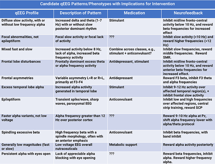
Johnstone, Gunkelman, and Lunt (2005) have proposed several EEG phenotypes that the raw EEG and qEEG can assess. Table 2 from Johnstone et al. (2005, p. 101) is reproduced below. As can be seen, these phenotypes have been clinically correlated with medication and neurofeedback protocol recommendations. Arns, Gunkelman, Breteler, and Spronk (2008) have reported that such phenotypes can be reliably assessed.
Koberda (2017) also reviews conditions for which qEEG is useful in a general neurology practice. These conditions include traumatic brain injury, cerebrovascular accident, pain and headache, depression and anxiety, cognitive dysfunction, Alzheimer's disease, autism spectrum disorder, ADHD, seizures, and epilepsy.
Kropotov (2016) reviews neuromarkers, including qEEG findings, that are helpful for the early identification, prevention, and personalized treatment of several mental disorders, such as ADHD and schizophrenia.
Diagnosis and Management
The qEEG has proved valuable in the diagnosis and management of ADHD, dementia, neurological disorders, Parkinson's disease, and refractory patients. It is a valuable tool in the differential diagnosis of dementia, monitoring neurological disorders, and assessing cognitive impairment in Parkinson's disease.
The effectiveness of the qEEG largely depends on the expertise of the clinicians and the quality of the laboratory performing the analysis. Therefore, a qEEG referral is appropriate in cases where detailed brainwave analysis can provide additional diagnostic or monitoring insights, particularly in complex neurological and psychiatric conditions.
Attention-Deficit/Hyperactivity Disorder
Evidence from multiple studies supports the appropriateness of qEEG as an auxiliary tool in the diagnosis and management of ADHD. It provides objective data that can complement traditional diagnostic methods, help identify ADHD subtypes, and guide treatment planning.
Research has identified distinct qEEG subtypes within the ADHD population, indicating that children with ADHD do not constitute a neurophysiologically homogeneous group. These subtypes are characterized by different patterns of brain wave activity, such as elevated delta power or increased alpha power, which can aid in more accurate diagnosis and understanding of ADHD (Byeon et al., 2020).
A systematic review highlighted that qEEG parameters could help characterize ADHD subtypes, contributing to personalized and more effective treatments (Galiana-Simal et al., 2020).
qEEG data have been shown to correlate with symptom severity in ADHD patients. For instance, theta-band power was positively correlated with inattention scores and reaction times, while gamma-band power was correlated with auditory continuous performance test results (Roh et al., 2015).
Differential Diagnosis of Dementia
The qEEG is useful for differentiating between various forms of dementia, such as Alzheimer's disease, Lewy body dementia, Parkinson's disease dementia, frontotemporal dementia, and vascular dementia. Specific qEEG patterns, like EEG slowing and coherence changes, help in distinguishing these conditions (Livint Popa et al., 2020).
Diagnosis and Monitoring of Neurological Disorders
The qEEG is valuable in diagnosing and monitoring neurological disorders, including epilepsy, vascular diseases, dementia, and encephalopathy. It provides detailed brain wave data that can complement traditional diagnostic methods and assessments (Kopańska et al., 2022).
Assessment of Cognitive Impairment in Parkinson's Disease
qEEG measures, particularly those reflecting EEG slowing such as decreased dominant frequency and increased theta power, correlate with cognitive impairment in Parkinson's disease and can predict future cognitive deterioration (Geraedts et al., 2018).
Across conditions the qEEG contributes in one of three ways. It subtypes a heterogeneous population, as with the delta-elevated and alpha-elevated presentations within ADHD. It differentiates conditions that look alike clinically, as with the slowing and coherence patterns that separate forms of dementia. Or it tracks and predicts change over time, as with the slowing measures that forecast cognitive deterioration in Parkinson's disease. Notice that none of these is a stand-alone diagnostic claim.
Refractory Patients
Swatzyna et al. (2016) proposed that isolated epileptiform discharges (IEDs) may be a transdiagnostic biomarker for refractory cases without seizures. Of 76 refractory patients presenting with IEDs and treated with anticonvulsants, 65, or 86%, improved; 6, or 8%, were unchanged; and 5, or 7%, deteriorated.
A systematic review by Swatzyna et al. (2022) concluded that IEDs were highly prevalent in children and adolescents without seizures diagnosed with ADHD, at more than 25%, and with autism spectrum disorder, at more than 59%. The prevalence was low, at 3%, in depression.
A 14-year-old arrives having failed three medication trials for what has been treated as ADHD with an irritable mood component. Nobody has ever recorded his EEG, because he has never had a seizure. On the raw record you see brief sharp transients over the left temporal region that do not evolve and do not recur in runs. That is not a seizure, and you are not diagnosing epilepsy. But given what Swatzyna and colleagues (2016, 2022) reported about isolated epileptiform discharges in refractory presentations, this is the finding that justifies a neurology referral, and it may be the reason three medication trials went nowhere.
General Clinical Utility
The qEEG is beneficial for detecting organic brain dysfunctions, categorizing clinical conditions, and localizing epileptic sources. However, its effectiveness depends on the competency of the clinicians and the quality of the laboratory performing the analysis (Duffy et al., 1994).
Check Your Understanding
- What is an EEG phenotype, and what did Arns and colleagues (2008) establish about phenotype assessment?
- Explain what the identification of qEEG subtypes within ADHD implies about the diagnosis as a category.
- Which qEEG patterns assist in the differential diagnosis of dementia, and what do they distinguish?
- Describe the relationship between EEG slowing measures and cognitive outcome in Parkinson's disease.
- Define isolated epileptiform discharges, state their reported prevalence in ADHD and autism spectrum disorder without seizures, and explain why the finding matters clinically.
Cutting-Edge Topics in qEEG Research
Isolated Epileptiform Discharges as a Transdiagnostic Marker
The proposal by Swatzyna and colleagues (2016) that IEDs mark refractory cases irrespective of diagnosis cuts across the usual categories, and their 2022 systematic review put prevalence figures on it: above 25% in ADHD and above 59% in autism spectrum disorder among young people who have never had a seizure, against 3% in depression. If those figures hold, a substantial number of treatment-resistant young patients are carrying an unrecognized electrophysiological finding that bears directly on medication choice.
Subtyping Heterogeneous Diagnoses
Byeon and colleagues (2020) described a high-alpha-power ADHD presentation and asked directly whether it is ADHD or a misdiagnosis, and Galiana-Simal and colleagues (2020) reviewed the broader subtyping literature. The shared premise is that diagnostic categories built from behavioral criteria may group together conditions that are physiologically distinct, which would explain why treatment response within a diagnosis is so variable.
Prediction Rather Than Description in Parkinson's Disease
Geraedts and colleagues (2018) showed that qEEG slowing measures do not merely correlate with current cognitive status in Parkinson's disease but predict future deterioration. Prediction is a higher bar than correlation and a more useful one clinically, because it creates a window for intervention. Watch for this framing to spread to other progressive conditions.
Assignment
Now that you have completed this unit, select one condition covered here and describe what a qEEG assessment would contribute to its evaluation that other methods would not. Then describe what the qEEG could not establish on its own, and name the other sources of information you would need.
Glossary
isolated epileptiform discharges (IEDs): brief, abnormal, paroxysmal spikes or sharp waves that stand out from the background EEG, typically last less than a second, and occur singly in people without clinical seizures.
Test Yourself on ClassMarker
Click the button below to take a 10-question exam over this entire unit. There is no password.
Review Flash Cards on Quizlet
Click the button below to review our chapter flash cards.
References
Arns, M., Gunkelman, J., Breteler, M., & Spronk, D. (2008). EEG phenotypes predict treatment outcome to stimulants in children with ADHD. Journal of Integrative Neuroscience, 7(3), 421-438. https://doi.org/10.1142/s0219635208001897
Byeon, J., Choi, T., Won, G., Lee, J., & Kim, J. (2020). A novel quantitative electroencephalography subtype with high alpha power in ADHD: ADHD or misdiagnosed ADHD? PLoS ONE, 15(12), e0242566. https://doi.org/10.1371/journal.pone.0242566
Duffy, F., Hughes, J., Miranda, F., Bernad, P., & Cook, P. (1994). Status of quantitative EEG (QEEG) in clinical practice, 1994. Clinical EEG and Neuroscience, 25, vi-xxii. https://doi.org/10.1177/155005949402500403
Galiana-Simal, A., Vecina-Navarro, P., Sánchez-Ruiz, P., & Vela-Romero, M. (2020). Quantitative electroencephalography as a tool for the diagnosis and follow-up of patients with attention-deficit/hyperactivity disorder. Revista de Neurologia, 70(6), 197-205. https://doi.org/10.33588/rn.7006.2019311
Geraedts, V., Boon, L., Marinus, J., Gouw, A., Hilten, J., Stam, C., Tannemaat, M., & Contarino, M. (2018). Clinical correlates of quantitative EEG in Parkinson disease. Neurology, 91, 871-883. https://doi.org/10.1212/WNL.0000000000006473
Johnstone, J., Gunkelman, J., & Lunt, J. (2005). Clinical database development: Characterization of EEG phenotypes. Clinical EEG and Neuroscience, 36(2), 99-107. https://doi.org/10.1177/155005940503600209
Koberda, J. L. (2017). QEEG (brain mapping) and LORETA Z-score neurofeedback in neuropsychiatric practice. In T. F. Collura & J. A. Frederick (Eds.), Handbook of clinical QEEG and neurotherapy (pp. 158-183). Routledge.
Kopańska, M., Ochojska, D., Dejnowicz-Velitchkov, A., & Banaś-Ząbczyk, A. (2022). Quantitative electroencephalography (QEEG) as an innovative diagnostic tool in mental disorders. International Journal of Environmental Research and Public Health, 19(4), 2465. https://doi.org/10.3390/ijerph19042465
Kropotov, J. D. (2016). Functional neuromarkers for psychiatry: Applications for diagnosis and treatment. Elsevier Academic Press.
Livint Popa, L., Dragos, H., Pantelemon, C., Verisezan Rosu, O., & Strilciuc, S. (2020). The role of quantitative EEG in the diagnosis of neuropsychiatric disorders. Journal of Medicine and Life, 13(1), 8-15. https://doi.org/10.25122/jml-2019-0085
Roh, S., Park, E., Park, Y., Yoon, S., Kang, J., Kim, D., & Lee, S. (2015). Quantitative electroencephalography reflects inattention, visual error responses, and reaction times in male patients with attention deficit hyperactivity disorder. Clinical Psychopharmacology and Neuroscience, 13, 180-187. https://doi.org/10.9758/cpn.2015.13.2.180
Swatzyna, R. J., Arns, M., Tarnow, J. D., Turner, R. P., Barr, E., MacInerney, E. K., Hoffman, A. M., & Boutros, N. N. (2022). Isolated epileptiform activity in children and adolescents: Prevalence, relevance, and implications for treatment. European Child & Adolescent Psychiatry, 31(4), 545-552. https://doi.org/10.1007/s00787-020-01597-2
Swatzyna, R. J., Tarnow, J. D., Turner, R. P., Roark, A. J., MacInerney, E. K., & Kozlowski, G. P. (2016). Isolated epileptiform discharges in psychiatry: Outcomes in an integrative practice. Neuropsychiatric Electrophysiology, 2(Suppl. 1), A-51. https://doi.org/10.1186/s40810-016-0021-4