
Brain Networks and Attention
What You Will Learn in This Chapter
Where should you place the active electrode? It is natural to assume the sensor belongs over the Brodmann area you want to up-train or down-train. That assumption works for some single-channel protocols, but it misses how the brain actually organizes itself.
This chapter replaces the one-site, one-function picture with a network picture. You will learn the difference between functional and structural networks, why a functional area is not a task area, and how Brodmann's century-old map fits alongside modern parcellations.
You will then tour the networks that matter most for training: oculomotor, motor, the newly described somato-cognitive action network, affective, central autonomic, social, executive, salience, and the default mode network. Along the way you will see how top-down and bottom-up attention cooperate, and why the salience network acts as the switch between them.
IQCB Blueprint Coverage: This unit addresses Functional and Structural Brain Networks (II. Neuroscience), Cortical Localization and Brodmann Areas (II. Neuroscience), and Attentional and Autonomic Network Function (II. Neuroscience).
Learning Objectives
After completing this section, you will be able to:
Distinguish functional from structural networks and explain why functional connectivity does not imply an anatomical connection.
Explain why a functional brain area is not a task area, and what that means for electrode placement.
Describe the strengths and limits of Brodmann's parcellation relative to modern cortical atlases.
Identify the principal components and functions of the oculomotor, motor, somato-cognitive action, affective, central autonomic, social, and executive networks.
Compare overt and covert attention, and endogenous and exogenous attention.
Explain how perceptual load determines whether stimulus selection occurs early or late.
Describe how the dorsal frontoparietal and temporoparietal systems divide top-down and bottom-up attentional control.
Explain the salience network's switching role between the executive and default mode networks.
Describe the default mode network's contributions to self-referential thought, social cognition, and creative fluency.
Justify a connectivity training protocol using network architecture rather than isolated scalp sites.
A Neurofeedback Paradigm Shift
With neurofeedback, we want to know where to place active electrodes and what brain activity to train. It is natural to assume that the sensor should sit over the part of the brain corresponding to a Brodmann area that needs to be up-trained or down-trained. That approach can work for some conditions that respond to single-channel training. However, one site connects to another, and so on, within networks.
Networks, which can be functional or structural, mediate connectivity. Connectivity usually serves a purpose, such as mediating a cognitive or emotional process like thinking or feeling. In functional networks, regions show correlated activity over time. Structural networks consist of axonal projections and pathways. Functional and structural networks can overlap, but functional connectivity does not automatically imply an underlying anatomical connection.
Fornito et al. (2016) emphasized the role of time scale in their convergence. As functional connectivity is averaged over longer periods, it may converge onto structural connectivity, although the two remain different measures that can yield connectomes with different topological values (p. 25).
Interventions as different as immobilizing a limb and neurofeedback can change functional connectivity. Newbold and Dosenbach (2021) placed a cast on the intact dominant hand of participants for two weeks and ran repeated fMRI scans to observe the effect on motor cortex connectivity. Within a day or two, functional connectivity between the left and right motor cortices had nearly disappeared, a faster timescale than many had predicted. Recovery was also rapid in two of the three participants.
The premise of connectivity training is that neurofeedback, using EEG and functional MRI, can increase or decrease functional connectivity to improve performance. Single-channel neurofeedback training may also alter connectivity within brain networks that are functionally or structurally linked to the trained cortical site.
Commonsense About Functional Brain Areas
Petersen and Fiez (1993) observed that a functional area of the brain is not a task area. There is no "tennis forehand area" waiting to be discovered, and no single area is devoted to a complex function, so attention and language are not localized to one Brodmann area or lobe. Any task or function draws on a complex, distributed set of brain areas. These networks, or coordinated sets of brain regions, likely activate and deactivate in intricate patterns that remain poorly understood.
The areas that perform a task are distributed across different locations, yet the processing is not diffusely spread among them. Each area makes a specific contribution that is determined by where it sits within its richly connected, parallel, distributed hierarchy.
Key Anatomical Terms
Long brain region names can be intimidating, and six descriptions can serve as a decoder ring. Anterior or rostral refers to toward the head end, whereas posterior or caudal means toward the tail. Dorsal means toward the top of the brain, while ventral means toward the bottom. A gyrus is a ridge of convoluted brain tissue, whereas a sulcus is a furrow (Breedlove & Watson, 2023).
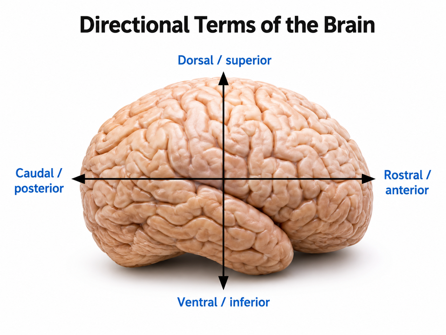
Networks carry both functional and anatomical names. For example, the dorsal attention network (DAN) corresponds to the dorsal frontoparietal network (D-FPN).
Brodmann Areas
The original Brodmann areas consisted of 47 numbered cytoarchitectural zones of the cerebral cortex based on Nissl staining. They require subdividing certain areas, and they vary in size and shape across individuals.
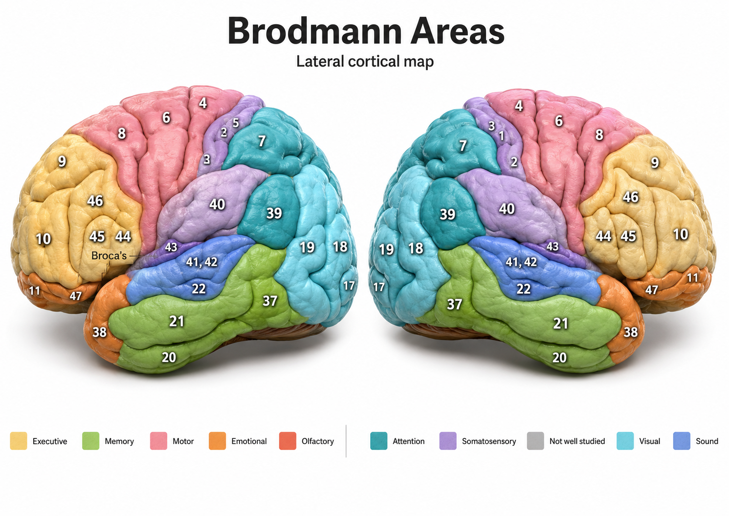
The numbered Brodmann areas of the cerebral cortex.
Gordon et al. (2016) compared the original Brodmann areas with several other brain atlases using fMRI. They concluded that Brodmann's parcellation captures real structure in the data but is too coarse to represent true cortical areas. Modern attempts to parcellate the cortex frequently reveal finer architectonic divisions than Brodmann reported. The more than 100-year-old Brodmann areas therefore lack some of the specificity needed for in vivo and functional studies.
Revised Brodmann maps reveal 180 regions per hemisphere, 97 of which were not previously identified (Glasser et al., 2016). Cognitive neuroscience tends to use modern brain atlases of defined regions (e.g., Yeo et al., 2011) and exact coordinates such as Montreal Neurological Institute (MNI) space and Talairach space (Chau & McIntosh, 2005). Researchers sometimes convert these coordinates to corresponding Brodmann areas because the areas are well known.
Brodmann areas participate in networks. They matter for neurofeedback because they help us target functional and structural networks rather than discrete, disconnected scalp sites. Neuroscience reveals the connections between Brodmann areas and how different networks become active or quiet together across diverse conditions.
For example, attention to an object's location engages the bilateral dorsal attention network (dorsal frontoparietal network), comprising the intraparietal sulcus and the frontal eye fields, while the largely right-lateralized ventral attention network (ventral frontoparietal network), comprising the temporoparietal junction and ventral frontal cortex, automatically processes unanticipated events. Flexible attention control involves the dynamic interaction of these top-down and bottom-up systems (Vossel et al., 2014).
Psychological disorders can be associated with shifts from normal activity in particular brain areas and their connections. After traumatic brain injury (TBI), neuronal connections change across the entire brain, and surviving long axonal projections no longer target inhibitory neurons (Frankowski et al., 2022).
Current neurofeedback protocols allow us to train networks involved in cognitive functions like attention or psychological disorders like depression using multiple electrodes simultaneously. Connectivity training enables us to increase or decrease communication between brain locations to treat symptoms and improve performance, and both activations and deactivations matter. Quantitative EEG (qEEG) normative databases can reveal the key parts of a network that need training and the required direction.
Networks, not isolated sites, mediate the cognitive and emotional processes we train. Functional networks show correlated activity over time, structural networks consist of axonal pathways, and the two overlap without being equivalent. A functional brain area is not a task area, so no single Brodmann area carries a complex function by itself. Brodmann's parcellation remains useful shorthand but is too coarse for modern work, which uses finer atlases and MNI or Talairach coordinates. Connectivity training targets communication between regions, and qEEG normative databases indicate which parts of a network to train and in which direction.
Check Your Understanding
- What is the difference between a functional network and a structural network, and why does correlated activity not prove an anatomical connection?
- What did the limb immobilization study by Newbold and Dosenbach (2021) demonstrate about how quickly functional connectivity can change?
- Why did Petersen and Fiez conclude that there is no "tennis forehand area" in the brain?
- On what grounds did Gordon and colleagues judge Brodmann's parcellation too coarse, and what do modern atlases offer instead?
- How does a network view change the way you decide where to place active electrodes?
Brain Organization and Dynamics
This section introduces the brain's network architecture, from large-scale systems to specific cortical regions. Understanding these systems is essential for neurofeedback practitioners, since many protocols target the networks described here.
The brain is organized into interactive, functional, distributed networks with spatial, temporal, and content-based relationships. These networks interact through feedback loops and transiently organized aggregates of neurons, all mediated by rhythmic, oscillatory electrical discharges that ultimately produce the EEG. This process is further shaped by selective attention to specific categories of interest.
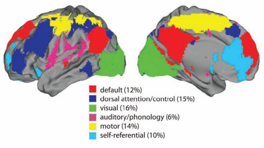
Each type of local cognitive, sensory processing, or emotional network produces oscillatory activity and contains internal stabilizing characteristics. These local networks exist within a global dynamic network system that links and provides interactive capacity to the smaller networks, also operating within an oscillatory framework.
A densely connected lateral prefrontal and posterior parietal cortical network orchestrates responses to novel cognitive tasks using flexible hubs. This frontoparietal network assigns tasks to the most appropriate brain regions and shares information among them to master new skills (Cole et al., 2013).
The central nervous system processes incoming content through separate regions that handle specialized input (e.g., auditory, kinesthetic, tactile, visual). Content is shared, integrated, compared to previous content, and analyzed, and decisions are made about memory and responses. All of this activity occurs within interacting networks linked by electrical and chemical signals, and the electrical discharges from this activity are recorded from the scalp surface as the EEG.
Network Overview
The networks most relevant to attention include the oculomotor, motor, affective, central autonomic, social, and executive circuits. Each plays a distinct role in how we focus, respond, and regulate behavior.
Oculomotor Network
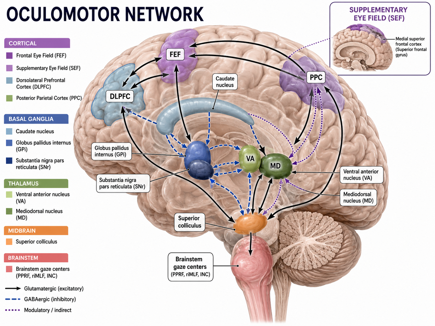
The frontal eye field (FEF), in concert with the dorsolateral prefrontal cortex, posterior parietal cortex, basal ganglia, and thalamus, programs and initiates voluntary eye movements, inhibits eye movements toward distracting stimuli, and allows us to return our focus to locations we have previously attended (Thompson & Thompson, 2015).
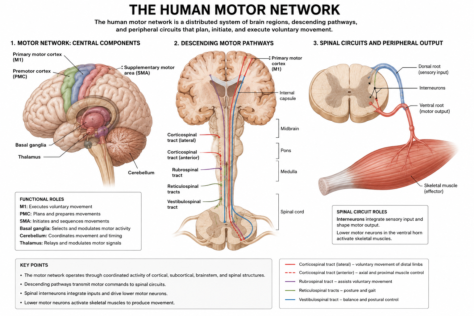
Motor Network
The supplementary motor area (SMA), in concert with the premotor cortex, primary motor cortex, sensorimotor cortex, and cerebellum, plans, initiates, and inhibits voluntary movements and muscle contractions (Breedlove & Watson, 2023; Thompson & Thompson, 2015). Understanding the motor network helps clinicians appreciate why surface EMG (SEMG) biofeedback and sensorimotor rhythm (SMR) neurofeedback can influence broader neural circuits.
Somato-Cognitive Action Network (SCAN)
Gordon and colleagues (2023) used precision fMRI from seven participants together with datasets from the Adolescent Brain Cognitive Development Study, the Human Connectome Project, and the UK Biobank, drawing on roughly 50,000 individuals. They identified three interconnected primary motor cortex (M1) regions that participate in the integrated movement of multiple body parts.
The somato-cognitive action network (SCAN) consists of three inter-effector regions of M1, the SMA, the dorsal anterior cingulate cortex, the centromedian and ventral intermediate nuclei of the thalamus, the posterior putamen, and the vermis and flocculonodular lobe of the cerebellum, which mediate posture and balance. Connectivity analysis showed that the SCAN communicates with the cingulo-opercular network, or salience network, which supports cognitive control and sustains task focus over extended periods.
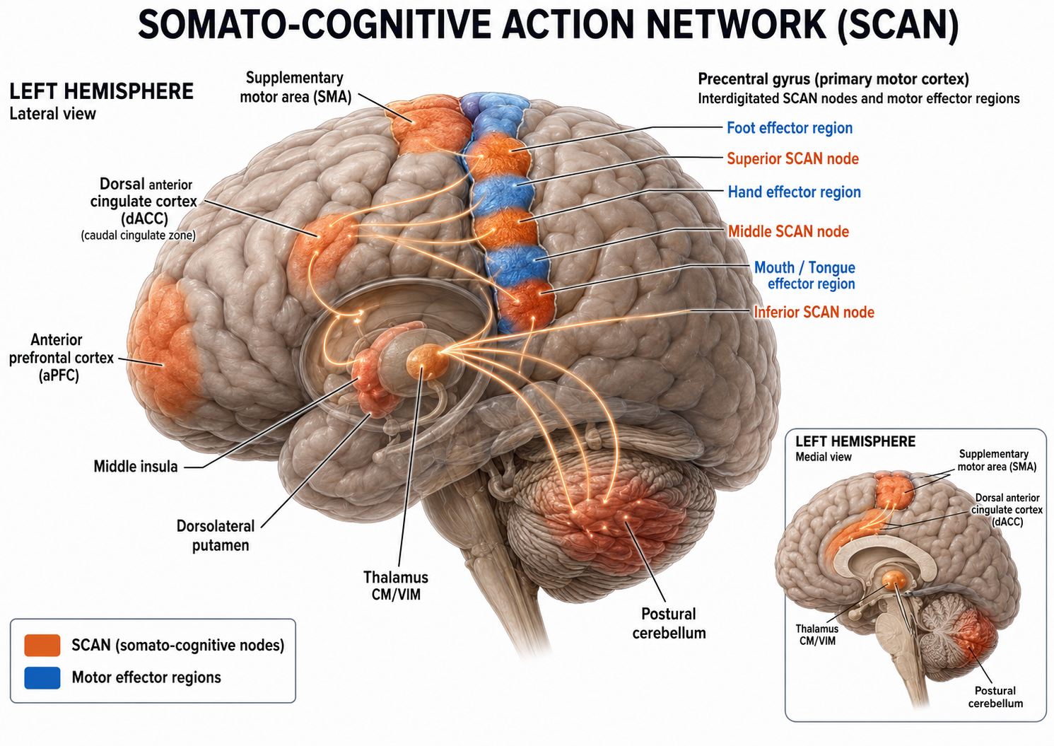
The somato-cognitive action network (SCAN).
M1's two interlacing systems establish a pattern of integration and isolation. Effector-specific regions for the foot, hand, and mouth support the isolation of fine motor control, while the SCAN integrates goals, body movement, and physiology.
The relative expansion of SCAN regions in humans may support uniquely human integrated actions, such as coordinating breathing for speech and combining hand, body, and eye movement for tool use. By enabling anticipatory postural, respiratory, cardiovascular, and arousal adjustments before action, such as shoulder tension, increased heart rate, or butterflies in the stomach, the SCAN provides a substrate that may help explain why mental and bodily states so often interact.
Mesa (2023) placed these findings in context. The dominant paradigm has treated the motor cortex as a simple relay that passes movement commands to muscles, with planning, cognition, and conscious initiation of movement occurring elsewhere in the brain.
The SCAN, in concert with the salience network, supports complex adaptations such as allostasis. These findings agree with primate studies showing that more M1 neurons govern movements independent of the specific muscles used than govern the contraction of particular muscles (Griffin et al., 2015; Kaufman et al., 2014). Together they challenge the cortical homunculus that Penfield and colleagues mapped in the 1930s, the distorted human figure whose body parts are sized by the amount of cortical area dedicated to them.
Affective Network
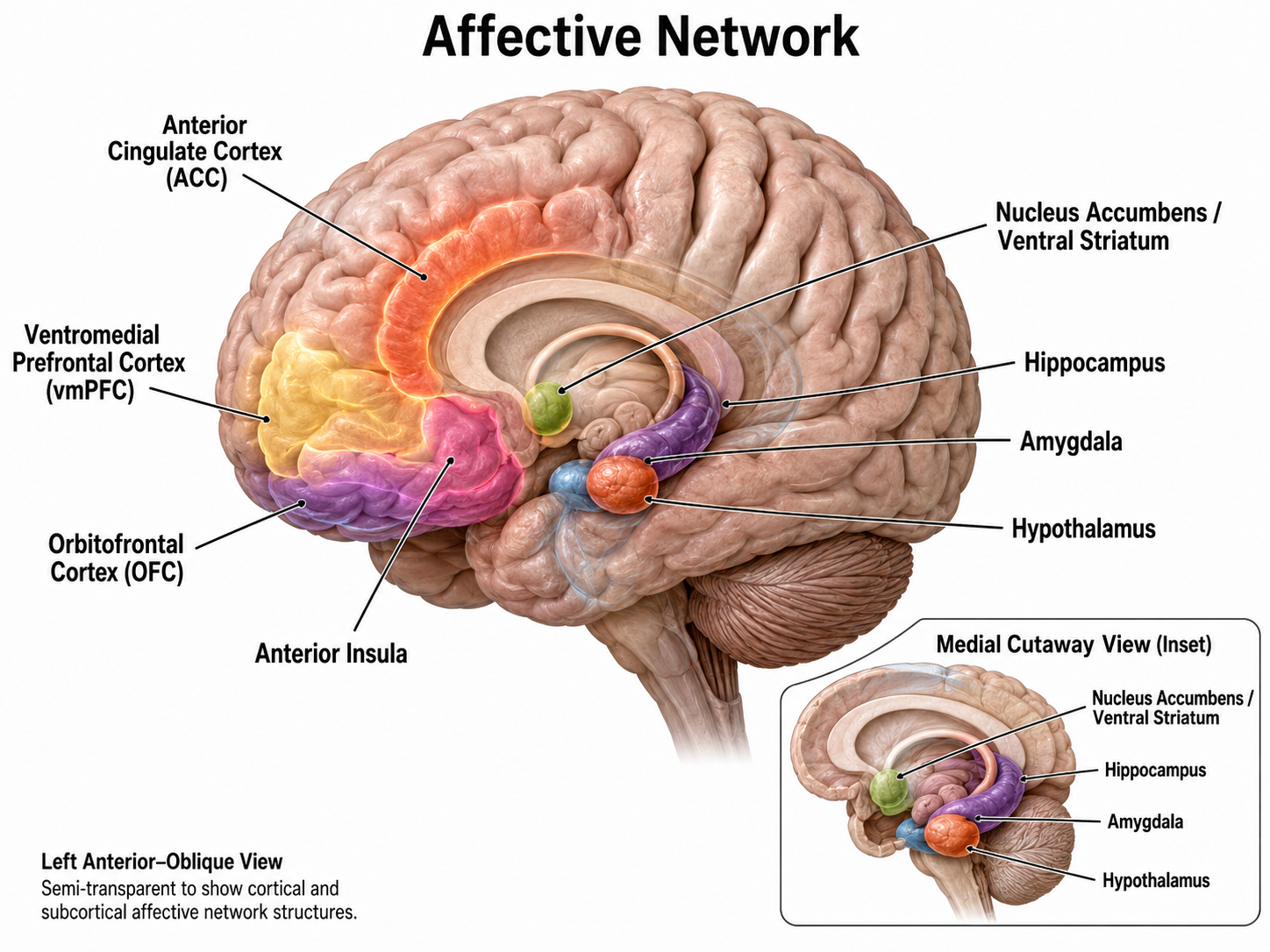
The pre- and subgenual (beneath the curved "knee") areas of the anterior cingulate cortex (ACC) participate in affective circuits triggered when we make mistakes (Arnsten, 2009). The dorsal rostral cingulate zone monitors cognitive activity to predict when errors are likely and greater executive control may be needed (Thompson & Thompson, 2015). The ventromedial prefrontal cortex projects to the amygdala, basal ganglia, hypothalamus, and brainstem arousal and reward pathways. This network is relevant to neurofeedback for anxiety and mood disorders.
Central Autonomic Network
The central autonomic network (CAN) consists of forebrain, limbic, and brainstem regions (Benarroch, 1993). Neuroimaging studies reveal a cortico-limbic network for autonomic control that includes the ventromedial prefrontal cortex, cingulate cortex, insula, mediodorsal thalamus, hypothalamus, and amygdala (Beissner et al., 2013; Schumann et al., 2021; Shoemaker et al., 2015).
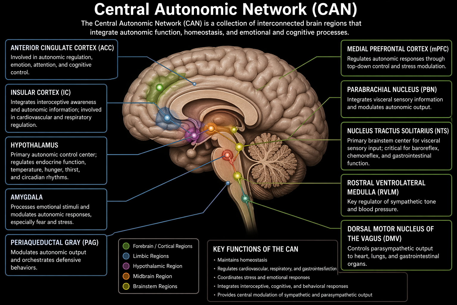
Thayer and Lane (2000) proposed a neurovisceral integration model that places the prefrontal cortex at the top of a hierarchy, with direct functional links to the insula and cingulate. The limbic system extends these connections through the amygdala to downstream subcortical regions, such as the hypothalamus and brainstem nuclei, that drive parasympathetic and sympathetic heart rate modulation. The model highlights how prefrontal control over subcortical structures links sympathovagal balance with cognitive and emotional processes. This framework is central to HRV biofeedback, which trains the prefrontal and autonomic circuitry that governs heart rate regulation.
Magnetic resonance imaging (MRI) and resting-state functional connectivity (RSFC) studies have reinforced the role of medial prefrontal and limbic interplay in heart rate control (Kumral et al., 2019; Sakaki et al., 2016). Individuals with slower heart rates show heightened RSFC within a network of central autonomic and sensorimotor regions compared with those who have faster heart rates (de la Cruz et al., 2019). Slower heart rates were associated with elevated RSFC between the ventromedial prefrontal cortex and the anterior insula.
Social Network
The orbitofrontal cortex (OFC), along with the basal ganglia and thalamus, orchestrates the highest level of emotional processing in the nervous system. The social network is responsible for socially responsible behavior, empathy, behavioral inhibition, emotional regulation, and sound judgment (Thompson & Thompson, 2015).
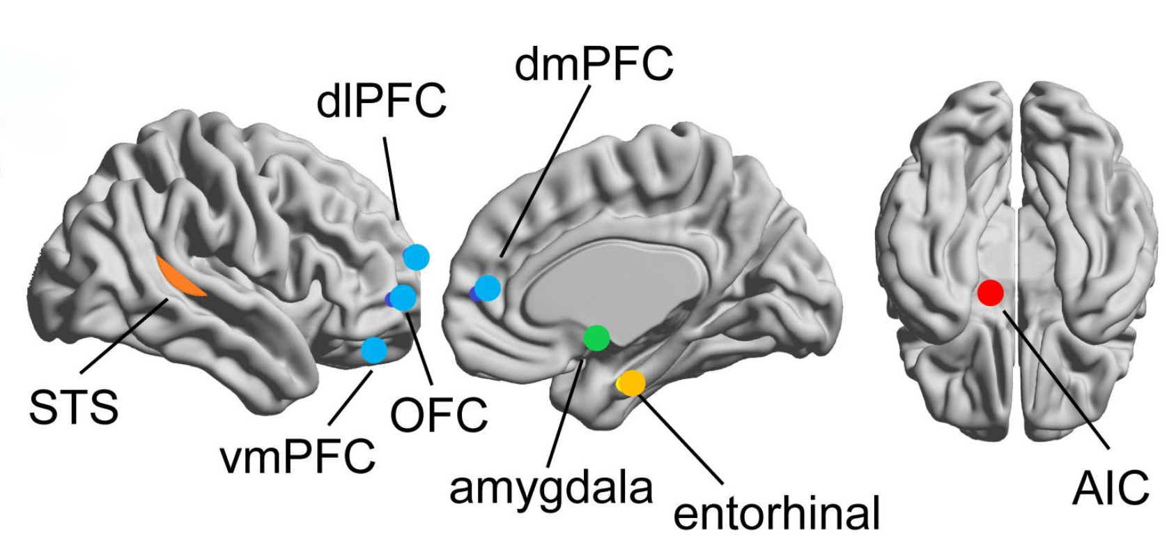
A mentalizing network that includes the ventromedial, orbitofrontal, dorsolateral, and dorsomedial prefrontal cortices lets us reason about our own and others' mental states (Hoskinson et al., 2019). A mirror network centered on the superior temporal sulcus is active during our own actions and our observation of others' actions, supporting observational learning and social cognition (Sadeghi et al., 2022). The amygdala detects salient stimuli, while the entorhinal cortex and anterior insular cortex contribute to this social circuitry.
Executive Network
The dorsolateral prefrontal cortex plays a critical role in executive functions, which Kropotov (2009) described as the coordination and control of motor and cognitive actions to attain specific goals. Executive functions include allocation of attention, cognitive inhibition, behavioral inhibition, working memory, and cognitive flexibility. The executive network focuses and maintains continuous attention (Faraone et al., 2006) and shows reduced activation and connectivity in ADHD, making it a primary target of neurofeedback protocols for ADHD and other attention-related conditions.
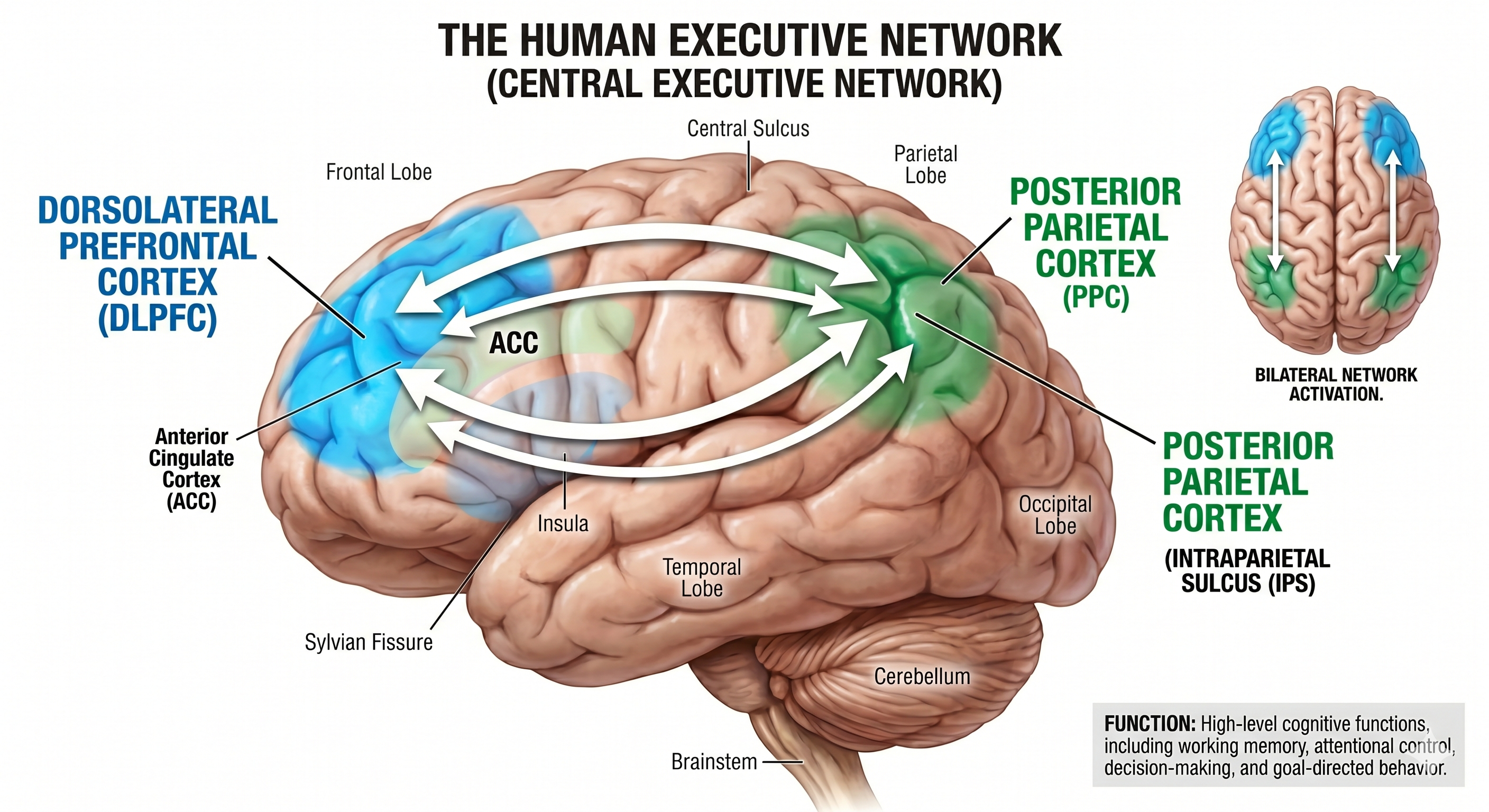
Local oscillatory networks sit inside a global dynamic system, and the electrical discharges from their interaction become the scalp EEG. The oculomotor network programs voluntary gaze and suppresses distraction, while the motor network plans and inhibits movement. The somato-cognitive action network interleaves with effector-specific motor regions and links goals, movement, and physiology, challenging the classic homunculus. The affective network monitors errors and predicts when executive control is needed, the central autonomic network governs sympathovagal balance through a prefrontal hierarchy, the social network supports empathy and judgment, and the executive network allocates attention and working memory.
Check Your Understanding
- How do local oscillatory networks relate to the global dynamic network system, and where does the EEG come from in this picture?
- What does the somato-cognitive action network integrate, and why does it challenge the cortical homunculus model?
- Which structures make up the central autonomic network, and what does the neurovisceral integration model add to that anatomy?
- What relationship did de la Cruz and colleagues find between resting heart rate and resting-state functional connectivity?
- Why is the executive network a primary target for ADHD neurofeedback protocols?
Attentional Processes
Attention is the selection of sensory information or cognition for enhanced processing. We can overtly or covertly attend to stimuli. In overt attention, our attentional focus and sensory orientation coincide. For example, you parse this sentence as you focus your gaze on it.
In covert attention, we shift our attentional focus from our sensory orientation. For example, you attend to a reminder on the corner of your screen while gazing at this sentence. While the midbrain superior colliculus is mainly implicated in overt attention, it may also regulate covert attention (Breedlove & Watson, 2023).
Attention is more selective than arousal, which is our overall level of alertness (Breedlove & Watson, 2023).
Research using divided attention tasks, where subjects simultaneously process multiple stimuli, shows that attentional resources are finite. The challenge of attending to more than one target increases when targets occupy different spatial locations. When we attend to a stimulus, we shift our attentional spotlight (focus) to select that stimulus for enhanced analysis (Bee & Micheyl, 2008).
Perceptual load (stimulus processing demands) determines the level at which an attentional bottleneck (stimulus selection) occurs. Complex stimuli involving a high perceptual load monopolize processing resources, resulting in early selection, where we filter out lower-priority competing stimuli before preliminary perceptual and semantic analysis.
Conversely, simple stimuli involving a low perceptual load leave free processing resources, resulting in late selection, where we filter out competing stimuli after performing extensive analysis (Lavie et al., 2004). This distinction has practical implications for neurofeedback, since training in high-demand environments may engage different attentional mechanisms than training in quiet clinical settings.
Cortical Regions That Guide Attention
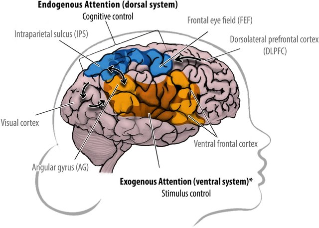
The dorsal frontoparietal system, comprising the intraparietal sulcus and frontal eye field, is responsible for the top-down direction of attention (Breedlove & Watson, 2023). The intraparietal sulcus (IPS), located in the parietal lobe, provides voluntary top-down steering of attention (Corbetta & Shulman, 1998).
The frontal eye field (FEF), found in the premotor region of the frontal lobes, directs gaze toward targets selected by the IPS (Paus et al., 1991). Target selection is guided by cognitive goals (top-down processing) rather than stimulus characteristics (bottom-up processing).
In contrast, the temporoparietal junction (TPJ), where the superior temporal gyrus and inferior parietal lobe intersect, mediates bottom-up shifts in attention in response to stimulus attributes (Corbetta & Shulman, 2002). The TPJ functions like a circuit breaker, overruling immediate attentional priorities and reallocating attentional resources to a new target (Breedlove & Watson, 2023).
Two Cortical Networks Regulate Attention
Two cortical networks cooperatively regulate subcortical and cortical systems to produce a coherent perceptual experience (Breedlove & Watson, 2023). A dorsal frontoparietal system provides top-down control of endogenous attention (voluntary attention), directing the attentional spotlight to support cognitive system priorities.
A right temporoparietal system provides bottom-up control of exogenous attention (involuntary reflexive attention), redirecting attention based on the novelty or importance of incoming stimuli.
Extensive interconnections between the two networks allow us to fluidly redirect attention from stimuli that are forebrain priorities (IPS) to those that are unexpected. This balance between top-down and bottom-up attention is a key consideration in neurofeedback training for attention disorders.
Salience Network
The salience network comprises structures that monitor our external and internal environments to determine which inputs are essential and require further processing and attention. The insula, primarily the anterior insula, is a crucial component because it facilitates bottom-up access to the brain's attentional and working memory resources (Menon & Uddin, 2010). The cingulate gyrus, particularly the right dorsal anterior cingulate cortex, is another crucial component (Thompson & Thompson, 2015).
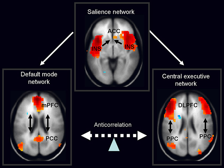
The salience network. Graphic shared by Nekovarova, Fajnerova, Horacek, and Spaniel under the Creative Commons Attribution 3.0 license.
The clinical literature on ADHD, depression, and schizophrenia has explored the role of the anterior cingulate cortex in these disorders. Deep brain stimulation of this region has successfully improved treatment-resistant depression (Mayberg et al., 2005).
The insula, a cortical region located within the lateral sulcus, functions as an integrative hub for the salience network. The insula integrates interoceptive awareness (perception of internal body signals), emotional experience, and external perception to facilitate an individual's global perception of the world. It directs specific networks in processing salient stimuli and generating appropriate responses (Wiebking & Northoff, 2014). For biofeedback practitioners, the insula's role in interoceptive awareness is particularly relevant, since biofeedback training fundamentally depends on clients learning to detect and interpret internal signals.
The insula appears to provide an interface between the brain's cognitive, homeostatic, and affective systems, linking the areas involved in monitoring internal signals with those engaged in processing incoming external sensory streams. The insula detects salient events via afferent pathways and switches between other large-scale networks when these events are recognized, thereby guiding attention and working memory.
The anterior and posterior insula interact to regulate autonomic responses to salient stimuli. Interactive communication between the insula and anterior cingulate cortex facilitates access to the motor system (Menon & Uddin, 2010).
This network appears to help us switch between the task-oriented (executive) network and the internally focused default mode network (Seeley et al., 2007; Shirer et al., 2012).
Default Mode Network (DMN)
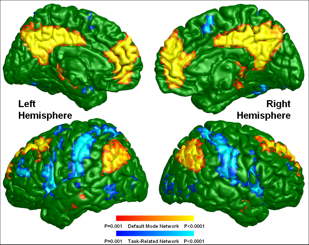
Brain regions are selectively active when we are conscious (Breedlove & Watson, 2023). The default mode network (DMN) consists of frontal, temporal, and parietal lobe circuits active during spontaneous cognition like introspection, daydreaming, and streams of consciousness. The DMN appears to contribute flexible memory retrieval and idea generation, critical elements of creativity, and is relatively inactive when pursuing external goals (Andrews-Hanna et al., 2010). In neurofeedback practice, understanding the DMN helps explain why some clients need to learn to suppress mind-wandering, as in ADHD, while others may benefit from cultivating it, as in creative performance training.
ADHD has been linked to irregular connectivity among brain regions, including within the DMN (Cao et al., 2014). The strength of specific brain connections can forecast variations in a person's capacity to sustain attention, and this holds true even at rest when the individual is not engaged in a specific task (Rosenberg et al., 2016, 2017).
There has been increasing discussion about whether some DMN regions belong to a distinct Parietal Memory Network that includes the precuneus, the mid-cingulate cortex, and the posterior inferior parietal lobule and dorsal angular gyrus (Gilmore et al., 2015; Hu et al., 2016).
Deactivations can be as important as activations. The degree of DMN deactivation appears critical for attentional control, since people who suppress it more readily can learn new material more easily (Nelson et al., 2016; Zerr et al., 2018). The DMN also helps synthesize details into single coherent events and supports envisioning the future (Gilmore et al., 2018).
The DMN may contribute to creative fluency, the ability to generate innovative ideas like alternative uses for everyday objects. A study of neurosurgical patients showed that left DMN stimulation reduced the number of uses generated but not their originality (Shofty et al., 2022).
Understanding Ourselves
The posterior cingulate cortex (PCC) and precuneus combine bottom-up attention with information from memory and perception. The ventral (lower) part of the PCC activates in all tasks involving the DMN, including those related to the self or others, remembering the past, thinking about the future, processing concepts, and spatial navigation. The dorsal (upper) part of PCC mediates involuntary awareness and arousal. The precuneus is concerned with visual, sensorimotor, and attentional information.
The medial prefrontal cortex (mPFC) participates in decisions about the self, such as personal information, autobiographical memories, future goals and events, and decision-making regarding those close to us like family members. The ventral (lower) part is involved in positive emotional information and reward.
The angular gyrus connects perception, attention, spatial cognition, and action and helps us recall episodic memories.
Understanding Others
The major functional hubs include the PCC, mPFC, and angular gyrus. The dorsal medial prefrontal cortex (dmPFC) participates in analyzing others' objectives. The temporoparietal junction (TPJ) contributes to theory of mind, models of others' cognitive processes, emotions, knowledge, and motivation. The lateral temporal cortex is concerned with short-term verbal memory, naming, and reading. Finally, the anterior temporal pole is part of a bilateral semantic system representing object concepts and a left hemisphere-dominant network concerned with naming and understanding object names.
Autobiography and Future Simulations
The major functional hubs include the PCC, mPFC, and angular gyrus. The hippocampus forms new declarative memories. The parahippocampal cortex (PHC) mediates spatial memory, navigation, and high-level visual processing like facial recognition. The retrosplenial cortex (RSC) is involved in episodic memory, navigation, predicting future events, and analyzing visual scenes. Finally, the posterior inferior parietal lobe (pIPL) integrates sensory information and participates in top-down attentional orienting.
The Pulvinar Mediates Attentional Shifts
The pulvinar nucleus, comprising the posterior quarter of the human thalamus, processes visual information and directs attention. The pulvinar shares widespread connections with the cingulate, parietal cortex, and superior colliculus, and is crucial for orienting, shifting attention, and filtering out irrelevant stimuli. Tasks that present subjects with more distracting stimuli increase pulvinar activation, as shown by functional MRI (fMRI) (Buchsbaum et al., 2006).
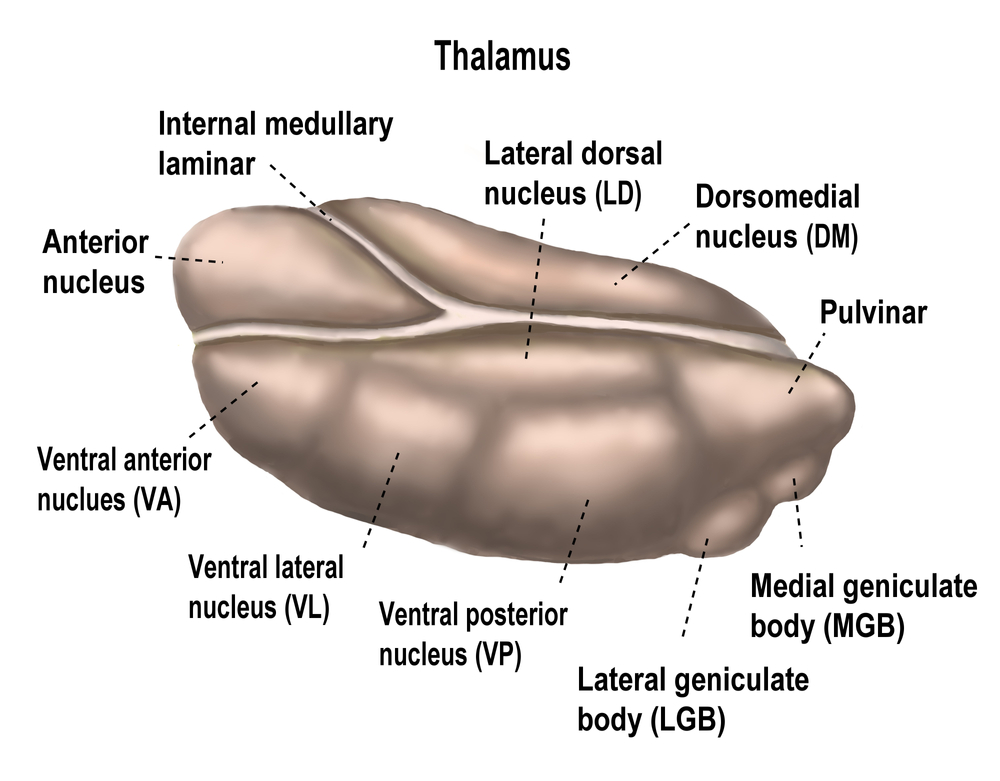
Overall, the pulvinar guides the processing of relevant information across wide-ranging cortical networks based on dynamically changing attentional priorities (Breedlove & Watson, 2023; Saalmann et al., 2012).
Attention selects information for enhanced processing and is more selective than arousal. Overt attention aligns focus with sensory orientation, while covert attention decouples them. Perceptual load determines whether stimulus selection happens early or late. A dorsal frontoparietal system steers endogenous, top-down attention through the intraparietal sulcus and frontal eye field, while a right temporoparietal system redirects exogenous, bottom-up attention. The salience network, anchored by the anterior insula and dorsal anterior cingulate, switches between the executive and default mode networks, and the pulvinar filters distraction across cortical networks.
Check Your Understanding
- How do overt and covert attention differ, and which midbrain structure is chiefly implicated in each?
- How does perceptual load determine whether early or late selection occurs, and why might that matter for training environments?
- What roles do the intraparietal sulcus, frontal eye field, and temporoparietal junction each play in directing attention?
- Why is the insula described as an integrative hub, and why is its interoceptive role especially relevant to biofeedback?
- Why can default mode network deactivation matter as much as activation for attentional control?
Training Networks Summary
Neurofeedback training increasingly monitors and trains network activity using qEEG normative databases. To employ these protocols effectively, neurofeedback professionals must thoroughly understand Brodmann areas and the functional and structural networks in which they participate. The dorsal frontoparietal system provides top-down attentional control while the temporoparietal system enables bottom-up redirection, with the salience network serving as the switch between them. This network architecture provides the rationale for targeting specific networks and frequencies to address attention disorders, optimize performance, and restore healthy brain function.
Assignment
Choose one client presentation you see often, such as inattention, anxiety, or performance under pressure. Identify which of the networks in this chapter you would expect to be involved, and explain how you would decide between a single-channel protocol and a connectivity protocol. What would a qEEG normative database need to show you before you committed to the connectivity approach?
Glossary
affective network: a network including the anterior cingulate, entorhinal, and temporal cortices, posterior parietal cortex, globus pallidus, substantia nigra pars reticulata, and mediodorsal thalamus that monitors performance and signals when greater executive control is needed.
allostasis: the process of achieving stability through anticipatory or reactive physiological and behavioral change.
amygdala: a group of medial temporal-lobe nuclei involved in salience evaluation, associative learning, affective memory, and coordination of defensive and autonomic responses.
anterior cingulate cortex (ACC): medial frontal cortex involved in cognitive control, motivation, affect, pain processing, and autonomic regulation, with functionally heterogeneous subdivisions.
arousal: a multidimensional state of wakefulness, alertness, and physiological activation supported by distributed brainstem, hypothalamic, and forebrain systems.
attention: the selection of sensory information or cognition for enhanced processing.
attentional bottleneck: a filter that limits enhanced processing to only the highest priority stimuli.
attentional spotlight: a shift of selective attention to choose stimuli for enhanced processing.
biofeedback: a learning process that uses measurements of physiological activity to provide feedback through which a person can acquire greater self-regulation.
central autonomic network (CAN): interconnected forebrain, hypothalamic, brainstem, and spinal regions that integrate internal-state information and regulate autonomic output.
covert attention: an attentional focus independent of sensory orientation.
creative fluency: the number of distinct usable ideas a person generates in a set time, as when listing alternative uses for everyday objects.
default mode network (DMN): a cortical network of sites located in frontal, temporal, and parietal regions that is most active during introspection and daydreaming and relatively inactive when pursuing external goals.
divided attention tasks: situations where subjects must simultaneously process two or more stimuli.
dorsal frontoparietal system: the network comprised of the intraparietal sulcus and frontal eye field responsible for the top-down direction of attention.
dorsolateral prefrontal cortex: lateral prefrontal cortex involved in working memory, cognitive control, planning, rule use, and goal-directed behavior; simple left-positive and right-negative mappings are inadequate.
early selection: the filtering out of lower-priority competing stimuli before preliminary perceptual and semantic analysis.
endogenous attention: a voluntary form of attention that directs the attentional spotlight to support cognitive system priorities.
executive network: a brain network involved in high-level cognitive functions such as working memory, problem solving, and attention, primarily including the dorsolateral prefrontal cortex and the posterior parietal cortex.
exogenous attention: an involuntary, reflexive form of attention redirected by the novelty or importance of incoming stimuli.
frontal eye field (FEF): a premotor region in the caudal middle frontal gyrus at its junction with the precentral gyrus, corresponding to Brodmann area 8, that controls voluntary eye movements and directs gaze toward attended targets.
functional networks: sets of brain regions whose activity is correlated over time, in contrast to structural networks defined by axonal projections and pathways.
hippocampus: a medial temporal-lobe structure essential to episodic-memory formation, spatial representation, and contextual regulation of stress and emotion.
insula: cortex buried within the lateral sulcus that integrates interoceptive, affective, sensory, cognitive, and autonomic information.
intraparietal sulcus (IPS): the region of the parietal lobe that provides voluntary top-down steering of attention.
late selection: filtering out competing stimuli after performing extensive analysis.
magnetic resonance imaging (MRI): a noninvasive imaging technology that uses a strong magnetic field and radio waves to produce detailed, high-resolution, three-dimensional images of the body.
neurofeedback: biofeedback in which measured brain activity is converted into contingent information to support learned modification of selected neural features.
neurovisceral integration model: Thayer and Lane's model linking prefrontal-subcortical regulation within a central autonomic network to cardiac vagal control and adaptive self-regulation.
oculomotor network: a network that programs and initiates voluntary eye movements, inhibits eye movements toward distracting stimuli, and allows us to return our focus to locations we've experienced in the past.
orbitofrontal cortex: prefrontal cortex above the orbits involved in valuation, outcome updating, emotion, sensory integration, and context-appropriate behavior.
overt attention: the agreement between attentional focus and sensory orientation.
perceptual load: the amount of sensory information a task requires a person to process, which determines how much capacity remains for irrelevant distractors.
prefrontal cortex (PFC): the anterior association cortex of the frontal lobes involved in executive control, planning, working memory, valuation, social cognition, and emotion regulation.
pulvinar nucleus: the posterior region of the thalamus that processes visual information and directs attention.
resting-state functional connectivity (RSFC): a neuroimaging method that identifies brain networks whose regions show synchronous activity when a person is not performing an explicit task.
salience network: a large-scale network centered on anterior insula and dorsal anterior cingulate cortex that detects behaviorally relevant events and coordinates network switching.
sensorimotor rhythm (SMR) neurofeedback: an operant conditioning protocol that trains the client to increase EEG amplitude in the 12 to 15 Hz band recorded over the sensorimotor cortex (typically C3, Cz, or C4 along the central strip).
social network: a set of social actors represented as nodes and their relationships represented as ties.
somato-cognitive action network (SCAN): a network of inter-effector regions in the primary motor cortex, together with the supplementary motor area, dorsal anterior cingulate cortex, centromedian and ventral intermediate thalamic nuclei, and posterior putamen, that integrates action goals, whole-body movement, and physiology.
structural networks: axonal projections and pathways.
superior colliculus: the dorsal midbrain structure that receives visual information and directs visual gaze and attention to selected stimuli.
supplementary motor area (SMA): the medial premotor region that, with the premotor cortex, primary motor cortex, sensorimotor cortex, and cerebellum, plans, initiates, and inhibits voluntary movements and muscle contractions.
surface-EMG biofeedback: biofeedback displaying surface-EMG activity to train reduction, activation, timing, or coordination of skeletal-muscle activity.
temporoparietal junction (TPJ): the intersection of the superior temporal gyrus and inferior parietal lobe that mediates bottom-up shifts in attention in response to stimulus attributes.
ventromedial prefrontal cortex: a medial and ventral prefrontal region involved in valuation, emotion regulation, extinction, decision-making, and integration of affective and autonomic information.
Test Yourself on ClassMarker
Click the button below to take a 10-question exam over this entire unit. There is no password.
Review Flash Cards on Quizlet
Click the button below to review our chapter flash cards.
References
Alexander, G. E., DeLong, M. R., & Strick, P. L. (1986). Parallel organization of functionally segregated circuits linking basal ganglia and cortex. Annual Review of Neuroscience, 9, 357-381. https://doi.org/10.1146/annurev.ne.09.030186.002041
Andrews-Hanna, J. R., Reidler, J. S., Huang, C., & Buckner, R. L. (2010). Evidence for the default network's role in spontaneous cognition. Journal of Neurophysiology, 104, 1664-1671. https://doi.org/10.1152/jn.00830.2009
Arnsten, A. F. (2009). Stress signaling pathways that impair prefrontal cortex structure and function. Nature Reviews Neuroscience, 10(6), 410-422. https://doi.org/10.1038/nrn2648
Bee, M. A., & Micheyl, C. (2008). The cocktail party problem: What is it? How can it be solved? And why should animal behaviorists study it? Journal of Comparative Psychology, 122(3), 235-251. https://doi.org/10.1037/0735-7036.122.3.235
Beissner, F., Meissner, K., Bär, K. J., & Napadow, V. (2013). The autonomic brain: An activation likelihood estimation meta-analysis for central processing of autonomic function. The Journal of Neuroscience, 33(25), 10503-10511. https://doi.org/10.1523/JNEUROSCI.1103-13.2013
Benarroch, E. E. (1993). The central autonomic network: Functional organization, dysfunction, and perspective. Mayo Clinic Proceedings, 68(10), 988-1001. https://doi.org/10.1016/s0025-6196(12)62272-1
Breedlove, S. M., & Watson, N. V. (2023). Behavioral neuroscience (10th ed.). Sinauer Associates, Inc.
Buchsbaum, M. S., Buchsbaum, B. R., Chokron, S., Tang, C., Wei, T. C., & Byne, W. (2006). Thalamocortical circuits: fMRI assessment of the pulvinar and medial dorsal nucleus in normal volunteers. Neuroscience Letters, 404(3), 282-287. https://doi.org/10.1016/j.neulet.2006.05.063
Cao, M., Shu, N., Cao, Q., Wang, Y., & He, Y. (2014). Imaging functional and structural brain connectomics in attention-deficit/hyperactivity disorder. Molecular Neurobiology, 50(3), 1111-1123. https://doi.org/10.1007/s12035-014-8685-x
Chau, W., & McIntosh, A. R. (2005). The Talairach coordinate of a point in the MNI space: How to interpret it. NeuroImage, 25(2), 408-416. https://doi.org/10.1016/j.neuroimage.2004.12.007
Cole, M. W., Reynolds, J. R., Power, J. D., Repovs, G., Anticevic, A., & Braver, T. S. (2013). Multi-task connectivity reveals flexible hubs for adaptive task control. Nature Neuroscience, 16, 1348-1355. https://doi.org/10.1038/nn.3470
Corbetta, M., & Shulman, G. I. (1998). Human cortical mechanisms of visual attention during orienting and search. Philosophical Transactions of the Royal Society of London. Series B: Biological Sciences, 353(1373), 1353-1362. https://doi.org/10.1098/rstb.1998.0289
Corbetta, M., & Shulman, G. I. (2002). Control of goal-directed and stimulus-driven attention in the brain. Nature Reviews: Neuroscience, 3(3), 201-215. https://doi.org/10.1038/nrn755
de la Cruz, F., Schumann, A., Köhler, S., Reichenbach, J. R., Wagner, G., & Bär, K. J. (2019). The relationship between heart rate and functional connectivity of brain regions involved in autonomic control. NeuroImage, 196, 318-328. https://doi.org/10.1016/j.neuroimage.2019.04.014
Faraone, S. V., Biederman, J., & Mick, E. (2006). The age-dependent decline of attention deficit hyperactivity disorder: A meta-analysis of follow-up studies. Psychological Medicine, 36(2), 159-165. https://doi.org/10.1017/S003329170500471X
Fornito, A., Zalesky, A., & Bullmore, E. T. (Eds.). (2016). Fundamentals of brain network analysis. Elsevier. https://doi.org/10.1016/C2012-0-06036-X
Frankowski, J. C., Tierno, A., Pavani, S., Cao, Q., Lyon, D. C., & Hunt, R. F. (2022). Brain-wide reconstruction of inhibitory circuits after traumatic brain injury. Nature Communications, 13, 3417. https://doi.org/10.1038/s41467-022-31072-2
Gilmore, A. W., Nelson, S. M., Chen, H. Y., & McDermott, K. B. (2018). Task-related and resting-state fMRI identify distinct networks that preferentially support remembering the past and imagining the future. Neuropsychologia, 110, 180-189. https://doi.org/10.1016/j.neuropsychologia.2017.06.016
Gilmore, A. W., Nelson, S. M., & McDermott, K. B. (2015). A parietal memory network revealed by multiple MRI methods. Trends in Cognitive Sciences, 19(9), 534-543. https://doi.org/10.1016/j.tics.2015.07.004
Glasser, M. F., Coalson, T. S., Robinson, E. C., Hacker, C. D., Harwell, J., Yacoub, E., Ugurbil, K., Andersson, J., Beckmann, C. F., Jenkinson, M., Smith, S. M., & Van Essen, D. C. (2016). A multi-modal parcellation of human cerebral cortex. Nature, 536(7615), 171-178. https://doi.org/10.1038/nature18933
Gordon, E. M., Chauvin, R. J., Van, A. N., Rajesh, A., Nielsen, A., Newbold, D. J., Lynch, C. J., Seider, N. A., Krimmel, S. R., Scheidter, K. M., Monk, J., Miller, R. L., Metoki, A., Montez, D. F., Zheng, A., Elbau, I., Madison, T., Nishino, T., Myers, M. J., . . . Dosenbach, N. U. F. (2023). A somato-cognitive action network alternates with effector regions in motor cortex. Nature, 617(7960), 351-359. https://doi.org/10.1038/s41586-023-05964-2
Gordon, E. M., Laumann, T. O., Adeyemo, B., Huckins, J. F., Kelley, W. M., & Petersen, S. E. (2016). Generation and evaluation of a cortical area parcellation from resting-state correlations. Cerebral Cortex, 26(1), 288-303. https://doi.org/10.1093/cercor/bhu239
Griffin, D. M., Hoffman, D. S., & Strick, P. L. (2015). Corticomotoneuronal cells are "functionally tuned." Science, 350(6261), 667-670. https://doi.org/10.1126/science.aaa8035
Hoskinson, K. R., Bigler, E. D., Abildskov, T. J., Dennis, M., Taylor, H. G., Rubin, K., Gerhardt, C. A., Vannatta, K., Stancin, T., & Yeates, K. O. (2019). The mentalizing network and theory of mind mediate adjustment after childhood traumatic brain injury. Social Cognitive and Affective Neuroscience, 14(12), 1285-1295. https://doi.org/10.1093/scan/nsaa006
Hu, Y., Wang, J., Li, C., Wang, Y. S., Yang, Z., & Zuo, X. N. (2016). Segregation between the parietal memory network and the default mode network: Effects of spatial smoothing and model order in ICA. Science Bulletin, 61(24), 1844-1854. https://doi.org/10.1007/s11434-016-1202-z
Kaufman, M. T., Churchland, M. M., Ryu, S. I., & Shenoy, K. V. (2014). Cortical activity in the null space: Permitting preparation without movement. Nature Neuroscience, 17(3), 440-448. https://doi.org/10.1038/nn.3643
Kropotov, J. D. (2009). Quantitative EEG, event-related potentials and neurotherapy. Academic Press, Elsevier.
Kumral, D., Schaare, H. L., Beyer, F., Reinelt, J., Uhlig, M., Liem, F., Lampe, L., Babayan, A., Reiter, A., Erbey, M., Roebbig, J., Loeffler, M., Schroeter, M. L., Husser, D., Witte, A. V., Villringer, A., & Gaebler, M. (2019). The age-dependent relationship between resting heart rate variability and functional brain connectivity. NeuroImage, 185, 521-533. https://doi.org/10.1016/j.neuroimage.2018.10.027
Lavie, N., Hirst, A., de Fockert, J. W., & Viding, E. (2004). Load theory of selective attention and cognitive control. Journal of Experimental Psychology: General, 133(3), 339-354. https://doi.org/10.1037/0096-3445.133.3.339
Mayberg, H. S., Lozano, A. M., Voon, V., McNeely, H. E., Seminowicz, D., Hamani, C., Schwalb, J. M., & Kennedy, S. H. (2005). Deep brain stimulation for treatment-resistant depression. Neuron, 45(5), 651-660. https://doi.org/10.1016/j.neuron.2005.02.014
Menon, V., & Uddin, L. Q. (2010). Saliency, switching, attention and control: A network model of insula function. Brain Structure and Function, 214(5-6), 655-667. https://doi.org/10.1007/s00429-010-0262-0
Mesa, N. (2023). New brain network connecting mind and body discovered. The Scientist.
Nelson, S. M., Savalia, N. K., Fishell, A. K., Gilmore, A. W., Zou, F., Balota, D. A., & McDermott, K. B. (2016). Default mode network activity predicts early memory decline in healthy young adults aged 18-31. Cerebral Cortex, 26(8), 3379-3389. https://doi.org/10.1093/cercor/bhv165
Newbold, D. J., & Dosenbach, N. U. F. (2021). Tracking plasticity of individual human brains. Current Opinion in Behavioral Sciences, 40, 161-168. https://doi.org/10.1016/j.cobeha.2021.04.018
Paus, T., Kalina, M., Patocková, L., Angerová, Y., Cerný, R., Mecir, P., Bauer, J., & Krabec, P. (1991). Medial vs lateral frontal lobe lesions and differential impairment of central-gaze fixation maintenance in man. Brain, 114, 2051-2067. https://doi.org/10.1093/brain/114.5.2051
Petersen, S. E., & Fiez, J. A. (1993). The processing of single words studied with positron emission tomography. Annual Review of Neuroscience, 16, 509-530. https://doi.org/10.1146/annurev.ne.16.030193.002453
Rosenberg, M. D., Finn, E. S., Scheinost, D., Constable, R. T., & Chun, M. M. (2017). Characterizing attention with predictive network models. Trends in Cognitive Sciences, 21(4), 290-302. https://doi.org/10.1016/j.tics.2017.01.011
Rosenberg, M. D., Finn, E. S., Scheinost, D., Papademetris, X., Shen, X., Constable, R. T., & Chun, M. M. (2016). A neuromarker of sustained attention from whole-brain functional connectivity. Nature Neuroscience, 19(1), 165-171. https://doi.org/10.1038/nn.4179
Saalmann, Y. B., Pinsk, M. A., Wang, L., Li, X., & Kastner, S. (2012). The pulvinar regulates information transmission between cortical areas based on attention demands. Science, 337(6095), 753-756. https://doi.org/10.1126/science.1223082
Sadeghi, S., Schmidt, S., Mier, D., & Hass, J. (2022). Effective connectivity of the human mirror neuron system during social cognition. Social Cognitive and Affective Neuroscience, 17(8), 732-743. https://doi.org/10.1093/scan/nsab138
Sakaki, M., Yoo, H. J., Nga, L., Lee, T. H., Thayer, J. F., & Mather, M. (2016). Heart rate variability is associated with amygdala functional connectivity with MPFC across younger and older adults. NeuroImage, 139, 44-52. https://doi.org/10.1016/j.neuroimage.2016.05.076
Schumann, A., de la Cruz, F., Köhler, S., Brotte, L., & Bär, K. J. (2021). The influence of heart rate variability biofeedback on cardiac regulation and functional brain connectivity. Frontiers in Neuroscience, 15, 691988. https://doi.org/10.3389/fnins.2021.691988
Seeley, W. W., Menon, V., Schatzberg, A. F., Keller, J., Glover, G. H., Kenna, H., . . . Greicius, M. D. (2007). Dissociable intrinsic connectivity networks for salience processing and executive control. Journal of Neuroscience, 27(9), 2349-2356. https://doi.org/10.1523/JNEUROSCI.5587-06.2007
Shirer, W. R., Ryali, S., Rykhlevskaia, E., Menon, V., Greicius, M. D. (2012). Decoding subject-driven cognitive states with whole-brain connectivity patterns. Cerebral Cortex, 22. https://doi.org/10.1093/cercor/bhr099
Shoemaker, J. K., Norton, K. N., Baker, J., & Luchyshyn, T. (2015). Forebrain organization for autonomic cardiovascular control. Autonomic Neuroscience: Basic & Clinical, 188, 5-9. https://doi.org/10.1016/j.autneu.2014.10.022
Shofty, B., Gonen, T., Bergmann, E., Mayseless, N., Korn, A., Shamay-Tsoory, S., Grossman, R., Jalon, I., Kahn, I., & Ram, Z. (2022). The default network is causally linked to creative thinking. Molecular Psychiatry. https://doi.org/10.1038/s41380-021-01403-8
Thayer, J. F., & Lane, R. D. (2000). A model of neurovisceral integration in emotion regulation and dysregulation. Journal of Affective Disorders, 61(3), 201-216. https://doi.org/10.1016/s0165-0327(00)00338-4
Thompson, M., & Thompson, L. (2015). The neurofeedback book (2nd ed.). Association for Applied Psychophysiology and Biofeedback.
Vossel, S., Geng, J. J., & Fink, G. R. (2014). Dorsal and ventral attention systems: Distinct neural circuits but collaborative roles. The Neuroscientist, 20(2), 150-159. https://doi.org/10.1177/1073858413494269
Wiebking, C., & Northoff, G. (2014). Interoceptive awareness and the insula - Application of neuroimaging techniques in psychotherapy. GSTF International Journal of Psychology, 1(1), 53-60. https://doi.org/10.5176/0000-0002_1.1.8
Yeo, B. T., Krienen, F. M., Sepulcre, J., Sabuncu, M. R., Lashkari, D., Hollinshead, M., Roffman, J. L., Smoller, J. W., Zöllei, L., Polimeni, J. R., Fischl, B., Liu, H., & Buckner, R. L. (2011). The organization of the human cerebral cortex estimated by intrinsic functional connectivity. Journal of Neurophysiology, 106(3), 1125-1165. https://doi.org/10.1152/jn.00338.2011
Zerr, C. L., Berg, J. J., Nelson, S. M., Fishell, A. K., Savalia, N. K., & McDermott, K. B. (2018). Learning efficiency: Identifying individual differences in learning rate and retention in healthy adults. Psychological Science, 29(9), 1436-1450. https://doi.org/10.1177/0956797618772540
Return to Top